

doi: 10.1128/JVI.78.17.9579-9583.2004.
Characterization of the human cytomegalovirus UL34 gene
Affiliations
- PMID: 15308752
- PMCID: PMC506942
- DOI: 10.1128/JVI.78.17.9579-9583.2004
Item in Clipboard
Characterization of the human cytomegalovirus UL34 gene
J Virol.
2004 Sep.
Abstract
UL34 encodes the transcriptional repressor of the human cytomegalovirus immune evasion gene, US3, and is essential for viral replication in tissue culture. Two different monocistronic transcripts originate from UL34 at early and late times postinfection and encode two predominant proteins and a third, minor protein. The UL34 proteins are differentially expressed throughout the viral replication cycle, with both proteins localizing to the nucleus and repressing expression of the US3 gene.
Figures
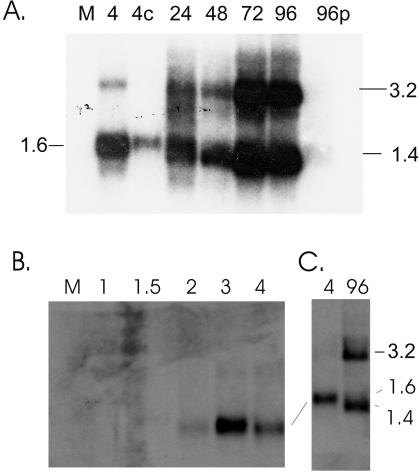
Northern blot analysis of UL34 transcription. Total cellular RNA was harvested from mock-infected cells (M) or infected cells at the indicated times postinfection. Following electrophoresis of equal amounts of RNA and transfer, blots were hybridized to a 32P-radiolabeled UL34 probe. (A) Expression of UL34 during a single-step replication cycle. 4c, RNA harvested at 4 hpi with infection and transcription occurring in the presence of cycloheximide; 96p, RNA harvested at 96 hpi in the presence of phosphonoformic acid. (B) Analysis of UL34 transcripts at early times of infection. (C) Comparison of RNA harvested 4 and 96 hpi. Equivalent amounts of RNA were electrophoresed in each lane, as demonstrated by ethidium bromide staining of rRNA (data not shown).
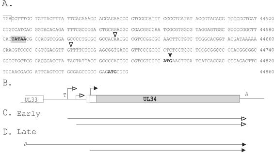
UL34 transcription initiation sites. (A) The nucleotide sequence of the 3′ end of UL33 and the 5′ end of UL34 is depicted (5). The UL33 stop codon is indicated by an open rectangle; the predicted early TATA box of UL34 is indicated with a gray rectangle. The early transcription initiation sites are indicated by open arrowheads; the late transcription initiation start site is indicated by a filled arrowhead. The first ATG codons in the early and late transcripts are indicated in bold; the ACG postulated to initiate translation of the 45-kDa UL34 protein is indicated by underlined letters. (B) Diagram of the UL34 gene with the TATA box indicated by a T, the early transcription initiation start sites indicated by open arrowheads, and the late transcription start site indicated by a filled arrowhead. The proposed open reading frames are depicted, with the dotted rectangle corresponding to translation initiation at ACG, the open rectangle corresponding to initiation of translation at the first methionine on the early UL34 transcripts, and the gray rectangle corresponding to the late open reading frame. All of the predicted proteins are in frame with each other and share the amino acid sequences indicated by the gray rectangle. T, TATA box; A, polyadenylation signal. (C) Schematic diagram of the early UL34 transcripts. (D) Schematic diagram of the late UL34 transcripts.
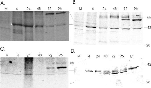
Analysis of UL34 protein expression. (A and B) Immunoprecipitation of [35S]methionine-labeled UL34 proteins. Mock-infected or HCMV-infected cells were radiolabeled with [35S]methionine at the indicated hours postinfection, immunoprecipitated using antisera to UL34, and analyzed by electrophoresis and autoradiography. Antisera from two different rabbits were used for the experiments illustrated. The three UL34 proteins are indicated by white circles in panel A; the 45-kDa UL34 protein is not easily visualized in panel B because of the exposure time. (C) Immunoprecipitation of [32P]-labeled proteins. (D) Western blot analysis of UL34 proteins. Mock or HCMV-infected cells were harvested at the indicated times postinfection and analyzed by Western blotting. The positions of the molecular weight markers are indicated; M mock-infected; 4, 24, 48, 72, and 96, hours postinfection; ivt, in vitro-translated pUL34.
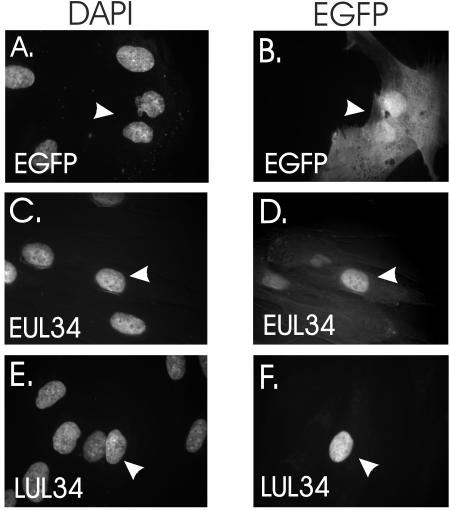
Analysis of the intracellular location of the early and late UL34 proteins. The early and late UL34 open reading frames were expressed as enhanced green fluorescent protein (EGFP) fusion proteins following transfection of human diploid fibroblasts with pBJ507 (9) or pBJ581, respectively. The late open reading frame was cloned using oligonucleotides 316 (5′ CCGGAATTCGAGATGCGTGACAACGTG 3′) and 317 (5′ CGCGTCGACTTATTGTTCTCCAGTGACG 3′), inserting the amplimer into pEGFP-C2 (Clontech). Constructs expressing fluorescent proteins were transfected into human diploid fibroblasts using Effectene (QIAGEN, Valencia, Calif.) as directed by the manufacturer. Following transfection, cells were stained with 4′,6′-diamidino-2-phenylindole. Fluorescence was visualized using a Nikon epifluorescence microscope. (A, C, and E) DAPI staining of cell nuclei; (B, D, and F) green fluorescence of EGFP; (A and B) EGFP; (C and D) the early pUL34-EGFP fusion protein; (E and F) the late pUL34-EGFP fusion protein. The transfected cells are indicated by arrows.
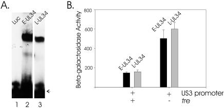
Comparison of the functions of the early and late UL34 proteins. (A) Electrophoretic mobility shift analysis of the DNA binding activity of in vitro-synthesized early UL34 (E-UL34) and late UL34 (L-UL34) proteins. The unbound DNA is indicated by an arrow; Luc is control luciferase protein. (B) Repression of the US3 promoter by the early and late UL34 proteins. Transient expression assays were used to measure the expression levels of a reporter construct expressing the lacZ gene under the control the US3 promoter and tre (1) or expression levels of a similar reporter construct that contains a mutant tre (pBJ214) (2) in the presence of a construct that expresses the early (E-UL34) (9) or late (L-UL34) form of the UL34 protein (pBJ575). pBJ575, which expresses the late UL34 open reading frame under the control of the HCMV major immediate-early promoter, was constructed by amplifying the late UL34 open reading frame using oligonucleotides 311 (5′ CGTCTAGAGGATCCACTTCTCCAACGACGATTC 3′) and 219 (5′ CTCGTCGACTTAAATACACAACGGGGTTATGG 3′) and inserting the amplimer into pBJ201 (2). This experiment was repeated multiple times with similar results; the results depicted are the averages from two experiments, with error bars representing one standard deviation. Background levels of beta-galactoside activity obtained from cells receiving the promoterless lacZ plasmid, pEQ3 (4), were subtracted from the experimental samples.
Similar articles
-
Identification of the functional domains of the essential human cytomegalovirus UL34 proteins.Virology. 2006 Sep 15;353(1):27-34. doi: 10.1016/j.virol.2006.05.019. Epub 2006 Jun 19. Virology. 2006. PMID: 16784766
-
Human cytomegalovirus UL34 early and late proteins are essential for viral replication.Viruses. 2014 Jan 28;6(2):476-88. doi: 10.3390/v6020476. Viruses. 2014. PMID: 24476753 Free PMC article.
-
Primate cytomegaloviruses encode and express an IL-10-like protein.Virology. 2000 Mar 15;268(2):272-80. doi: 10.1006/viro.2000.0195. Virology. 2000. PMID: 10704336
-
The proteins of human cytomegalovirus.Birth Defects Orig Artic Ser. 1984;20(1):49-62. Birth Defects Orig Artic Ser. 1984. PMID: 6329373 Review. No abstract available.
-
Human cytomegalovirus: host immune modulation by the viral US3 gene.Int J Biochem Cell Biol. 2009 Mar;41(3):503-6. doi: 10.1016/j.biocel.2008.10.012. Epub 2008 Oct 18. Int J Biochem Cell Biol. 2009. PMID: 18992841 Review.
Cited by
-
Dual analysis of the murine cytomegalovirus and host cell transcriptomes reveal new aspects of the virus-host cell interface.PLoS Pathog. 2013;9(9):e1003611. doi: 10.1371/journal.ppat.1003611. Epub 2013 Sep 26. PLoS Pathog. 2013. PMID: 24086132 Free PMC article.
-
Decoding human cytomegalovirus.Science. 2012 Nov 23;338(6110):1088-93. doi: 10.1126/science.1227919. Science. 2012. PMID: 23180859 Free PMC article.
-
pUL34 binding near the human cytomegalovirus origin of lytic replication enhances DNA replication and viral growth.Virology. 2018 May;518:414-422. doi: 10.1016/j.virol.2018.03.017. Epub 2018 Apr 5. Virology. 2018. PMID: 29626748 Free PMC article.
-
Insights into the Transcriptome of Human Cytomegalovirus: A Comprehensive Review.Viruses. 2023 Aug 8;15(8):1703. doi: 10.3390/v15081703. Viruses. 2023. PMID: 37632045 Free PMC article. Review.
-
UL34 Deletion Restricts Human Cytomegalovirus Capsid Formation and Maturation.Int J Mol Sci. 2022 May 21;23(10):5773. doi: 10.3390/ijms23105773. Int J Mol Sci. 2022. PMID: 35628580 Free PMC article.
References
-
- Chee, M. S., A. T. Bankier, S. Beck, R. Bohni, C. M. Brown, R. Cerny, T. Horsnell, C. A. Hutchison III, T. Kouzarides, J. A. Martignetti, E. Preddie, S. C. Satchwell, P. Tomlinson, K. M. Weston, and B. G. Barrell. 1990. Analysis of the protein-coding content of the sequence of human cytomegalovirus strain AD169. Curr. Top. Microbiol. Immunol. 154:125-170. - PubMed
Publication types
MeSH terms
Substances
LinkOut - more resources
Full Text Sources
