. 2004 Sep;16(9):2448-62.
doi: 10.1105/tpc.104.022608. Epub 2004 Aug 12.

Evidence for a direct link between glutathione biosynthesis and stress defense gene expression in Arabidopsis

Affiliations

Evidence for a direct link between glutathione biosynthesis and stress defense gene expression in Arabidopsis

Louise Ball et al. Plant Cell. 2004 Sep.

Abstract

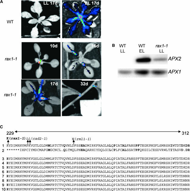
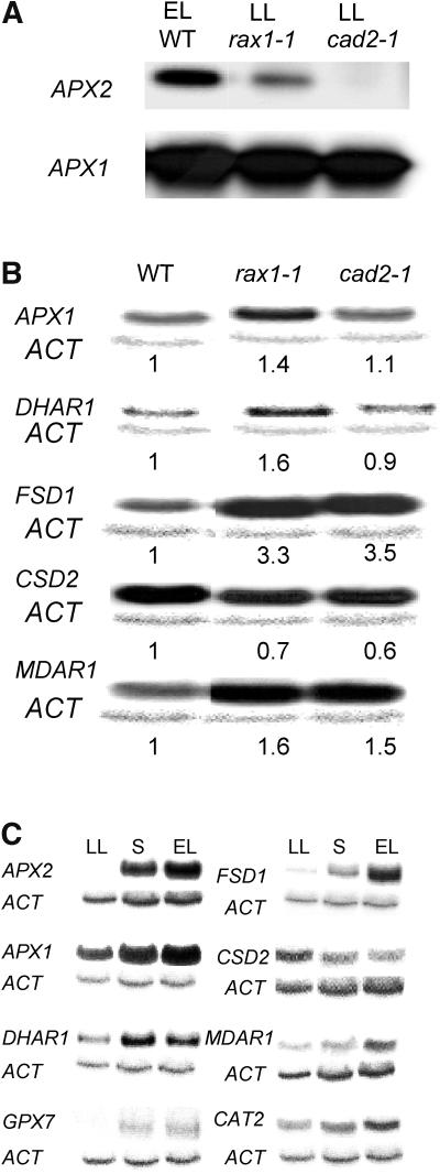
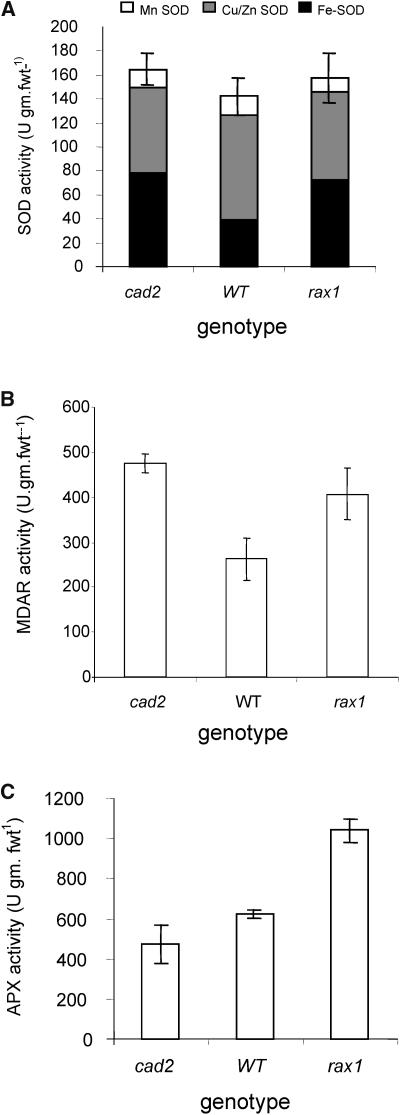
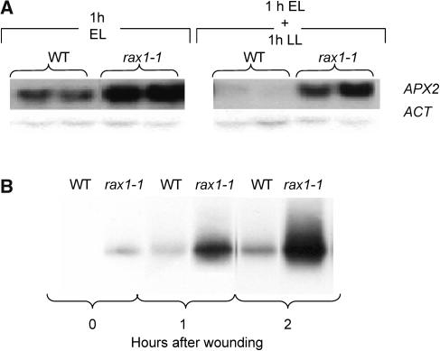
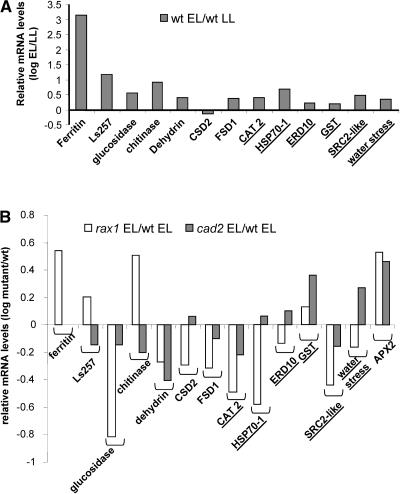
The mutant regulator of APX2 1-1 (rax1-1) was identified in Arabidopsis thaliana that constitutively expressed normally photooxidative stress-inducible ASCORBATE PEROXIDASE2 (APX2) and had >/=50% lowered foliar glutathione levels. Mapping revealed that rax1-1 is an allele of gamma-GLUTAMYLCYSTEINE SYNTHETASE 1 (GSH1), which encodes chloroplastic gamma-glutamylcysteine synthetase, the controlling step of glutathione biosynthesis. By comparison of rax1-1 with the GSH1 mutant cadmium hypersensitive 2, the expression of 32 stress-responsive genes was shown to be responsive to changed glutathione metabolism. Under photo-oxidative stress conditions, the expression of a wider set of defense-related genes was altered in the mutants. In wild-type plants, glutathione metabolism may play a key role in determining the degree of expression of defense genes controlled by several signaling pathways both before and during stress. This control may reflect the physiological state of the plant at the time of the onset of an environmental challenge and suggests that changes in glutathione metabolism may be one means of integrating the function of several signaling pathways.


Figures

Figure 1.
Figure 1.
Constitutive APX2 Expression in rax1-1, a Lesion in GSH1. (A) Typical false color image from a CCD camera of luciferase activity in a long day/low light–grown, 17-d-old wild-type APX2LUC Arabidopsis rosette before and after exposure to a 10-fold excess light stress for 45 min (LL 17d and EL 17d, respectively) and in long day–grown APX2LUC/rax1-1 plants at 10, 16, 17, and 32 d after germination. The background image of rosettes was taken when the plants were first placed under the camera, and the luciferase image was taken, after 3 min in the dark, for one minute with an aperture setting of 1.8. (B) PCR-based detection of APX2 transcript under nonstress conditions in rax1-1. Rapid amplification of cDNA 3′ends (3′RACE) PCR-amplified APX2 and APX1 cDNA, equivalent to 3 μg of total RNA, was separated by agarose gel electrophoresis, blotted, and hybridized to 32P-labeled gene-specific probes. In the lane with wild-type excess light (EL) APX2-specific PCR products 100-fold less volume was loaded as for the low light (LL) samples from wild-type or rax1-1 plants. The RNA was pooled from three separate plants harvested on two occasions (n = 6). Detection of APX1 was used here as a control for the PCR. (C) Alignment of the derived amino acid sequences of γ-ECS residues 229 to 312 from Arabidopsis (1) with that from trypanosome (2) and eight other plant species (3 to 10). This region includes the putative catalytic domain as defined by Leuder and Phillips (1996). The alignment between Arabidopsis and trypanosome with conserved residues in bold is from the same article. The asterisks indicate where the trypanosome sequence shows no homology with those from rat, yeast, and nematode. The rax1-1 (R229K) mutation is shown as well as cad2-1 (deletion P238, K239; Cobbett et al., 1998) and rml1-1 (D259N; Vernoux et al., 2000). The plant γ-ECS sequences are from Indian mustard (3; Y10848), Medicago truncatula (4; AF041340), pea (Pisum sativum) (5; AF128455), dwarf bean (Phaseolus vulgaris) (6; AF128454), rice (Oryza sativa) (7; AJ508916), maize (Zea mays) (8; AJ302783), tomato (Lycopersicon esculentum) (9; AF017983), and onion (Allium cepa) (10; AF401621).
Figure 2.
Figure 2.
Comparison of Antioxidant Defense Gene Expression in rax1-1 and cad2-1. (A) PCR-based detection of APX2 transcript under nonstress conditions in rax1-1 and cad2-1. 3′RACE PCR-amplified APX2 and APX1 cDNAs were detected by autoradiography as in the legend of Figure 1B. The wild-type control (EL WT) is from 10-fold excess light (EL) stressed plants, and loading of the APX2-specific product was 1% that of the volume loaded for the mutants. The APX1 is shown here as a PCR control for all cDNA samples. Each sample is pooled from six plants. LL, low light. (B) Steady state levels of transcripts of antioxidant defense genes in nonstressed wild type, cad2-1, and rax1-1. RNA gel blots loaded with 20 μg of total RNA were hybridized to specific probes and washed at high stringency, and images were developed using a phosphoimager (see Methods). The RNA preparations of rax1-1 and cad2-1 were the same as those in (A). The numbers below each row are values of signal relative to the wild type from this experiment, calculated from densitometer readings derived from the digitized images (Fryer et al., 2003). The band below each group is actin mRNA used as a loading control (see Methods). (C) rax1-1–responsive gene expression in excess light–exposed and systemic leaves. Steady state levels of transcripts encoded by antioxidant defense genes in the wild type (n = 3) partially exposed to excess light (15-fold). RNA was prepared from directly exposed leaves (EL) and nonexposed leaves that responded systemically to this stress (S; Karpinski et al., 1999), compared with low light nonstressed controls (LL). The amounts of RNA used and the processing of the blots was as in (B).
Figure 3.
Figure 3.
Antioxidant Enzyme Activities Are Altered in Nonstressed rax1-1 and cad2-1 Plants. Enzyme activities in cell-free extracts of leaves pooled from six plants, combined from two separate analyses. The assays were performed according to Jimenez et al. (1997). (A) Mn-, Cu/Zn-, and Fe-SOD. (B) MDAR. (C) APX.
Figure 4.
Figure 4.
Induction of APX2 in Stressed rax1-1 Plants. (A) Autoradiograph of RNA gel blot of APX2 transcript levels in two pools (n = 3 for each pool) of rax1-1 and wild-type plants after exposure to a fivefold excess light stress for 1 h (1h EL). The same number of excess-light stressed plants were left for another 1 h under ambient light conditions before harvesting (1h EL + 1h LL). Actin mRNA (ACT) is used here as a loading control. The protocols for producing the RNA gel blot autoradiographs were as described in the legend of Figure 2B. (B) Induction of APX2 expression in wounded wild-type and rax1-1 plants. Autoradiograph of APX2 cDNA amplified by 3′RACE PCR (see Methods), blotted, and hybridized to a gene-specific probe. Under the same conditions, APX3 transcript was constant in all samples (data not shown). Samples were taken before wounding (0 h) and then at 1 and 2 h after wounding. RNA was pooled from three wounded leaves from six plants.
Figure 5.
Figure 5.
Excess Light Stress Responses of APX2 and Other Stress-Responsive Genes in Wild-Type and Mutant Plants. (A) Transcript levels of stress defense genes in light-stressed (fivefold excess light for 45 minutes) wild-type plants relative to those in low light. Genes with altered expression in nonstressed rax1-1 and cad2-1 (Table 3) are shown. These are compared with light stress–responsive genes (underlined) whose transcripts were not affected in the mutants under nonstress conditions. All measurements were made using qRT-PCR (see Methods) using three determinations from each of two separate experiments (n = 6). The mean threshold cycle (Ct) values used to calculate the log ratio of excess light (EL)/low light (LL) transcript levels had standard deviations from the mean for each cDNA no greater than ±10%. The locus identifiers for the genes are given in Supplemental Table 2S online. (B) Transcript levels of stress defense genes after fivefold excess light exposure for 45 min in cad2-1 and rax1-1 relative to the wild type. The same genes were assayed by qRT-PCR as in (A) in two separate sets of determinations. The mean threshold cycle (Ct) values used to calculate the log ratio of mutant/wild-type transcript levels had standard deviations from the mean for each cDNA no greater than ±5%.
Figure 6.
Figure 6.
Growth of Avirulent Bacteria in rax1-1 and cad2-1 Plants. In two experiments on separate batches of plants, avirulent P. syringae pv tomato DC3000avrRpm1 was vacuum infiltrated into whole rosettes at day 0 (0 d postinoculation [dpi]). Note that two different inoculation densities were used in experiments 1 and 2. Bacteria were recovered from leaves 0 and 2 d (dpi) later and the increase in colony-forming units (cfu) per cm2 determined. The data shown are mean values (±se, n = 3).

Similar articles

Cited by

References

    1. Allan, A.C., and Fluhr, R. (1997). Two distinct sources of elicited reactive oxygen species in tobacco epidermal cells. Plant Cell 9, 1559–1572. - PMC - PubMed
    1. Asada, K. (1999). The water-water cycle in chloroplasts: Scavenging of active oxygen species and dissipation of excess photons. Annu. Rev. Plant Physiol. Mol. Biol. 50, 601–639. - PubMed
    1. Ball, L. (2001). Identification of Arabidopsis Mutants with Altered ASCORBATE PEROXIDASE II Gene Expression. PhD dissertation (Norwich, UK: University of East Anglia).
    1. Bechtold, U., Murphy, D.J., and Mullineaux, P.M. (2004). Arabidopsis peptide methionine sulfoxide reductase2 prevents cellular oxidative damage in long nights. Plant Cell 16, 908–919. - PMC - PubMed
    1. Berger, S., Mitchell-Olds, T., and Stotz, H.U. (2002). Local and differential control of vegetative storage protein expression in response to herbivore damage in Arabidopsis thaliana. Physiol. Plant 114, 85–91. - PubMed

Publication types

MeSH terms