

Transcranial magnetic stimulation (TMS) of the human frontal cortex: implications for repetitive TMS treatment of depression
- PMID: 15309043
- PMCID: PMC446221
Transcranial magnetic stimulation (TMS) of the human frontal cortex: implications for repetitive TMS treatment of depression
Abstract
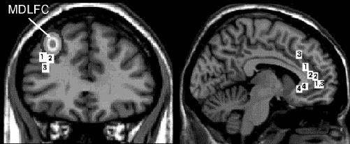
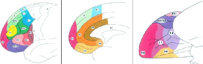
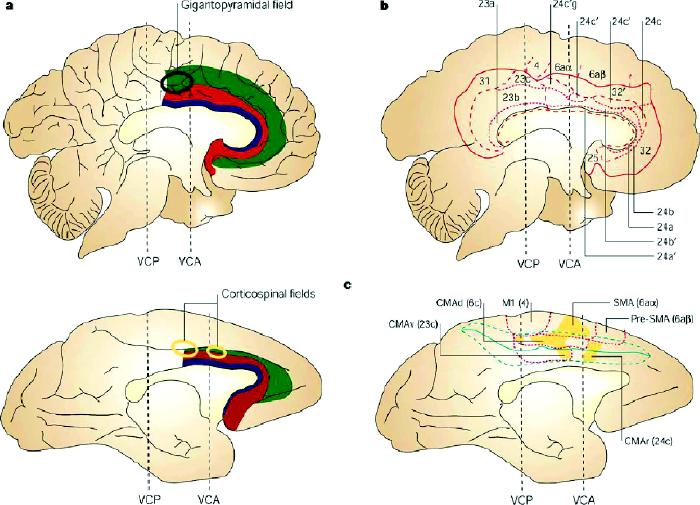
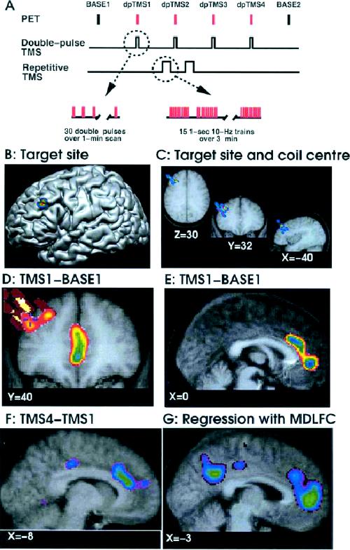
Repetitive transcranial magnetic stimulation (rTMS) is a noninvasive tool used to manipulate activity in specific neural circuits of the human brain. Clinical studies suggest that, in some patients with major depression, rTMS has the potential to alleviate symptoms that may be related to functional abnormalities in a frontocingulate circuit. This paper reviews the rationale for the use of rTMS in this context. The following topics are discussed: symptoms and cognition in major depression, with special emphasis on the initiation of speech; neuroimaging studies of depression; rTMS as treatment for depression; structure and function of the mid-dorsolateral frontal and anterior cingulate cortices; and combined TMS/positron emission tomography studies of frontocortical connectivity.
La stimulation magnétique transcrânienne répétitive (SMTr) est un outil non effractif utilisé pour manipuler l'activité de circuits neuraux précis du cerveau humain. Les études cliniques indiquent que chez certains patients aux prises avec une dépression majeure, la SMTr peut atténuer les symptômes pouvant être reliés à des anomalies fonctionnelles dans un circuit frontocingulaire. Cette communication passe en revue la justification de l'utilisation de la SMTr dans ce contexte. On aborde les sujets suivants : symptômes et cognition en cas de dépression majeure, avec accent sur l'initiation de la parole; étude de la dépression par neuro-imagerie; SMTr comme traitement de la dépression; structure et fonction des cortex cingulaires frontal et antérieur mésodorsolatéral; et étude de la connectivité frontocorticale par SMT et tomographie par émission de positrons combinées.
Figures
Similar articles
-
[Repetitive transcranial magnetic stimulation in major depression: response factor].Encephale. 2012 Sep;38(4):360-8. doi: 10.1016/j.encep.2011.08.004. Epub 2011 Oct 11. Encephale. 2012. PMID: 22980479 Review. French.
-
High (15 Hz) and low (1 Hz) frequency transcranial magnetic stimulation have different acute effects on regional cerebral blood flow in depressed patients.Psychol Med. 2003 Aug;33(6):997-1006. doi: 10.1017/s0033291703007955. Psychol Med. 2003. PMID: 12946084 Clinical Trial.
-
Changes in brain connectivity during a sham-controlled, transcranial magnetic stimulation trial for depression.J Affect Disord. 2018 May;232:143-151. doi: 10.1016/j.jad.2018.02.019. Epub 2018 Feb 21. J Affect Disord. 2018. PMID: 29494898 Free PMC article.
-
Default mode network mechanisms of transcranial magnetic stimulation in depression.Biol Psychiatry. 2014 Oct 1;76(7):517-26. doi: 10.1016/j.biopsych.2014.01.023. Epub 2014 Feb 5. Biol Psychiatry. 2014. PMID: 24629537 Free PMC article.
-
Neuroimaging and neuromodulation approaches to study eating behavior and prevent and treat eating disorders and obesity.Neuroimage Clin. 2015 Mar 24;8:1-31. doi: 10.1016/j.nicl.2015.03.016. eCollection 2015. Neuroimage Clin. 2015. PMID: 26110109 Free PMC article. Review.
Cited by
-
Antidepressant-like effects of nicotine and transcranial magnetic stimulation in the olfactory bulbectomy rat model of depression.Brain Res Bull. 2008 Sep 5;77(1):13-8. doi: 10.1016/j.brainresbull.2008.05.007. Epub 2008 Jun 26. Brain Res Bull. 2008. PMID: 18582540 Free PMC article.
-
Transcranial magnetic stimulation over the right temporoparietal junction influences the sense of agency in healthy humans.J Psychiatry Neurosci. 2020 Jul 1;45(4):271-278. doi: 10.1503/jpn.190099. J Psychiatry Neurosci. 2020. PMID: 32329986 Free PMC article.
-
Comparative effects of 10-Hz rTMS and iTBS on cortico-striatal connectivity in major depressive disorder: a sham-controlled study.Mol Psychiatry. 2025 Jun 28. doi: 10.1038/s41380-025-03091-0. Online ahead of print. Mol Psychiatry. 2025. PMID: 40581658
-
Hypomanic shift observed during rTMS treatment of patients with unipolar depressive disorder: four case reports.Ann Gen Psychiatry. 2013 Apr 24;12(1):12. doi: 10.1186/1744-859X-12-12. Ann Gen Psychiatry. 2013. PMID: 23618105 Free PMC article.
-
Efficacy of transcranial magnetic stimulation targets for depression is related to intrinsic functional connectivity with the subgenual cingulate.Biol Psychiatry. 2012 Oct 1;72(7):595-603. doi: 10.1016/j.biopsych.2012.04.028. Epub 2012 Jun 1. Biol Psychiatry. 2012. PMID: 22658708 Free PMC article.
References
-
- Blazer DG, Kessler RC, McGonagle KA, Swartz MS. The prevalence and distribution of major depression in a national community sample: the National Comorbidity Survey. Am J Psychiatry 1994;151:979-86. - PubMed
-
- Hirschfeld RM. Efficacy of SSRIs and newer antidepressants in severe depression: comparison with TCAs. J Clin Psychiatry 1999; 60:326-35. - PubMed
-
- American Psychiatric Association. The practice of electroconvulsive therapy. New York: Oxford University Press; 1990.
-
- Murphy FC, Sahakian BJ, Carroll RE. Cognitive impairment in depression: psychological models and clinical issues. In: Ebert D, Ebmeier KP, editors. Advances in biological psychiatry. Vol 19 of New models for depression series. New York : Karger; 1998. p. 1-33.
-
- Sobin C, Sackheim HA. Psychomotor symptoms of depression. Am J Psychiatry 1997;154:4-17. - PubMed
Publication types
MeSH terms
LinkOut - more resources
Full Text Sources
Other Literature Sources