

Review
doi: 10.1016/j.micinf.2004.05.002.
Endocytosis of influenza viruses
Affiliations
- PMID: 15310470
- PMCID: PMC2715838
- DOI: 10.1016/j.micinf.2004.05.002
Item in Clipboard
Review
Endocytosis of influenza viruses
Microbes Infect.
2004 Aug.
Abstract
Receptor-mediated endocytosis is known to play an important role in the entry of many viruses into host cells. However, the exact internalization mechanism has, until recently, remained poorly understood for many medically important viruses, including influenza. Developments in real-time imaging of single viruses as well as the use of dominant-negative mutants to selectively block specific endocytic pathways have improved our understanding of the influenza infection process.
Figures
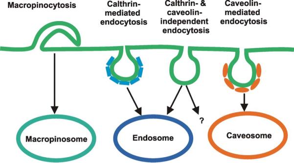
Possible endocytic pathways exploited by viruses for infection.
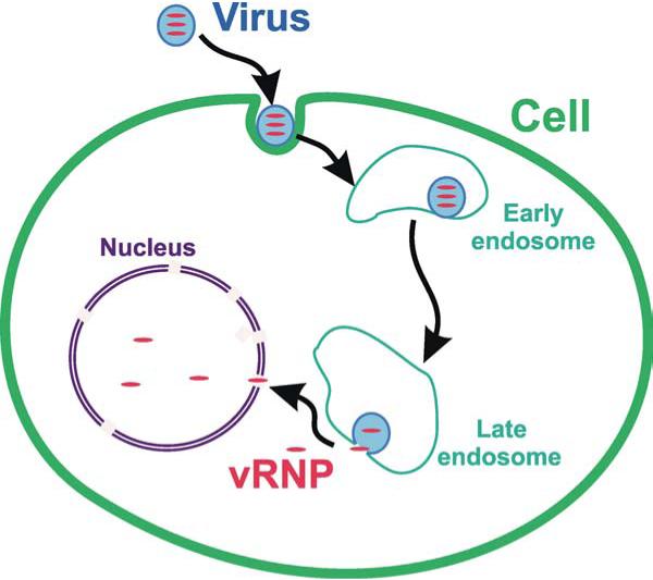
The influenza entry pathway. Influenza viruses bind to receptors containing sialic acid on the cell surface. Virus particles are then endocytosed and enter early endosomes. Subsequently the viruses are trafficked to late endosomes where the low pH triggers viral fusion. Viral ribonucleoproteins (vRNP) escape into the cytosol and are imported into the nucleus, where replication occurs.
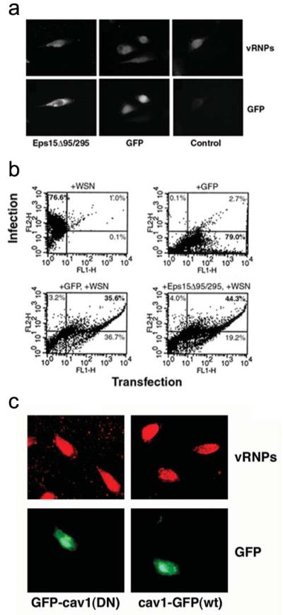
Influenza infection can occur independent of clathrin and caveolin. Cells were infected with ∼0.1 PFU of WSN and incubated at 37°C for 4 h. Infection was monitored with immunofluorescence using antibody against the influenza NP. Transfection was monitored with GFP fluorescence. (a,b) HeLa cells were transfected with GFP, or GFP-Eps15Δ95/295, or left untransfected before infection. (a) Immunofluorescence and GFP images of cells infected with WSN. (b) Fluorescence activated cell sorter analysis. Infection was monitored on the y-axis and transfection on the x-axis. Labels indicate the percentage of cells within each quadrant. (c) Immunofluorescence and GFP images of cells infected with WSN. HeLa cells were transfected with cav1-GFP(wt) (a fusion protein of GFP with wildtype caveolin-1) or GFP-cav1(DN) (a fusion protein of GFP with the dominant negative caveolin-1). (Figure adopted from Ref.[19])
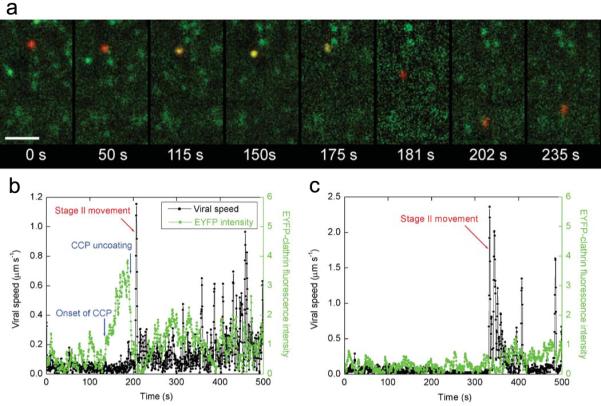
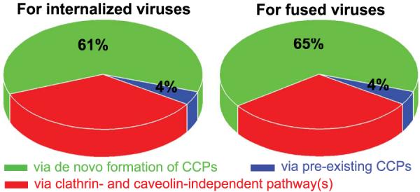
Endocytosis of individual viruses. (a) Snapshots of a virus internalized via a CCP. Scale bar: 10 μm. t = 0 s: the virus (red) binds to the cell. t = 115 s: a CCP labeled with EYFP (green) begins to form at the virus site. t = 175 s: the clathrin coat rapidly disassembles. t = 181 s, 202 s, and 235 s: transport of the virus on microtubules. (b) Time-trajectory of a virus internalized via de novo formation of a CCP. Black symbols are the instant velocity of the virus. Green symbols are the EYFP fluorescence intensity associated with the virus. Red arrow indicates the onset of microtubule-dependent movement, used as the signal for internalization. (c) The time-trajectories of a virus internalized without association with a clathrin-coated structure. Symbols are as defined in b. (Figure adopted from Ref. [29])
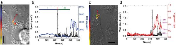
Post-endocytic trafficking and fusion of individual viruses. (a) The trajectory of a DiD-labeled virus inside a cell. The color of the trajectory codes time with the colored bar indicating a uniform time axis from 0 s (black) to 500 s (yellow). The red star indicates the fusion site. (b) Time trajectories of the velocity (black) and the DiD fluorescence intensity (blue) of the virus. The labels I, II, and III indicate stage I (actin-dependent), II (unidirectional, microtubule- and dynein-dependent), and III (bidirectional, microtubule-dependent) movement, respectively. The fluorescence dequenching signal of the lipophilic dye, DiD, near 400 s indicates viral fusion. (c) The trajectory of a Cy3/CypHer5-labeled virus inside a cell. Color coding of the trajectory is similar to that in (b). The green star indicates the initial acidification site (to pH ∼6). (d) Time trajectories of the velocity (black) and the fluorescent emission ratio of CypHer5 (a pH-dependent dye) and Cy3 (a pH-independent dye) (red) of the virus. The pH scale is labeled according to calibration measurements performed on virus particles in vitro. Scale bars: 10 μm. (Figure adopted from Ref.[28])
References
-
- Sieczkarski SB, Whittaker GR. Dissecting virus entry via endocytosis. J. Gen. Virol. 2002;83:1535–1545. - PubMed
-
- Pelkmans L, Helenius A. Insider information: what viruses tell us about endocytosis. Curr. Opin. Cell. Biol. 2003;15:414–422. - PubMed
-
- Nichols BJ, Lippincott-Schwartz J. Endocytosis without clathrin coats. Trends Cell Biol. 2001;11:406–412. - PubMed
-
- Conner SD, Schmid SL. Regulated portals of entry into cells. Nature. 2003;422:37–44. - PubMed
-
- Pearse BMF, Smith CJ, Owen DJ. Clathrin coat construction in endocytosis. Curr. Opin. Struct. Biol. 2000;10:220–228. - PubMed
Publication types
MeSH terms
Substances
Grants and funding
LinkOut - more resources
Full Text Sources
Other Literature Sources
Medical
