

FRD3 controls iron localization in Arabidopsis
- PMID: 15310833
- PMCID: PMC523319
- DOI: 10.1104/pp.104.045633
FRD3 controls iron localization in Arabidopsis
Abstract
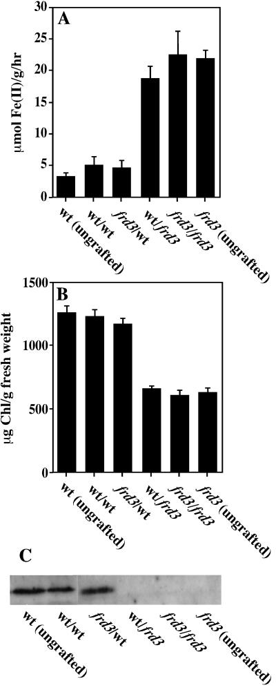
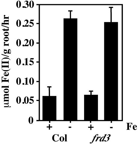
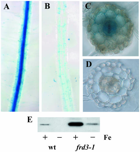
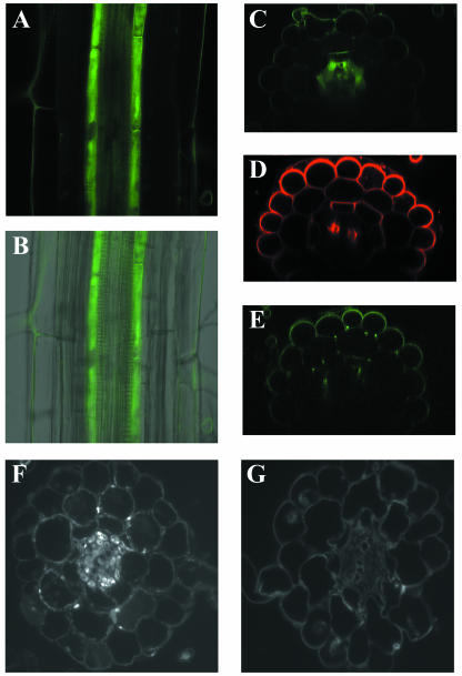
The frd3 mutant of Arabidopsis exhibits constitutive expression of its iron uptake responses and is chlorotic. These phenotypes are consistent with defects either in iron deficiency signaling or in iron translocation and localization. Here we present several experiments demonstrating that a functional FRD3 gene is necessary for correct iron localization in both the root and shoot of Arabidopsis plants. Reciprocal grafting experiments with frd3 and wild-type Arabidopsis plants reveal that the phenotype of a grafted plant is determined by the genotype of the root, not by the genotype of the shoot. This indicates that FRD3 function is root-specific and points to a role for FRD3 in delivering iron to the shoot in a usable form. When grown under certain conditions, frd3 mutant plants overaccumulate iron in their shoot tissues. However, we demonstrate by direct measurement of iron levels in shoot protoplasts that intracellular iron levels in frd3 are only about one-half the levels in wild type. Histochemical staining for iron reveals that frd3 mutants accumulate high levels of ferric iron in their root vascular cylinder, the same tissues in which the FRD3 gene is expressed. Taken together, these results clearly indicate a role for FRD3 in iron localization in Arabidopsis. Specifically, FRD3 is likely to function in root xylem loading of an iron chelator or other factor necessary for efficient iron uptake out of the xylem or apoplastic space and into leaf cells.
Figures

Similar articles
-
The FRD3-mediated efflux of citrate into the root vasculature is necessary for efficient iron translocation.Plant Physiol. 2007 May;144(1):197-205. doi: 10.1104/pp.107.097162. Epub 2007 Mar 9. Plant Physiol. 2007. PMID: 17351051 Free PMC article.
-
FRD3, a member of the multidrug and toxin efflux family, controls iron deficiency responses in Arabidopsis.Plant Cell. 2002 Aug;14(8):1787-99. doi: 10.1105/tpc.001495. Plant Cell. 2002. PMID: 12172022 Free PMC article.
-
The FRD3 citrate effluxer promotes iron nutrition between symplastically disconnected tissues throughout Arabidopsis development.Plant Cell. 2011 Jul;23(7):2725-37. doi: 10.1105/tpc.111.088088. Epub 2011 Jul 8. Plant Cell. 2011. PMID: 21742986 Free PMC article.
-
BYPASS1: how a tiny mutant tells a big story about root-to-shoot signaling.J Integr Plant Biol. 2010 Jan;52(1):77-85. doi: 10.1111/j.1744-7909.2010.00902.x. J Integr Plant Biol. 2010. PMID: 20074142 Review.
-
Directing iron transport in dicots: regulation of iron acquisition and translocation.Curr Opin Plant Biol. 2017 Oct;39:106-113. doi: 10.1016/j.pbi.2017.06.014. Epub 2017 Jul 6. Curr Opin Plant Biol. 2017. PMID: 28689052 Review.
Cited by
-
How a microbial drug transporter became essential for crop cultivation on acid soils: aluminium tolerance conferred by the multidrug and toxic compound extrusion (MATE) family.Ann Bot. 2010 Jul;106(1):199-203. doi: 10.1093/aob/mcq115. Epub 2010 May 28. Ann Bot. 2010. PMID: 20511585 Free PMC article.
-
Plant Frataxin in Metal Metabolism.Front Plant Sci. 2018 Nov 21;9:1706. doi: 10.3389/fpls.2018.01706. eCollection 2018. Front Plant Sci. 2018. PMID: 30519254 Free PMC article.
-
Facing the challenges of Cu, Fe and Zn homeostasis in plants.Nat Chem Biol. 2009 May;5(5):333-40. doi: 10.1038/nchembio.166. Nat Chem Biol. 2009. PMID: 19377460 Free PMC article. Review.
-
Large expression differences in genes for iron and zinc homeostasis, stress response, and lignin biosynthesis distinguish roots of Arabidopsis thaliana and the related metal hyperaccumulator Thlaspi caerulescens.Plant Physiol. 2006 Nov;142(3):1127-47. doi: 10.1104/pp.106.082073. Epub 2006 Sep 22. Plant Physiol. 2006. PMID: 16998091 Free PMC article.
-
The bHLH transcription factor POPEYE regulates response to iron deficiency in Arabidopsis roots.Plant Cell. 2010 Jul;22(7):2219-36. doi: 10.1105/tpc.110.074096. Epub 2010 Jul 30. Plant Cell. 2010. PMID: 20675571 Free PMC article.
References
-
- Ausubel FM, Brent R, Kingston RE, Moore DD, Seidman JG, Smith JA, Struhl K (2004) Current Protocols in Molecular Biology. John Wiley & Sons, New York
-
- Clough SJ, Bent AF (1998) Floral dip: a simplified method for Agrobacterium-mediated transformation of Arabidopsis thaliana. Plant J 16: 735–743 - PubMed
-
- Curie C, Panaviene Z, Loulergue C, Dellaporta SL, Briat J-F, Walker EL (2001) Maize yellow stripe1 encodes a membrane protein directly involved in Fe(III) uptake. Nature 409: 346–349 - PubMed
Publication types
MeSH terms
Substances
LinkOut - more resources
Full Text Sources
Medical
Molecular Biology Databases
