. 2004 Aug 24;101(34):12543-8.
doi: 10.1073/pnas.0404700101. Epub 2004 Aug 13.

Derivation of midbrain dopamine neurons from human embryonic stem cells

Affiliations

Derivation of midbrain dopamine neurons from human embryonic stem cells

Anselme L Perrier et al. Proc Natl Acad Sci U S A. .

Abstract

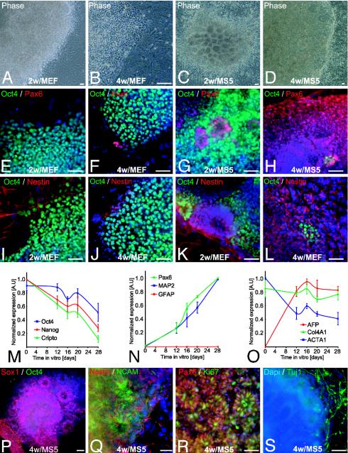
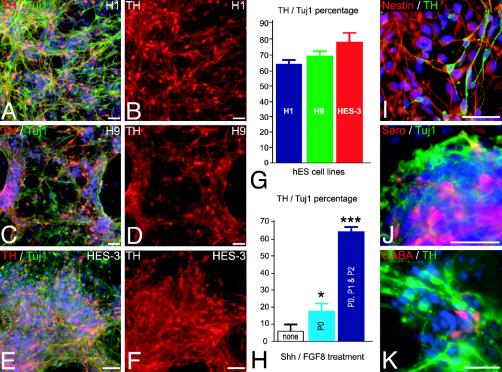
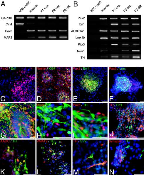
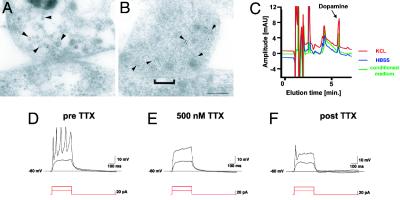
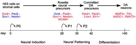
Human embryonic stem (hES) cells are defined by their extensive self-renewal capacity and their potential to differentiate into any cell type of the human body. The challenge in using hES cells for developmental biology and regenerative medicine has been to direct the wide differentiation potential toward the derivation of a specific cell fate. Within the nervous system, hES cells have been shown to differentiate in vitro into neural progenitor cells, neurons, and astrocytes. However, to our knowledge, the selective derivation of any given neuron subtype has not yet been demonstrated. Here, we describe conditions to direct hES cells into neurons of midbrain dopaminergic identity. Neuroectodermal differentiation was triggered on stromal feeder cells followed by regional specification by means of the sequential application of defined patterning molecules that direct in vivo midbrain development. Progression toward a midbrain dopamine (DA) neuron fate was monitored by the sequential expression of key transcription factors, including Pax2, Pax5, and engrailed-1 (En1), measurements of DA release, the presence of tetrodotoxin-sensitive action potentials, and the electron-microscopic visualization of tyrosinehydroxylase-positive synaptic terminals. High-yield DA neuron derivation was confirmed from three independent hES and two monkey embryonic stem cell lines. The availability of unlimited numbers of midbrain DA neurons is a first step toward exploring the potential of hES cells in preclinical models of Parkinson's disease. This experimental system also provides a powerful tool to probe the molecular mechanisms that control the development and function of human midbrain DA neurons.


Figures

Fig. 1.
Fig. 1.
Stromal feeder-induced neural differentiation of hES cells. (AD) Representative phase contrast images of hES cells (line H1) cocultured for 2 or 4 weeks on MEF (A and B) or MS5 (C and D). (EH) Pax6 and Oct4 expression in hES cells differentiated on MEF or MS5. (IL) Oct4 and nestin expression in hES cells on MEF or MS5. (MO) Semiquantitative RT-PCR analysis for genes characteristic of undifferentiated ES cells (M); neural lineage (N); and non-neural, endodermal, and mesodermal differentiation (O). Data are presented as normalized values (see Materials and Methods) and derived from three independent experiments, line H1, days 0–28. GFAP, glial fibrillary acidic protein; AFP, α-fetoprotein; Col4A1, collagen type IV α; ACTA1, α1-actin. (PS) Characterization of hES-derived rosettes on MS5. Expression of Sox1 (P) and nestin and NCAM (Q) confirmed the neural identity of the rosettes. (R) Cell proliferation was assessed by Ki67 labeling. (S) Tuj1-positive cells were observed surrounding rosettes. Dapi, 4′,6-diamidino-2-phenylindole. (Scale bars, 50 μm.)
Fig. 2.
Fig. 2.
High-yield derivation of TH-positive neurons from hES-derived rosettes. (AF) Representative images of TH/Tuj1-positive (red, TH; green, Tuj1) neurons derived from the hES cell lines H1 (A and B), H9 (C and D), and HES-3 (E and F). (G) Percentage of hES-derived TH neurons for each of the hES cell lines upon differentiation at passage 2. (H) Percentage of TH neurons upon differentiation of rosettes obtained in the absence of SHH and FGF8 (none, white bar), in the presence of SHH and FGF8 during passage 0 (P0, light blue bar), or in the presence of SHH and FGF8 during passages 0, 1, and 2 (P0, P1 and P2, dark blue bar). *, P < 0.05; ***, P < 0.001 compared to none. (I) Most cells negative for TH (green) expressed the neural precursor marker nestin (red). Other neuronal subtypes present were serotonergic (Sero, red) (J) and GABAergic neurons (red) (K). (Scale bars, 50 μm.)
Fig. 3.
Fig. 3.
Phenotypic characterization of midbrain dopaminergic differentiation. (A and B) RT-PCR analyses of undifferentiated hES cells, passage 0 cells (day 28, Rosette), passage 1 cells during cell expansion (day 35, P1 exp.), passage 2 cells during cell expansion (day 42, P2 exp.), and passage 2 cells during cell differentiation (day 50, P2 diff.). Shown are ES-cell, neural, and neuronal markers (A) and markers of midbrain DA neuron development (B). ALDH1A1, aldehyde dehydrogenase 1; Nurr1, nuclear orphan receptor 1. (C) Pax2-positive cells (red) at passage 1 did not coexpress En1 (green). (D and E) Cells at passage 2 expressed Ki67 (green) and nestin (red) (D) and coexpressed Pax2 (red) and En1 (green) (E). (F) Pax6 (red) and Lmx (green) expression in passage 2 expansion cells. (GN) Differentiated cells at passage 2: TH/Tuj1-positive (red, TH; green, Tuj1) neurons with radially oriented processes emanating from a rosette (G); Pax2- or Pax5-positive (red) immature precursors next to TH-positive (green) neurons (H and I); En1-p (green) cells coexpressing TH (red) (J); TH-positive (red) cells coexpressing AADC (green) (K) or VMAT2 (L); TH-positive neurons expressing the synaptic markers SV2 (green) (M) or synapsin (red) (N). Similar results were obtained with three independent hES lines. CE show H9-derived cells; GI and M show H1-derived cells; and F, JL, and N show HES-3-derived cells. (Scale bars, 50 μm.)
Fig. 4.
Fig. 4.
Ultrastructural and functional characterization of hES-derived DA neurons. (A and B) Electron micrographs of TH ImmunoGold-labeling (5 nm) at synaptic contacts formed between neurons derived from hES cells. Gold particles are associated with small vesicles presumably containing neurotransmitters located at the presynaptic terminal (arrowheads). (A and B) Two examples of synapses are shown: A shows recently formed immature contact with a faint membrane density and few synaptic vesicles, and B shows a more differentiated synaptic contact with a distinct postsynaptic density (bracket) and a larger number of vesicles clustered in the proximity of the cell membrane. (Scale bar, 125 nm.) (C) Representative HPLC chromatogram showing high levels of DA in the medium of rosette-derived neurons (green line: medium conditioned for 24 h, 751 ± 239 pg/ml). Relatively low levels of basal DA release were detected (blue line: exposure to buffer for 15 min, 147 ± 78 pg/ml) in contrast to very high levels of DA after 15 min of KCL-evoked depolarization (red line: 1,283 ± 421 pg/ml). Data were derived from three independent experiments. Rosette-derived neurons were patched in the whole-cell configuration in current clamp mode. (D) Action potentials were evoked by depolarizing currents at a threshold of –40 mV. (E and F) Action potentials were tetrodotoxin (TTX)-sensitive.
Fig. 5.
Fig. 5.
Developmental progression, in vitro neural induction, neural patterning, and differentiation were monitored by the expression of specific lineage markers in hES-derived progeny as indicated. Neural induction was achieved under serum-free conditions on stromal feeder cells. At passages 1 and 2, hES-derived rosettes were plated on a polyornithine/laminin substrate in the absence of feeder cells. Neural patterning occurred in the presence of SHH/FGF8 (days 28–42). Neural differentiation required withdrawal of SHH/FGF8 and exposure to various factors that increase dopaminergic differentiation and survival (see text for details).

Similar articles

Cited by

References

    1. Thomson, J. A., Itskovitz-Eldor, J., Shapiro, S. S., Waknitz, M. A., Swiergiel, J. J., Marshall, V. S. & Jones, J. M. (1998) Science 282, 1145–1147. - PubMed
    1. Lee, S.-H., Lumelsky, N., Studer, L., Auerbach, J. M. & McKay, R. D. G. (2000) Nat. Biotechnol. 18, 675–679. - PubMed
    1. Kawasaki, H., Mizuseki, K., Nishikawa, S., Kaneko, S., Kuwana, Y., Nakanishi, S., Nishikawa, S. I. & Sasai, Y. (2000) Neuron 28, 31–40. - PubMed
    1. Wichterle, H., Lieberam, I., Porter, J. A. & Jessell, T. M. (2002) Cell 110, 385–397. - PubMed
    1. Brustle, O., Jones, K. N., Learish, R. D., Karram, K., Choudhary, K., Wiestler, O. D., Duncan, I. D. & McKay, R. G. (1999) Science 285, 754–756. - PubMed

Publication types