. 2004 Dec 1;384(Pt 2):295-305.
doi: 10.1042/BJ20040955.

Kinase-independent transcriptional co-activation of peroxisome proliferator-activated receptor alpha by AMP-activated protein kinase

Affiliations

Kinase-independent transcriptional co-activation of peroxisome proliferator-activated receptor alpha by AMP-activated protein kinase

Myriam Bronner et al. Biochem J. .

Abstract

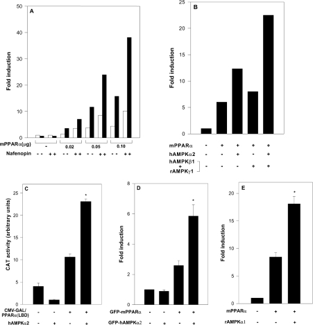
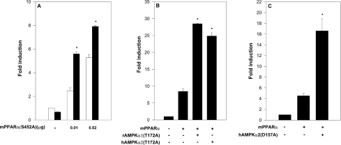
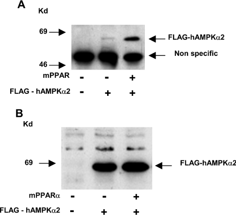
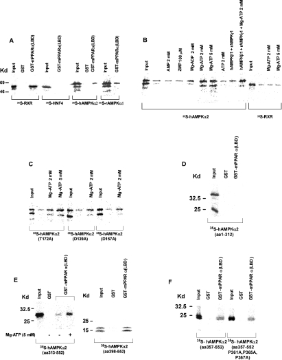
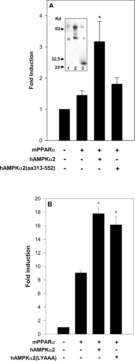
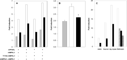
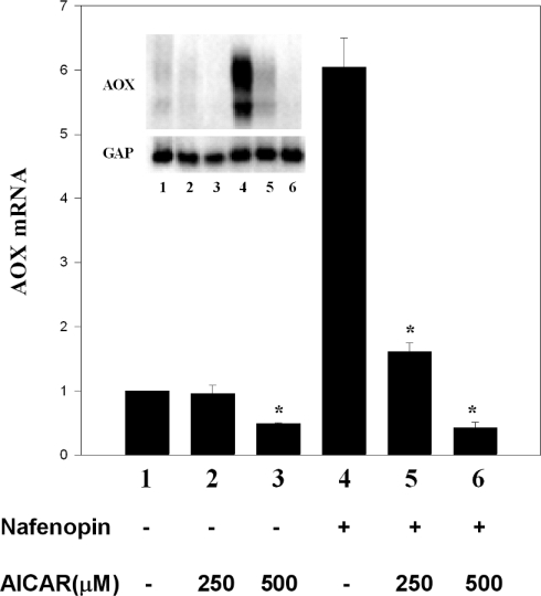
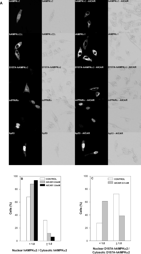
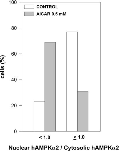
AMPK (AMP-activated protein kinase) responds to intracellular ATP depletion, while PPARalpha (peroxisome proliferator-activated receptor alpha) induces the expression of genes coding for enzymes and proteins involved in increasing cellular ATP yields. PPARalpha-mediated transcription is shown here to be co-activated by the alpha subunit of AMPK, as well as by kinase-deficient (Thr172Ala) and kinase-less (Asp157Ala, Asp139Ala) mutants of AMPKalpha. The Ser452Ala mutant of mPPARalpha mutated in its putative consensus AMPKalpha phosphorylation site is similarly co-activated by AMPKalpha. AMPKalpha or its kinase-less mutants bind to PPARalpha; binding is increased by MgATP, to a lesser extent by MgADP, but not at all by AMP or ZMP [AICAR (5-aminoimidazole-4-carboxamide ribonucleoside) monophosphate]. ATP-activated binding of AMPKalpha to PPARalpha is mediated primarily by the C-terminal regulatory domain of AMPKalpha. PPARalpha co-activation by AMPKalpha may, however, require its secondary interaction with the N-terminal catalytic domain of AMPKalpha, independently of its kinase activity. While AMPK catalytic activity is activated by AICAR, PPARalpha co-activation and PPARalpha-controlled transcription are robustly inhibited by AICAR, with concomitant translocation of nuclear AMPKalpha or its kinase-less mutants to the cytosol. In conclusion, AMPKalpha, independently of its kinase activity, co-activates PPARalpha both in primary rat hepatocytes and in PPARalpha-transfected cells. The kinase and transcriptional co-activation modes of AMPKalpha are both regulated by the cellular ATP/AMP ratio. Co-activation of PPARalpha by AMPKalpha may transcriptionally complement AMPK in maintaining cellular ATP status.


Figures

Figure 1
Figure 1. Activation of PPARα by AMPK
Cells were transfected with expression plasmids for mPPARα and AMPK subunits as described in the Experimental section. Fold induction represents CAT activity (means±S.E.M. for triplicate plates, or means of duplicate plates differing by no more than 10%) normalized by CMV-β-galactosidase and further by CAT activity of pSG5-transfected cells. (A) 293 cells were transfected with the reporter plasmid rAOX(PPRE)-CAT and with the expression plasmid for mPPARα (0.02–0.1 μg) as indicated, and co-transfected with the expression plasmid for hAMPKα2 (2.0 μg) (■) or the control plasmid pcDNA3 (□). Following transfection, cells were incubated in the absence or presence of 20 μM nafenopin as indicated. (B) 293 cells were transfected with the reporter plasmid rAOX(PPRE)-CAT and the expression plasmid for mPPARα (0.1 μg), and co-transfected with expression plasmids for hAMPKα2 (2.0 μg), hAMPKβ1 (1.0 μg) and/or rAMPKγ1 (1.0 μg) as indicated. Following transfection, cells were incubated in the presence of 20 μM nafenopin. (C) HeLa cells were transfected with the reporter plasmid pG5-CAT and the expression plasmid for GAL4 (0.4 μg) or GAL4-mPPARα(LBD) (0.4 μg), and co-transfected with the expression plasmid for hAMPKα2 (2.0 μg) as indicated. Following transfection, cells were incubated in the presence of 100 μM nafenopin. (D) HeLa cells were transfected with the reporter plasmid rAOX(PPRE)-CAT and co-transfected with the expression plasmid for GFP–mPPARα (0.02 μg) with or without the expression plasmid for GFP–hAMPKα2 (2.0 μg) as indicated. Following transfection, cells were incubated in the presence of 10 μM nafenopin. (E) 293 cells were transfected with the reporter plasmid rAOX(PPRE)-CAT and co-transfected with the expression plasmids for mPPARα (0.05 μg) and/or rAMPKα1 (2.0 μg) as indicated. Following transfection, cells were incubated in the presence of 10 μM nafenopin. Significance of differences: *P<0.05 compared with no added AMPKα.
Figure 2
Figure 2. Activation of PPARα by AMPKα is independent of its kinase activity
Cells were transfected with the reporter plasmid rAOX(PPRE)-CAT and co-transfected with the indicated expression plasmids for mPPARα and AMPKα as described in the Experimental section. Fold induction represents CAT activity (means±S.E.M. for triplicate plates, or means of duplicate plates differing by no more than 10%) normalized by CMV-β-galactosidase and normalized further by CAT activity of pSG5-transfected cells. (A) 293 cells were transfected with the expression plasmid for mPPARα(S452A) (0–0.02 μg) as indicated, in the absence (□) or presence (■) of transfected hAMPKα2 (2.0 μg). Following transfection, cells were incubated in presence of 10 μM nafenopin. Significance: *P<0.05 compared with no added AMPKα. (B) 293 cells were transfected with the expression plasmid for mPPARα (0.05 μg), and co-transfected with expression plasmids for rAMPKα1(T172A) (2.0 μg) or hAMPKα2(T172A) (2.0 μg) as indicated. Following transfection, cells were incubated in the presence of 10 μM nafenopin. Significance: *P<0.05 compared with no added rAMPKα(T172A). (C) COS-7 cells were transfected with the expression plasmid for hAMPKα2(D157A) (2.0 μg) as indicated. Following transfection, cells were incubated in the presence of 10 μM nafenopin. Significance: *P<0.05 compared with no added hAMPKα2(D157A).
Figure 3
Figure 3. PPARα–AMPKα interaction in vivo
293 cells were transfected with expression plasmids for mPPARα (0.2 μg) and FLAG–hAMPKα2 (1.0 μg) as indicated. (A) mPPARα complexes were immunoprecipitated with anti-mPPARα antiserum, immunoprecipitates were resolved by SDS/PAGE and FLAG–hAMPKα2 complexes were characterized by Western blotting using anti-FLAG M2 monoclonal antiserum as described in the Experimental section. (B) Expression of FLAG–hAMPKα2 was monitored by Western blotting of cell lysates using anti-FLAG antisera. Kd, kDa.
Figure 4
Figure 4. PPARα–AMPKα interaction in vitro
GST or GST–mPPARα(LBD) was tethered to glutathione–agarose beads and incubated in the presence of the indicated 35S-labelled proteins as described in the Experimental section. Input represents 20% of the respective 35S-labelled proteins subjected to pull-down. (A) Interaction of PPARα with hAMPKα2 and rAMPKα1. (B) Activation by MgATP of the PPARα–hAMPKα2 interaction. (C) PPARα interaction with hAMPKα2 mutants. (D) PPARα interaction with AMPKα2-(1–312). (E) PPARα interaction with hAMPKα2-(313–552) and hAMPKα2-(398–552). (F) PPARα interaction with hAMPKα2-(357–552) and hAMPKα2-(357–552)(P361A, P365A, P367A). Kd, kDa.
Figure 5
Figure 5. PPARα co-activation by AMPKα is independent of the LXXLL domain of AMPKα
(A) 293 cells were transfected with the reporter plasmid rAOX(PPRE)-CAT and the expression plamid for RXRα (0.03 μg), and co-transfected with the expression plasmid for mPPARα (0.03 μg), hAMPKα2 (5.0 μg) or hAMPKα2-(313–552) (5.0 μg) as indicated. Following transfection, cells were incubated for 17 h in the presence of 20 μM nafenopin. Fold induction represents CAT activity (means±S.E.M. of duplicate plates differing by no more than 10% from five independent experiments) normalized by CMV-β-galactosidase and further normalized by CAT activity of pSG5-transfected cells. Significance: *P<0.05 compared with no added hAMPKα2. Expression of hAMPKα2 and hAMPKα2-(313–552) was verified by Western blot analysis of the cell lysate using sheep anti-hAMPKα2 antibody (inset): lane 1, pcDNA3 (5.0 μg); lane 2, hAMPKα2 (5.0 μg); lane 3, hAMPKα2-(313–552) (5.0 μg). Kd, kDa. (B) COS-7 cells were transfected with the reporter plasmid rAOX(PPRE)-CAT and the expression plasmid for mPPARα (0.05 μg), and co-transfected with hAMPKα2 (2.0 μg) or hAMPKα2(L204YAAA) (2.0 μg) as indicated. Following transfection, cells were incubated in the presence of 10 μM nafenopin. Fold induction represents CAT activity (means±S.E.M. for triplicate plates or means of duplicate plates differing by no more than 10%) normalized by CMV-β-galactosidase and further normalized by CAT activity of pSG5-transfected cells. Significance: *P<0.05 compared with no added AMPKα.
Figure 6
Figure 6. Inhibition of PPARα by AICAR
Cells were transfected with the reporter plasmid rAOX(PPRE)-CAT and co-transfected with the indicated expression plasmids for mPPARα and AMPKα mutants as described in the Experimental section. Fold induction represents CAT activity (means+S.E.M. for triplicate plates or means of duplicate plates differing by no more than 10%) normalized by CMV-β-galactosidase and further normalized by CAT activity of pSG5-transfected cells. (A) 293 cells were transfected with the reporter plasmid rAOX(PPRE)-CAT and the expression plasmids for mPPARα (0.1 μg) or mPPARα(S452A) (0.1 μg), and co-transfected with expression plasmids for rAMPKα1 (1.0 μg), rAMPKα1(T172A) (1.0 μg) and/or hAMPKβ1 (1.0 μg) plus rAMPKγ1 (1.0 μg) as indicated. CAT activity was determined in cells incubated overnight in the absence of added ligand (grey bars), as well as in cells incubated for an additional 7 h with 30 μM nafenopin in the absence (□) or presence (■) of added 250 μM AICAR. Note the nafenopin-dependent activity during the 7 h incubation period in the absence and presence of AICAR. (B) 293 cells were transfected with the reporter plasmid rAOX(PPRE)-CAT and the expression plasmid for mPPARα (0.02 μg), and co-transfected with expression plasmid for hAMPKα2(D157A) (2.0 μg). CAT activity was determined in cells incubated overnight in the absence of added ligand (grey bar), as well as in cells incubated for an additional 6 h with 10 μM nafenopin in the absence (□) or presence (■) of added 500 μM AICAR. Note the nafenopin-dependent activity during the 6 h incubation period in the absence and presence of AICAR. Significance: *P<0.05 compared with no added AICAR. (C) COS-7 cells were transfected with the reporter plasmid rAOX(PPRE)-CAT and the expression plasmid for mPPARα (0.05 μg). CAT activity was determined in cells incubated overnight in the absence of added ligands and incubated for an additional 7 h in the presence of DMSO vehicle, 50 nM iloprost, 10 μM Wy-14,643 or 10 μM nafenopin as indicated, in the absence (white bars) or presence of 200 (hatched bars), 250 (black bars) or 600 (cross-hatched bars) μM AICAR.
Figure 7
Figure 7. Inhibition by AICAR of the expression of PPARα-responsive genes
Rat primary hepatocytes were prepared and cultured in triplicate on collagen gels as described in the Experimental section. Overnight-cultured cells were treated with 250 μM or 500 μM AICAR for 1 h and then incubated for a further 24 h in the absence or presence of 30 μM nafenopin as indicated. Peroxisomal AOX was probed using a 1756 bp peroxisomal AOX cDNA. Bars represent the AOX transcript normalized by glyceraldehyde-3-phosphate dehydrogenase (GAP) mRNA. Values are means±S.E.M. of triplicate plates; *P<0.05 compared with no added AICAR. The results are representative of three similar experiments.
Figure 8
Figure 8. Displacement by AICAR of transfected nuclear AMPKα
(A) HeLa cells were cultured on glass coverslips and transfected overnight with expression plasmids for GFP–rAMPKα1 (6.0 μg), GFP–hAMPKα2 (6.0 μg), hAMPKβ1 (4.0 μg), rAMPKγ1 (4.0 μg), GFP–hAMPKα2(D157A) (6.0 μg), GFP–mPPARα (4.0 μg) or GFP–human p53 (2.0 μg) as indicated. Following transfection, cells were incubated for 6 h in the absence or presence of 0.5 mM AICAR as indicated, fixed, permeabilized, further incubated with the respective antibody and analysed by bright-field illumination (right panel of each pair) or by confocal microscopy (left panel of each pair) as described in the Experimental section. Representative micrographs are shown. (B, C) HeLa cells were transfected overnight with expression plasmids for GFP–hAMPKα2 (6.0 μg) (B) or GFP–hAMPKα2(D157A) (6.0 μg) (C) Following transfection, cells were incubated for 6 h in the absence or presence of 0.5 or 1.0 mM AICAR as indicated. Following treatment, cells were fixed, permeabilized, further incubated with anti-AMPKα2 antibody and analysed by confocal microscopy as described in the Experimental section. The ratio of nuclear hAMPKα2/cytosolic hAMPKα2 (B) or of nuclear hAMPKα2(D157A)/cytosolic hAMPKα2(D157A) (C) was determined for 100–200 cells.
Figure 9
Figure 9. Displacement by AICAR of endogenous nuclear AMPKα
INS cells were cultured on glass coverslips and incubated for 4 h in the absence or presence of 0.5 mM AICAR. Following treatment, cells were fixed, permeabilized, further incubated with anti-AMPKα2 antibody and analysed by confocal microscopy as described in the Experimental section. The ratio of nuclear hAMPKα2/cytosolic hAMPKα2 was determined for 100 cells.

Similar articles

Cited by

References

    1. Desvergne B., Wahli W. Peroxisome proliferator-activated receptors: nuclear control of metabolism. Endocr. Rev. 1999;20:649–688. - PubMed
    1. Lazennec G., Canaple L., Saugy D., Wahli W. Activation of peroxisome proliferator-activated receptors (PPARs) by their ligands and protein kinase A activators. Mol. Endocrinol. 2000;14:1962–1975. - PMC - PubMed
    1. Latruffe N., Malki M. C., Nicolas-Frances V., Clemencet M. C., Jannin B., Berlot J. P. Regulation of the peroxisomal beta-oxidation-dependent pathway by peroxisome proliferator-activated receptor alpha and kinases. Biochem. Pharmacol. 2000;60:1027–1032. - PubMed
    1. Hardie D. G., Carling D., Carlson M. The AMP-activated/SNF1 protein kinase subfamily: metabolic sensors of the eukaryotic cell? Annu. Rev. Biochem. 1998;67:821–855. - PubMed
    1. Kemp B. E., Mitchelhill K. I., Stapleton D., Michell B. J., Chen Z. P., Witters L. A. Dealing with energy demand: the AMP-activated protein kinase. Trends Biochem. Sci. 1999;24:22–25. - PubMed

MeSH terms