. 2004 Dec 1;384(Pt 2):327-36.
doi: 10.1042/BJ20040622.

Interactions among p22, glyceraldehyde-3-phosphate dehydrogenase and microtubules

Affiliations

Interactions among p22, glyceraldehyde-3-phosphate dehydrogenase and microtubules

Josefa Andrade et al. Biochem J. .

Abstract

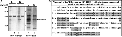
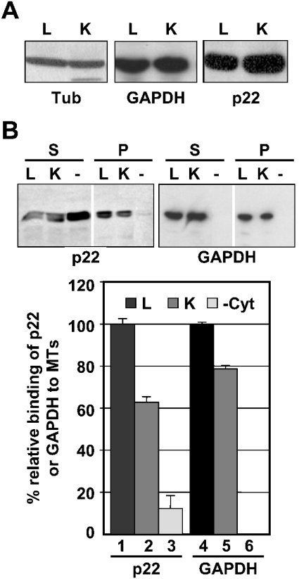
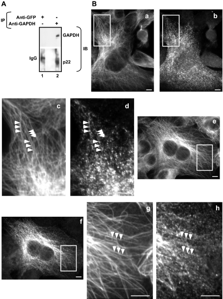
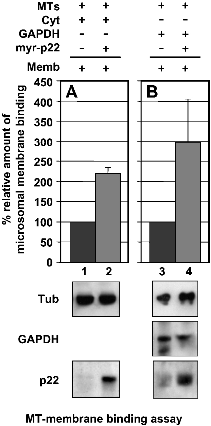
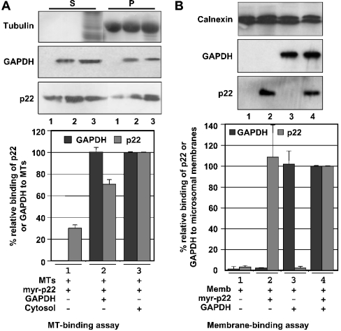
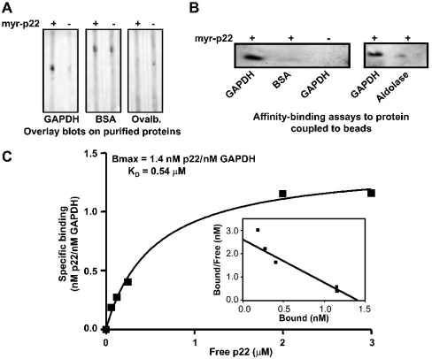
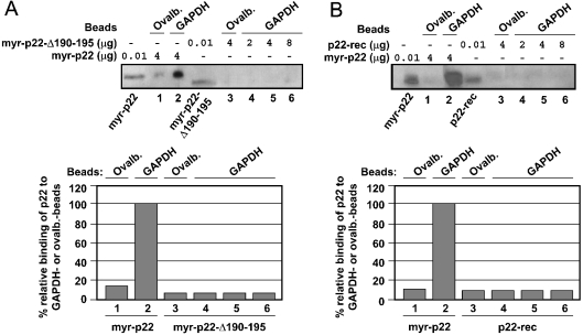
Previously, we have shown that p22, an EF-hand Ca2+-binding protein, interacts indirectly with microtubules in an N-myristoylation-dependent and Ca2+-independent manner. In the present study, we report that N-myristoylated p22 interacts with several microtubule-associated proteins within the 30-100 kDa range using overlay blots of microtubule pellets containing cytosolic proteins. One of those p22-binding partners, a 35-40 kDa microtubule-binding protein, has been identified by MS as GAPDH (glyceraldehyde-3-phosphate dehydrogenase). Several lines of evidence suggest a functional relationship between GAPDH and p22. First, endogenous p22 interacts with GAPDH by immunoprecipitation. Secondly, p22 and GAPDH align along microtubule tracks in analogous punctate structures in BHK cells. Thirdly, GAPDH facilitates the p22-dependent interactions between microtubules and microsomal membranes, by increasing the ability of p22 to bind microtubules but not membranes. We have also shown a direct interaction between N-myristoylated p22 and GAPDH in vitro with a K(D) of approximately 0.5 microM. The removal of either the N-myristoyl group or the last six C-terminal amino acids abolishes the binding of p22 to GAPDH and reduces the ability of p22 to associate with microtubules. In summary, we report that GAPDH is involved in the ability of p22 to facilitate microtubule-membrane interactions by affecting the p22-microtubule, but not the p22-membrane, association.


Figures

Figure 1
Figure 1. p22 interacts with microtubule-associated GAPDH
(A) Left panel: blot overlays of microtubule pellets from co-sedimentation assays performed in the presence of either liver (L) or kidney (K) cytosolic proteins were probed with (+) or without (−) myr-p22. Bands 1–4 indicate p22-interacting partners. Values on the left are molecular masses in kDa. Right panel: microtubule pellets from co-sedimentation assays performed in the presence of either liver (L) or kidney (K) cytosol were analysed by SDS/PAGE and silver staining. (B) MS was used to identify band 2 as GAPDH. Alignment of GAPDH sequence (accession no. NP_058704) with MS peptide sequences derived from band 2 (shaded boxes).
Figure 2
Figure 2. p22 and GAPDH associate with microtubule pellets in the presence of liver or kidney cytosolic proteins
(A) Rat liver (L) and kidney (K) cytosolic proteins were subjected to immunoblots using tubulin, GAPDH and p22 antibodies. (B) Upper panels: microtubule co-sedimentation assays were incubated with myr-p22 in the absence (lane-) or presence of rat liver (lane L) or kidney (lane K) cytosols and processed as described previously [22]. Supernatants (S) and microtubule pellets (P) were analysed by immunoblotting using anti-p22 and anti-GAPDH. (B) Lower panel: p22 and GAPDH immunoblots were quantified as described in the Experimental section, and the percentage binding of p22 or GAPDH to microtubule pellets in the presence of liver cytosol was normalized to 100% respectively. Results represent means±S.D. for three experiments. MTs, microtubules.
Figure 3
Figure 3. Characterization of p22–GAPDH interaction
(A) p22 co-immunoprecipitates with GAPDH. Rat liver cytosolic proteins (500 μg) were incubated with anti-GAPDH (lane 2) or anti-GFP as a negative control (lane 1). Immunoprecipitates (IP) were analysed by immunoblotting (IB) using anti-GAPDH or anti-p22. (B) BHK cells were processed for double-label immunofluorescence microscopy using anti-tubulin (panels a, c, e and g) and anti-GAPDH (panels b and d) or anti-p22 APpep2 antibodies (panels f and h). Panels c, d, g and h are magnifications of the regions of interest (white squares) in panels a, b, e and f respectively. Arrowheads indicate co-localization between microtubule and GAPDH or p22 tracks. Scale bars, 10 μm.
Figure 4
Figure 4. Role of GAPDH in the p22-dependent interactions between microsomal membranes and microtubules
(A) Lower panels: a two-step microtubule–membrane-binding assay [14] was used to assay the role of GAPDH in p22-dependent microtubule–membrane interactions. First, DYNABEADS® M-280 Tosylactivated were covered with taxol-polymerized microtubules and incubated with rat liver cytosol in the presence (lane 2) or absence of myr-p22 (lane 1). Secondly, beads were incubated with microsomal membranes. Equal amounts of reaction mixtures were analysed by SDS/PAGE and immunoblotting using antibodies against tubulin, calnexin and p22. (A) Upper panel: a value of 100% was assigned to the relative binding of microsomal membranes to microtubule-covered beads after incubation with cytosol in the absence of myr-p22 (lane 1). Results represent means±S.D. for three experiments. (B) Lower panels: DYNABEADS® M-280 Tosylactivated were covered with taxol-polymerized microtubules and incubated with purified rabbit muscle GAPDH in the presence (lane 2) or absence of myr-p22 (lane 1). Then, beads were incubated with microsomal membranes. Equal amounts of reaction mixtures were analysed by SDS/PAGE and immunoblotting using antibodies against tubulin, calnexin, p22 and GAPDH. (B) Upper panel: a value of 100% was assigned to the relative binding of microsomal membranes to microtubule-covered beads after incubation with GAPDH in the absence of myr-p22 (lane 1). Results represent means±S.D. for three experiments.
Figure 5
Figure 5. The association of p22 with microtubules, but not with microsomal membranes, is stimulated by GAPDH
(A) Upper panels: microtubule co-sedimentation assays were performed in the presence of myr-p22 (lane 1), p22-myr plus rabbit muscle GAPDH (lane 2) or myr-p22 plus rat liver cytosol (lane 3). Equal amounts of supernatants (S) and microtubule pellets (P) were analysed by immunoblotting using anti-p22, anti-tubulin and anti-GAPDH. Lower panel: the percentage of p22 or GAPDH binding to microtubule pellets in the presence of rat liver cytosol was normalized to 100%. Results represent means±S. D. for three experiments. (B) Upper panels: microsomal membranes were incubated in the presence or absence (lane 1) of rabbit muscle GAPDH (lanes 3 and 4) and/or myr-p22 (lanes 2 and 4) and re-pelleted. Equal amounts of membrane pellets were analysed by SDS/PAGE and immunoblotting using p22, GAPDH and calnexin antibodies. Lower panel: immunoblots were quantified as described in the Experimental section. A value of 100% was assigned to the binding of p22 or GAPDH to microsomal membrane pellets on incubation of microsomal membranes in the presence of myr-p22 and GAPDH (lane 4). Results represent means±S. D. for three experiments.
Figure 6
Figure 6. p22 interacts directly with GAPDH in vitro
(A) Blot overlays were performed on 5 μg of purified rabbit muscle GAPDH, BSA or ovalbumin (Ovalb.) in the presence (+) or absence (–) of myr-p22. (B) Affigel-10 beads coupled with GAPDH, BSA or aldolase were incubated in the presence (+) or absence (–) of myr-p22. The amount of myr-p22 bound to the beads was detected by immunoblotting using anti-p22. (C) KD analysis of the binding of myr-p22 to GAPDH was performed by incubating the indicated concentrations of myr-p22 (molar amounts) with 50 μg of GAPDH-coupled with beads. The amount of bound myr-p22 was detected by immunoblotting with anti-p22. The values for KD and Bmax were obtained from three repeat experiments as described in the Experimental section.
Figure 7
Figure 7. The last six C-terminal amino acids and the N-myristoyl moiety are required for the binding of p22 to GAPDH
(A) Upper panel: Affigel-10 beads coupled with GAPDH (lanes 2, 4–6) were incubated in the presence of 4 μg of myr-p22 (lanes 2) or 2, 4 or 8 μg of myr-p22-Δ190-195 (lanes 4–6). Affigel-10 beads coupled with ovalbumin (lanes 1 and 3) were incubated in the presence of 4 μg of myr-p22 (lanes 1) or myr-p22-Δ190-195 (lanes 3). Equal amounts of beads were assayed by immunoblotting with anti-p22. myr-p22 or myr-p22-Δ190-195 (0.01 μg each) was used as the control. (B) Upper panel: Affigel-10 beads coupled with ovalbumin (lanes 1 and 3) were incubated in the presence of 4 μg of myr-p22 (lanes 1) or p22-rec (lanes 3). Affigel-10 beads coupled with GAPDH (lanes 2, 4–6) were incubated in the presence of 4 μg of myr-p22 (lanes 2) or 2, 4 or 8 μg of p22-rec (lanes 4–6). Equal amounts of beads were assayed by immunoblotting with anti-p22. myr-p22 (0.05 μg) or p22-rec (0.01 μg) was used as the control. (A, B) Lower panels: quantification of relative percentage of myr-p22, myr-p22-Δ190-195 or p22-rec binding to GAPDH- or ovalbumin-beads. In (A, B) lanes 1–6 in upper panels correspond to lanes 1–6 in lower panels. In (A, B) lower panels, the amount of myr-p22 associated with GAPDH-coupled beads when 4 μg of myr-p22 was incubated with 50 μg of GAPDH is considered as 100%.
Figure 8
Figure 8. p22 associates with microtubule pellets in a N-myristoylation- and C-terminal-dependent manner
Upper panel: microtubule co-sedimentation assays were performed in the presence (lanes +Cyt) or absence (lanes −Cyt) of rat liver cytosol and myr-p22-Δ190-195, myr-p22 and/or p22-rec. Equal amounts of supernatants (S) and pellets (P) were analysed by immunoblotting using anti-p22. Lower panel: quantification of percentage relative binding of myr-p22, myr-p22-Δ190-195 or p22-rec to microtubule pellets in the presence or absence of liver cytosol. The percentage of p22 binding to microtubule pellets in the presence of cytosol was normalized to 100%. Results represent means±S.D. for four experiments. MTs, microtubules.

References

    1. Bloom G. S., Goldstein L. S. Cruising along microtubule highways: how membranes move through the secretory pathway. J. Cell Biol. 1998;140:1277–1280. - PMC - PubMed
    1. Allan V. J., Schroer T. A. Membrane motors. Curr. Opin. Cell Biol. 1999;11:476–482. - PubMed
    1. Karcher R. L., Deacon S. W., Gelfand V. I. Motor-cargo interactions: the key to transport specificity. Trends Cell Biol. 2002;12:21–27. - PubMed
    1. Cassimeris L., Spittle C. Regulation of microtubule-associated proteins. Int. Rev. Cytol. 2001;210:163–226. - PubMed
    1. Schuyler S. C., Pellman D. Microtubule ‘plus-end-tracking proteins’: the end is just the beginning. Cell (Cambridge, Mass.) 2001;105:421–424. - PubMed

MeSH terms