Review
. 2004 Sep;113(1):1-14.
doi: 10.1111/j.1365-2567.2004.01959.x.

The macrophage and the apoptotic cell: an innate immune interaction viewed simplistically?

Affiliations
Review

The macrophage and the apoptotic cell: an innate immune interaction viewed simplistically?

Christopher D Gregory et al. Immunology. 2004 Sep.

Abstract

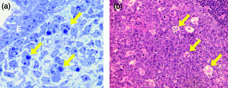
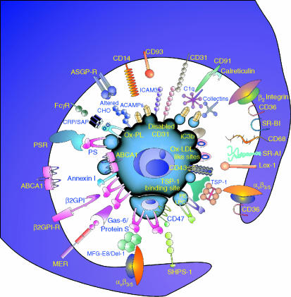
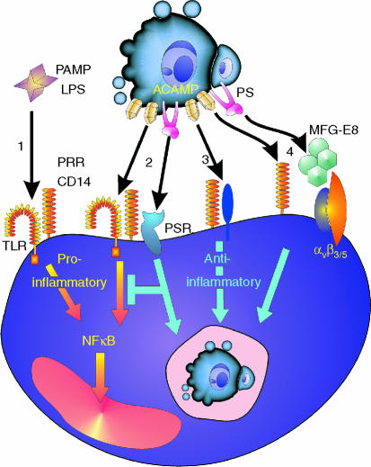
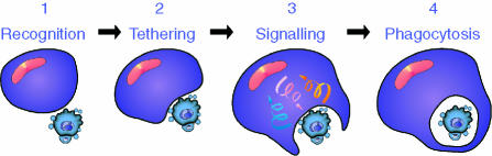
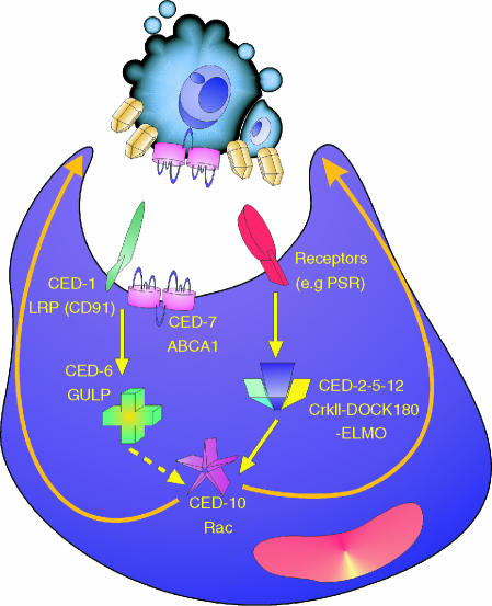
Macrophages play important roles in the clearance of dying and dead cells. Typically, and perhaps simplistically, they are viewed as the professional phagocytes of apoptotic cells. Clearance by macrophages of cells undergoing apoptosis is a non-phlogistic phenomenon which is often associated with actively anti-inflammatory phagocyte responses. By contrast, macrophage responses to necrotic cells, including secondarily necrotic cells derived from uncleared apoptotic cells, are perceived as proinflammatory. Indeed, persistence of apoptotic cells as a result of defective apoptotic-cell clearance has been found to be associated with the pathogenesis of autoimmune disease. Here we review the mechanisms by which macrophages interact with, and respond to, apoptotic cells. We suggest that macrophages are especially important in clearing cells at sites of histologically visible, high-rate apoptosis and that, otherwise, apoptotic cells are removed largely by non-macrophage neighbours. We challenge the view that necrotic cells, including persistent apoptotic cells are, of necessity, proinflammatory and immunostimulatory and suggest that, under appropriate circumstances, persistent apoptotic cells can provide a prolonged anti-inflammatory stimulus.


Figures

Figure 1
Figure 1
Engulfment of apoptotic cells in situ. (a) Clearance of apoptotic cells in the mouse footplate during the dissolution of the interdigital web at embryonic day 14·5. Arrows show examples of macrophages that have phagocytosed apoptotic mesenchymal cells with classical morphological features of the death programme. E, epidermis. Toluidine blue stain. (b) Standard haematoxylin- and eosin-stained section of lymph node showing tingible body macrophages (arrows) of a germinal centre containing engulfed remnants of apoptotic cells.
Figure 2
Figure 2
Close encounters between apoptotic cells and phagocytes. Schematic view of molecules implicated in interactions mediating recognition, binding and engulfment of a theoretical, ‘consummate’ apoptotic cell with a similarly theoretical phagocyte. Molecules used by nonmacrophages as well as macrophages are included (see Table 1 for comparison). αvβ3, αvβ5 vitronectin receptor integrins; ABCA1, ATP-binding cassette transporter A1; ACAMPs, apoptotic cell-associated molecular patterns; ASGP-R, asialoglycoprotein receptor; β2GPI, β2 glycoprotein I; β2GPI-R, β2GPI-receptor; β2 integrins include CR3 and CR4; C1q, first component of complement; CHO, carbohydrate; CRP, C-reactive protein; Del-1, developmental endothelial locus-1; Gas-6, growth arrest specific gene-6; iC3b, inactivated complement fragment C3b; ICAM-3 (CD50), intercellular adhesion molecule-3; Lox-1, oxidised low density lipoprotein receptor 1; LPC, lysophosphatidylcholine; MER, myeloid epithelial reproductive tyrosine kinase; MFG-E8, milk fat globule epidermal growth factor-8; Ox-PL oxidised phospholipids; PE, phosphatidylethanolamine; PS, phosphatidylserine; PSR, PS-receptor; SAP, serum amyloid protein, SHPS-1, Src homology 2 domain-bearing protein tyrosine phosphatase substrate-1; SR-AI, scavenger receptor AI; SR-BI, scavenger receptor BI; TSP-1, thrombospondin-1.
Figure 3
Figure 3
Outline of possible scenarios for common mechanisms of pattern recognition of PAMPs and ACAMPs with differential macrophage responses illustrated by the pattern recognition receptor (PRR), CD14. 1: Proinflammatory response to a prototypic PAMP, LPS, in which the GPI-anchored CD14 cooperates with TLRs to activate NFκB and elicit proinflammatory responses. 2: engagement of CD14 by ACAMPs may engage a similar default inflammatory signalling pathway but here additional receptor–ligand interactions, e.g. the PS/PSR interaction shown, dominantly suppress the proinflammatory response and may also engage anti-inflammatory signalling pathway(s). 3: Alternatively, in a ligand-dependent manner, CD14 may associate with different signal-transduction partners that activate anti-inflammatory rather than proinflammatory responses in the phagocyte. 4: A further scenario is that CD14–ACAMP interactions merely tether the apoptotic cell to the phagocyte and that additional receptor/ligand interactions such as PSR/PS or, as illustrated, vitronectin receptor/MFG-E8/PS, activate anti-inflammatory responses in the macrophage.
Figure 4
Figure 4
Phases of interaction between apoptotic cell and macrophage. (1) Initial contact events (e.g. CD31/CD31) precede (2), the tethering stage (involving, e.g. CD14, CD47) which facilitates the initiation of (3) ‘outside-in’ and ‘inside-out’ signal transduction events (involving, e.g. PS/PSR, integrins) that activate the phagocyte. The phagocyte responses include reinforcement of intercellular adhesion, production of anti-inflammatory cytokines and (4) phagocytosis.
Figure 5
Figure 5
Apoptotic-cell clearance pathways conserved between worms and mammals. Mutagenesis studies in Caenorhabditis elegans have identified seven genes involved in clearance that comprise two partially redundant pathways, one involving ced-1, -6, and -7, the other involving ced-2, -5, -10 and -12. Mammalian homologues of the CED-1, -6 and -7 proteins are LDL-related receptor protein (LRP, also known as CD91), engulfment adaptor protein (GULP), and the ATP-binding cassette transporter, ABCA1, respectively. The homologous proteins in the ced-2, -5, -10, -12 pathway are the adapter proteins CrkII and Dock180, the Rho family GTP-binding protein Rac and ELMO (engulfment and motility), respectively. The latter, in complex with Dock180. acts as a guanine nucleotide exchange factor for Rac. The ced-2, -5, -10, -12 pathway is functional not only in mobilizing the actin cytoskeleton in extending the cytoplasm to surround the apoptotic cell during engulfment but also in cell migration.

References

    1. Savill J, Fadok V. Corpse clearance defines the meaning of cell death. Nature. 2000;407:784–8. - PubMed
    1. Fadok VA, Bratton DL, Henson PM. Phagocyte receptors for apoptotic cells: recognition, uptake, and consequences. J Clin Invest. 2001;108:957–62. - PMC - PubMed
    1. Hengartner MO. Apoptosis: corralling the corpses. Cell. 2001;104:325–8. - PubMed
    1. Henson PM, Bratton DL, Fadok VA. The phosphatidylserine receptor: a crucial molecular switch? Nat Rev Mol Cell Biol. 2001;2:627–33. - PubMed
    1. Henson PM, Bratton DL, Fadok VA. Apoptotic cell removal. Curr Biol. 2001;11:R795–805. - PubMed

Publication types

LinkOut - more resources