Enhancement of the synthetic ligand-mediated function of liver NK1.1Ag+ T cells in mice by interleukin-12 pretreatment
- PMID: 15312134
- PMCID: PMC1782545
- DOI: 10.1111/j.1365-2567.2004.01932.x
Enhancement of the synthetic ligand-mediated function of liver NK1.1Ag+ T cells in mice by interleukin-12 pretreatment
Abstract
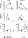
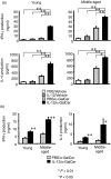
We previously reported that mouse NK1.1 Ag+ T (NKT) cells activated by interleukin-12 (IL-12) act as anti-tumour/anti-metastatic effectors. However, IL-12 reportedly induces a rapid disappearance of liver NKT cells by activation-induced apoptosis. In the present study, however, we show that injection of IL-12 into mice merely down-regulates the NK1.1 expression of liver NKT cells and Vbeta8+ intermediate T-cell receptor cells and CD1d/alpha-galactosylceramide (alpha-GalCer)-tetramer reactive cells in the liver remained and did not decrease. Furthermore, when IL-12-pretreated (24 hr before) mice were injected with alpha-GalCer, not only serum interferon-gamma but also serum IL-4 concentrations increased several-fold in comparison to the control alpha-GalCer-injected mice. However, IL-12 pretreatment markedly up-regulated serum ALT levels and Fas-ligand expression on NKT cells after alpha-GalCer injection in middle-aged mice only. Consistently, the liver mononuclear cells (MNC) from IL-12-pretreated mice stimulated with alpha-GalCer in vitro produced much greater amounts of interferon-gamma and IL-4, and also showed a more potent cytotoxicity against tumour targets than those from mice pretreated with phosphate-buffered saline. Liver MNC from middle-aged mice, but not from young mice pretreated with IL-12, also showed increased cytotoxicity following in vitro alpha-GalCer stimulation against cultured hepatocytes. Furthermore, IL-12 treatment of middle-aged mice enhanced tumour necrosis factor receptor 1 mRNA expression in liver Vbeta8+ T cells, and in vitro experiments also revealed that IL-12 pretreatment of liver MNC from middle-aged mice enhanced their tumour necrosis factor-alpha production after alpha-GalCer stimulation. Synthetic ligand-mediated functions of NKT cells, including IL-4 production, are thus enhanced by IL-12 pretreatment.
Figures


Similar articles
-
Age-associated augmentation of the synthetic ligand- mediated function of mouse NK1.1 ag(+) T cells: their cytokine production and hepatotoxicity in vivo and in vitro.J Immunol. 2002 Dec 1;169(11):6127-32. doi: 10.4049/jimmunol.169.11.6127. J Immunol. 2002. PMID: 12444115
-
Activation of mouse liver natural killer cells and NK1.1(+) T cells by bacterial superantigen-primed Kupffer cells.Hepatology. 1999 Aug;30(2):430-6. doi: 10.1002/hep.510300209. Hepatology. 1999. PMID: 10421651
-
Cytotoxic NK1.1 Ag+ alpha beta T cells with intermediate TCR induced in the liver of mice by IL-12.J Immunol. 1995 May 1;154(9):4333-40. J Immunol. 1995. PMID: 7722291
-
Innate Valpha14(+) natural killer T cells mature dendritic cells, leading to strong adaptive immunity.Immunol Rev. 2007 Dec;220:183-98. doi: 10.1111/j.1600-065X.2007.00561.x. Immunol Rev. 2007. PMID: 17979847 Review.
-
CD1d-restricted NKT regulatory cells: functional genomic analyses provide new insights into the mechanisms of protection against Type 1 diabetes.Novartis Found Symp. 2003;252:146-60; discussion 160-4, 203-10. Novartis Found Symp. 2003. PMID: 14609217 Review.
Cited by
-
Activated natural killer T cells in mice induce acute kidney injury with hematuria through possibly common mechanisms shared by human CD56+ T cells.Am J Physiol Renal Physiol. 2018 Sep 1;315(3):F618-F627. doi: 10.1152/ajprenal.00160.2018. Epub 2018 Jul 11. Am J Physiol Renal Physiol. 2018. PMID: 29993279 Free PMC article.
-
Analysis of the susceptibility of CD57 T cells to CD3-mediated apoptosis.Clin Exp Immunol. 2005 Feb;139(2):268-78. doi: 10.1111/j.1365-2249.2004.02687.x. Clin Exp Immunol. 2005. PMID: 15654825 Free PMC article.
-
Suppressive role of hepatic dendritic cells in concanavalin A-induced hepatitis.Clin Exp Immunol. 2011 Nov;166(2):258-68. doi: 10.1111/j.1365-2249.2011.04458.x. Clin Exp Immunol. 2011. PMID: 21985372 Free PMC article.
-
Safety levels of systemic IL-12 induced by cDNA expression as a cancer therapeutic.Immunotherapy. 2022 Feb;14(2):115-133. doi: 10.2217/imt-2021-0080. Epub 2021 Nov 16. Immunotherapy. 2022. PMID: 34783257 Free PMC article.
-
Infections, Reactions of Natural Killer T Cells and Natural Killer Cells, and Kidney Injury.Int J Mol Sci. 2022 Jan 1;23(1):479. doi: 10.3390/ijms23010479. Int J Mol Sci. 2022. PMID: 35008905 Free PMC article. Review.
References
-
- Makino Y, Koseki H, Adachi Y, Akasaka T, Tsuchida K, Taniguchi M. Extrathymic differentiation of a T cell bearing invariant V alpha 14J alpha 281 TCR. Int Rev Immunol. 1994;11:31–46. - PubMed
-
- Hashimoto W, Takeda K, Anzai R, Ogasawara K, Sakihara H, Sugiura K, Seki S, Kumagai K. Cytotoxic NK1.1 Ag+ alpha beta T cells with intermediate TCR induced in the liver of mice by IL-12. J Immunol. 1995;154:4333–40. - PubMed
-
- Takeda K, Seki S, Ogasawara K, et al. Liver NK1.1+ CD4+ alpha beta T cells activated by IL-12 as a major effector in inhibition of experimental tumor metastasis. J Immunol. 1996;156:3366–73. - PubMed
MeSH terms
Substances
LinkOut - more resources
Full Text Sources
Research Materials
Miscellaneous
