. 2004 Sep;113(1):44-56.
doi: 10.1111/j.1365-2567.2004.01928.x.

Thymocytes induced by antigen injection into the anterior chamber activate splenic CD8+ suppressor cells and enhance the antigen-induced production of immunoglobulin G1 antibodies

Affiliations

Thymocytes induced by antigen injection into the anterior chamber activate splenic CD8+ suppressor cells and enhance the antigen-induced production of immunoglobulin G1 antibodies

Xingya Li et al. Immunology. 2004 Sep.

Abstract

Injection of antigen into the ocular anterior chamber (AC) of a mouse eye (an immunologically privileged site) induces the activation of immunoregulatory NK1.1+, CD4- CD8-, T-cell receptor (TCR) alphabeta+ thymocytes. These thymocytes transfer the suppression of delayed-type hypersensitivity (DTH) when injected into mice sensitized to the same antigen but do not effect the suppression of DTH. On the other hand, the immunized recipients of these transferred thymocytes produce splenic CD8+ T cells that effect the suppression of DTH. However, it is unclear whether the thymocytes transferred from the AC-injected donor differentiate into and/or activate CD8+ T-splenic suppressor cells. We therefore sought to determine the origin of splenic suppressor cells produced in the recipients of immunoregulatory thymocytes transferred from donors that receive an injection of antigen into the AC. CD45.1+ thymocytes from mice that received an AC injection of 2,4,6-trinitrobenzene sulphonic acid (TNP)-bovine serum albumin (BSA) were transferred to congenic CD45.2+ TNP-BSA-immune recipients. Spleen cells from the recipients were then sorted based on anti-CD45.1 or -CD45.2 antibody binding and assayed for suppressor cells. This was done by the injection of separated spleen cells into the footpad of TNP-BSA-immunized mice, concurrent with the induction of footpad swelling (contact sensitivity) of the footpad elicited by an epicutaneous application of picryl chloride. The systemic distribution of antigen after the injection of antigen into the AC was demonstrated by the injection of fluorescein or 125I-labelled TNP-BSA into the AC. The results demonstrate that (i) splenic CD8+ T-suppressor cells produced in the immunized recipients of immunoregulatory thymocytes are derived from the CD45.2 recipient of the CD45.1+ thymocytes; (ii) the induction of recipient splenic suppressor T cells by the transferred immunoregulatory thymocytes requires that the recipient be immunized to the same antigen as that used to induce immunoregulatory thymocytes; (iii) antigen is introduced to the thymus after an injection of antigen into the AC; (iv) although the transfer of the suppression of DTH by regulatory thymocytes was not dependent on interleukin-4 (IL-4), CD4+ NK1.1- regulatory thymocytes from AC-injected donors enhanced the production of immunoglobulin G1 antibodies to TNP-BSA by an IL-4-dependent mechanism. These observations suggest that the adult thymus plays an active role in the induction and maintenance of anterior chamber-associated immune deviation as manifested by the generation of the suppression of cell-mediated immunity to exogenous antigen and the antigen-induced production of IgG1 antibodies.


Figures
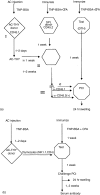
Figure 1
Figure 1
Adoptive transfers and assays (a) Activation of splenic suppressor cells by regulatory thymocytes from AC-injected donors. To prepare regulatory thymocytes (AC-Thy) (I) naïve mice receive an injection of TNP-BSA into an AC. (II) Two days later thymocyte suspensions are prepared and thymocytes injected i.v. into CD45.2+, TNP-BSA-immune receipients. One week after the mice received AC-Thy (III) spleen cells (SPL) are prepared, separated on the basis of the CD45 allele and/or CD8 are injected into the footpads of TNP-BSA immunized mice immediately after epicutaneous challenge with PC1. Footpad swelling is measured 24 hr later and serum collected at the time of challenge, 7, 14, 21 days later to assay for IgG1 antibodies to TNP-BSA. (b) Assay of immunoregulatory thymocytes. One-2 days after thymocyte donors received an injection of TNP-BSA into the AC thymocytes (unseparated) NK1.1+, CD4+ were injected i.v. into TNP-BSA-immunized mice. One week later, the mice received epicutaneous PC1 to a footpad. Swelling of the footpad was measured 24 hr later and the mice were bled 1 or 2 weeks after challenge.
Figure 2
Figure 2
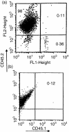
Spleen cells of recipients of CD45.1+ thymocytes contain CD45.1+ cells. Naïve CD45.1+ thymocyte donor mice received an injection of TNP-BSA into the AC. Twenty-four h later thymocytes were prepared and injected i.v. into CD45.2+ recipient mice. (a) One week later, spleen cells from the recipient C57Bl/6 mice (CD45.2+) were stained with PE-anti-CD45.2 and FITC anti-CD45.1 antibodies. (b) Spleen cells from C57Bl/6 mice that did not receive CD45.1+ thymocytes were stained with anti-CD45.2 and CD45.1 antibodies.
Figure 3
Figure 3
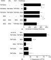
Thymocytes from AC-injected donors activate splenic suppressor cells.Three CD45.1+ thymocyte donors received an injection of TNP-BSA into an AC. Twenty-four hr later thymocyte suspensions were prepared and injected i.v. into CD45.2+, TNP-BSA-immunized recipients. After 1 week, recipient spleen cells (SPL) were incubated with biotinylated anti-CD45.1 or anti-CD45.2 and separated with biotin-paramagnetic beads. CD45.2+ cells were released enzymatically from the anti-CD45.2 beads and incubated with anti-CD8 paramagnetic beads. 2·5 × 104 positively or negatively selected-recipient cells or naïve C57 BL/6j (CD45.2+) spleen cells were injected into the footpad of TNP-BSA-immunized mice immediately after epicutaneous PCl challenge. Footpad swelling was measured 24 hr later. (a) One representative experiment (of four). Data shows the mean ± SEM swelling of four mice/group. *P < 0·02. (b) The data represents the mean percentage suppression ± SEM of 12 mice/group in four separate experiments. *P = <0·0.01. % suppression =1 − (µm injected with SPL − µm naïve control) × (100 µm no SPL − µm naïve control). Footpad swelling of immunized mice that were challenged but were injected with naïve spleen cells or no spleen cells ranged from 600 to 1100 µm. Swelling of naïve mice ranged from 50 to 250 µm.
Figure 4
Figure 4
Thymocytes from AC-injected donors activate splenic suppressor cells in mice immunized to the same antigen that activated the thymocytes. Thymocyte (THY) donor BALB/c mice (three to four/group) received an injection of TNP-BSA or BSA into an AC. Two days later thymocytes were prepared and injected i.v. into naïve spleen cell donors or donors immunized 1 week previously with 400 µg TNP-BSA/FCA or OVA/FCA. One week after the injection of the thymocytes spleen cell suspensions were prepared and 2·5 × 104 spleen cells (SPL) were injected into a footpad concurrently with an epicutaneous challenge of PCl. Swelling was measured 24 hr later. The reduction of swelling (in µm) is shown as a percentage of the swelling obtained in footpads that did not receive spleen cells (Fig. 3 for calculation) and represents the mean ± SE of 8–12 mice in three separate experiments. *P < 0·001 compared to spleen cells from TNP-BSA-immunized mice that received thymocytes from mice injected with TNP-BSA into the AC.
Figure 5
Figure 5
The generation of splenic suppressor cells by AC injection depends on immunization of the donor of the spleen cells. BALB/c mice (four/group/experiment) were immunized s.c. with 400 µg TNP-BSA ± CFA or 300 µg poly AU. (a) One week after immunizing a footpad was challenged with epicutaneous PCl and swelling measured 24 hr later. The data represents the mean swelling ± SEM of 12 mice/group mice, three separate experiments. *P < 0·001. The swelling of naïve mice (30 µm) has been subtracted from the swelling of immunized mice.(b) The mice were bled seven days after challenge and the serum of each mouse assayed for IgG1 anti-TNP-BSA antibodies. Data represents the geometric mean titre (1/dilution) of four sera/group in three separate experiments. *P < 0·01. (c) Spleen cell donors from AC-injected mice were immunized with TNP-BSA as shown. One week after immunizing the donors received TNP-BSA in an injection into the AC. One week after the AC injection, spleen cell suspensions from each of four of the donors were pooled and 2·5 × 104 spleen cells were injected into the footpad of a TNP-BSA-immunized mouse concurrent with an epicutaneous challenge with PCl. Swelling was measured 24 hr later. Swelling of naïve mice challenged with PCl was 65 ± 15 µm. The data represents the mean swelling ± SEM of four mice/group. *P < 0·01.
Figure 6
Figure 6
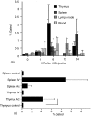
Distribution of FITC-antigen after injection of TNP-BSA into the anterior chamber. Three BALB/c naïve mice received an injection of four µg FITC-TNP-BSA into an anterior chamber. (a) 1–24 hr after injection, blood and organs were removed. Cells suspensions were prepared and analysed by flow cytometry. Data represents the mean percent of fluorescent cells minus the mean percentage of fluorescent cells from non-injected mice ± SEM of six to eight separate experiments. (b) Cell suspensions from organs obtained 6 hr after the injection of antigen i.v. or into the AC were compared. Data represents the mean ± SEM percent of fluorescent cells of six separate experiments. *P < 0·05.
Figure 7
Figure 7
Distribution of 125I-labelled TNP-BSA after injection into the anterior chamber. Four µg 125I-labelled TNP-BSA was injected into the anterior chamber of 3 naïve BALB/c mice and 1–168 hr after injection organs or blood were removed. (a,b) Whole individual organs were counted for 125I. Peripheral blood cells were prepared from whole blood by centrifugation of 1 ml of blood/mouse. The cell pellet was washed three times and counted for 125I. The data represents the mean counts/10 min of 15 samples in five separate experiments (three samples/experiment). (c) 125I-TNP-BSA in the eye of each of four mice.
Figure 8
Figure 8
Thymocytes from AC-injected mice promote the antigen-induced production of IgG1 antibodies. C57BL/6j mice immunized 1 week previously with TNP-BSA received i.v. thymocytes prepared 24 hr after the naïve donor mice received an injection of TNP-BSA into the AC (AC-thymocytes). One week after the injection of the thymocytes, the recipient mice were bled to collect sera and challenged with epicutaneous PCl to a footpad. (a) Footpad swelling was measured 24 hr later. Data represents the mean ± SEM of swelling of eight TNP-BSA-immunized mice, four mice/group, two experiments. Footpad swelling of naïve mice was 110 ± 17 µm. *P < 0·01 (b). The mice were bled at the time of challenge and 7 and 14 days after challenge. IgG1 antibodies to TNP-BSA were assayed by ELISA using doubling dilutions of antiserum and biotinylated anti-IgG1 and alkaline phosphatase-conjugated streptavidin added to microtitre trays coated with 1 µg/well of antigen. The data represents the geometric mean titre ± SEM of the pooled sera in duplicate assays of three mice/group in two separate experiments. The titre of naïve mice was 40 ± 20. *P < 0·01 IMM-immunized mice that did not receive thymocytes.
Figure 9
Figure 9
Phenotype of IgG1-enhancing thymocytes from AC-injected mice. Thymocytes from C57BL/6 mice were prepared by AC injection of TNP-BSA and were separated by immunomagnetic beads into NK1.1+ and NK1.1 cells. 1 × 10⁵ NK1.1+or 5 × 10⁶ NK1.1 thymocytes were injected i.v. into TNP-BSA-immunized mice. One week after the injection of the thymocytes, the mice were challenged by epicutaneous PCl to a footpad. (a) Swelling of the footpad was measured 24 hr later. Footpad swelling of naïve mice challenged with PCl was 35 ± 5 µm. The data represents the mean swelling ± SEM of four mice/group. *P < 0·01. (b) 7 days after the challenge the mice were bled and the sera assayed individually for IgG1 antibodies to TNP-BSA. The data represents the geometric mean titre ± SEM of three to four mice/group in two separate experiments. Unseparated, unseparated thymocytes. (c) CD4+ thymocytes promote antigen-induced IgG1 antibody production. Thymocytes prepared 2 days after AC injection of TNP-BSA were separated by immunomagnetic beads into CD4+ and CD4 cell populations. 5 × 10⁵ or 1 × 10⁷ thymocytes were injected i.v. into TNP-BSA-immunized recipients. The mice were challenged 7 days after the injection of AC-Thy and bled 1 week after the challenge. Data represents the geometric mean titre ± SEM of four individual sera/group *P < 0·01.
Figure 10
Figure 10
Thymocytes from AC-injected, IL-4–/– mice do not enhance the production of IgG1 antibodies. BALB/c IL-4–/– and +/+ mice received an injection of TNP-BSA into the AC. Two days later, thymocytes from these donors were injected into TNP-BSA-immunized mice. One week after the injection of the thymocytes, the recipients were challenged on one footpad with epicutaneous PCl. Swelling of the footpad was measured 24 hr later and 7 days after the challenge the mice were bled intracardially. Sera were assayed by ELISA individually for IgG1 anti-TNP-BSA antibodies. IL-4+/+: sera from recipients of IL-4+/+ thymocytes, IL-4–/–: sera from recipients of IL-4–/– thymocytes, Naïve: sera from mice that received naïve thymocytes from donors that did not receive an AC injection of TNP-BSA. The data are expressed as the geometric mean titre (one/dilution) of eight sera, four mice/group in two experiments. *P < 0·05.

Similar articles

Cited by

References

    1. Streilein JW. Immunoregulation and the eye. A dangerous compromise. FASEB J. 1987;1:199–208. - PubMed
    1. Streilein JW, Stein-Streilein J. Does innate immune privilege exist? J Leukoc Biol. 2000;67:479–87. - PubMed
    1. Wilbanks GA, Streilein JW. Distinctive humoral responses following anterior chamber and intravenous administration of soluble antigen. Evidence for active suppression of IgG2a-secreting B cells. Immunology. 1990;71:566–72. - PMC - PubMed
    1. Streilein JW, Niederkorn JY. Induction of anterior chamber-associated immune deviation requires an intact, functional spleen. J Exp Med. 1981;153:1058–67. - PMC - PubMed
    1. Whittum JA, Niederkorn JY, McCulley JP, Streilein JW. The role of suppressor T cells in herpes simplex-induced immune deviation. J Virol. 1984;51:556–8. - PMC - PubMed

Publication types

MeSH terms

LinkOut - more resources