

Case Reports
Segmental agenesis of the internal carotid artery distal to the posterior communicating artery leading to the definition of a new embryologic segment
Affiliations
- PMID: 15313707
- PMCID: PMC7976531
Item in Clipboard
Case Reports
Segmental agenesis of the internal carotid artery distal to the posterior communicating artery leading to the definition of a new embryologic segment
AJNR Am J Neuroradiol.
2004 Aug.
Abstract
We report a case of segmental agenesis of the right internal carotid artery (ICA) in a 53-year-old woman, associated with a saccular aneurysm of the left anterior cerebral artery (A1 segment) presenting with a right hemispheric transient ischemic attack. The agenetic segment was located distal to the origin of the posterior communicating artery (PComA), as documented both angiographically and by direct surgical inspection. This observation suggests the existence of a previously unrecognized embryologic segment of the ICA, located between the PComA origin and the terminal bifurcation of the ICA into the anterior and middle cerebral arteries.
Figures
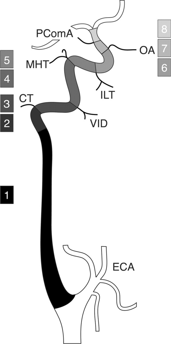
Schematic representation of the ICA segmentation. The ICA is divided into eight segments: 1) cervical, 2) ascending petrous, 3) horizontal petrous, 4) ascending cavernous, 5) horizontal cavernous, 6) clinoid, 7) ophthalmic, and 8) terminal. In the classification proposed by Lasjaunias and Santoyo-Vazquez (1), segments 7 and 8 are grouped as a single terminal segment, for a total of seven segments. In the nomenclature proposed by Fischer in 1938 (10), C1, C2, C3, C4, and C5 correspond to the segments 8, 7, 6, 5, and 4, respectively. CT, caroticotympanic artery; ECA, external carotid artery; ILT, inferolateral trunk; MHT; meningohypophyseal trunk; OA, ophthalmic artery; PcomA, posterior communicating artery; VID, vidian artery.
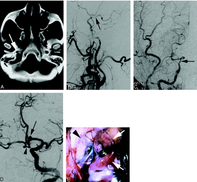
A 53-year-old patient with segmental agenesis of the right ICA. A, Axial CT scan showing asymmetry of the carotid canal in disfavor of the right side (arrow) B, DSA image (right common carotid injection, lateral view) documenting the termination of the right ICA into the right posterior communicating artery (arrow). The right carotid bifurcation is normal and the right ICA below the agenetic segment is decreased in caliber but otherwise unremarkable. C, DSA image (right common carotid injection, anteroposterior view) showing the same as above. D, DSA image (left common carotid artery, left anterior oblique view) showing collateral flow to the right anterior circulation via a large anterior communicating artery. Note the presence of a saccular aneurysm (arrow) on the left proximal anterior cerebral artery (A1 segment). The arrowhead identifies the prominent loop of the right A1 segment that is retracted on Fig 2E. E, Intraoperative microsurgical view of the distal right ICA (arrowhead). A prominent loop of the right A1 segment (arrows) is retracted to uncover the right ICA termination. Note the absence of fibrotic connection between the ICA and the right anterior and middle cerebral arteries.
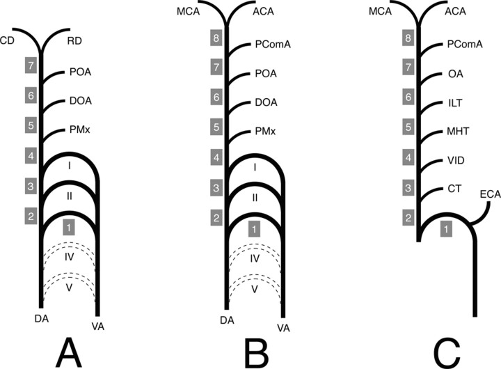
Schematic representation of the ICA developmental anatomy. I, II, III, IV, and V, aortic arch I–V; ACA, anterior cerebral artery; CD, caudal division of the ICA; CT, caroticotympanic artery; DA, dorsal aorta; DOA, dorsal ophthalmic artery; ECA, external carotid artery; ILT, inferolateral trunk; MCA, middle cerebral artery; MHT, meningohypophyseal trunk; OA, ophthalmic artery; PComA, posterior communicating artery; POA, primitive ophthalmic artery; PMx, primitive maxillary artery; RD, rostral division of the ICA; VA, ventral aorta; VID, vidian artery. A, ICA embryology according to Lasjaunias and Santoyo-Vazquez (1). The ICA is constituted of seven segments separated by embryonic vessels. Segment 1 is derived for the third aortic arch, whereas all the other segments (2–7) come from the DA. The distal ICA bifurcates into the RD and CD. B, Modified ICA embryology. A new segment (8) has been added owing to reported observation. The PComA is now considered as an embryonic branch separating two segments (7 and 8) and no longer as the CD of the ICA. The ICA termination corresponds to its bifurcation into the ACA and MCA. C, Schematic representation of the adult ICA based on the developmental anatomy described above. The ICA is derived from eight embryologic segments. The segment labeling (1–8) corresponds to the labeling in Figure 1.
Comment in
-
Segmental agenesis of the internal carotid artery distal to the posterior communicating artery leading to the definition of a new embryologic segment.AJNR Am J Neuroradiol. 2006 Feb;27(2):246-7; author reply 247. AJNR Am J Neuroradiol. 2006. PMID: 16484382 Free PMC article. No abstract available.
References
-
- Lasjaunias P, Santoyo-Vazquez A. Segmental agenesis of the internal carotid artery: angiographic aspects with embryological discussion. Anat Clin 1984;6:133–141 - PubMed
-
- Meder JF, Blustajn J, Trystram D, et al. Radiologic anatomy of segmental agenesis of the internal carotid artery. Surg Radiol Anat 1997;19:385–394 - PubMed
-
- Dilenge D. Bilateral agenesis of internal carotid artery. J Can Assoc Radiol 1975;26:91–94 - PubMed
-
- Lasjaunias P, Moret J, Mink J. The anatomy of the inferolateral trunk (ILT) of the internal carotid artery. Neuroradiology 1977;13:215–220 - PubMed
Publication types
MeSH terms
LinkOut - more resources
Full Text Sources
Medical
Miscellaneous