

Warthin tumor of the parotid gland: diagnostic value of MR imaging with histopathologic correlation
- PMID: 15313720
- PMCID: PMC7976549
Warthin tumor of the parotid gland: diagnostic value of MR imaging with histopathologic correlation
Abstract
Background and purpose: The purpose of our study was to describe the MR imaging appearance of Warthin tumors multiple MR imaging techniques and to interpret the difference in appearance from that of malignant parotid tumors.
Methods: T1-weighted, T2-weighted, short inversion time inversion recovery, diffusion-weighted, and contrast-enhanced dynamic MR images of 19 Warthin tumors and 17 malignant parotid tumors were reviewed. MR imaging results were compared with those of pathologic analysis.
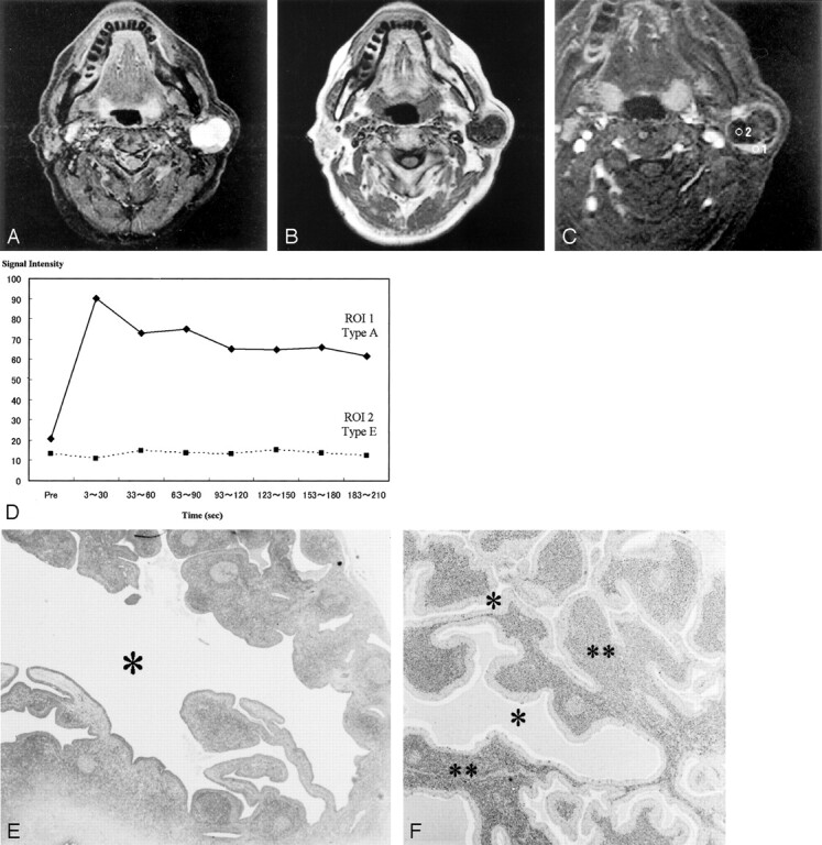
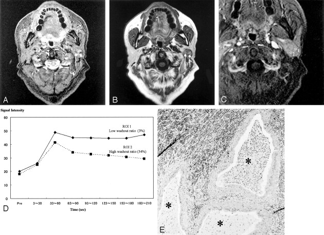
Results: Epithelial stromata and lymphoid tissue with slitlike small cysts in Warthin tumors showed early enhancement and a high washout rate (> or =30%) on dynamic contrast-enhanced images, and accumulations of complicated cysts showed early enhancement and a low washout ratio (< 30%). The areas containing complicated cysts showed high signal intensity on T1-weighted images, whereas some foci in those areas showed low signal intensity on short tau inversion recovery images. The mean minimum signal intensity ratios (SIRmin) of Warthin tumor on short tau inversion recovery (0.29 +/- 0.22 SD) (P < .01) and T2-weighted images (0.28 +/- 0.09) (P < .05) were significantly lower than those of malignant parotid tumors (0.53 +/- 0.19, 0.48 +/- 0.19). The average washout ratio of Warthin tumors (44.0 +/- 20.4%) was higher than that of malignant parotid tumors (11.9 +/- 11.6%). The mean apparent diffusion coefficient of Warthin tumors (0.96 +/- 0.13 x 10(-3)mm2/s) was significantly lower (P < .01) than that of malignant tumors (1.19 +/- 0.19 x 10(-3)mm2/s).
Conclusion: Detecting hypointense areas of short tau inversion recovery and T2-weighted images or low apparent diffusion coefficient values on diffusion-weighted images was useful for predicting whether salivary gland tumors were Warthin tumors. The findings of the dynamic contrast-enhanced study also were useful.
Figures
Similar articles
-
Texture-based and diffusion-weighted discrimination of parotid gland lesions on MR images at 3.0 Tesla.NMR Biomed. 2013 Nov;26(11):1372-9. doi: 10.1002/nbm.2962. Epub 2013 May 23. NMR Biomed. 2013. PMID: 23703801
-
Parotid gland tumors: can addition of diffusion-weighted MR imaging to dynamic contrast-enhanced MR imaging improve diagnostic accuracy in characterization?Radiology. 2008 Dec;249(3):909-16. doi: 10.1148/radiol.2493072045. Epub 2008 Oct 21. Radiology. 2008. PMID: 18941162
-
Characterization of Parotid Tumors With Dynamic Susceptibility Contrast Perfusion-Weighted Magnetic Resonance Imaging and Diffusion-Weighted MR Imaging.J Comput Assist Tomogr. 2017 Jan;41(1):131-136. doi: 10.1097/RCT.0000000000000486. J Comput Assist Tomogr. 2017. PMID: 27636248
-
Parotid tumors: MR imaging with pathological correlation.Eur Radiol. 2003 Dec;13 Suppl 4:L25-33. doi: 10.1007/s00330-003-1999-0. Eur Radiol. 2003. PMID: 15018162 Review.
-
[Bilateral Warthin tumors. Case report and review of literature].Acta Otorrinolaringol Esp. 1996 Mar-Apr;47(2):157-9. Acta Otorrinolaringol Esp. 1996. PMID: 8695208 Review. Spanish.
Cited by
-
Diffusion kurtosis imaging and dynamic contrast-enhanced MRI for the differentiation of parotid gland tumors.Eur Radiol. 2022 Apr;32(4):2748-2759. doi: 10.1007/s00330-021-08312-y. Epub 2021 Oct 12. Eur Radiol. 2022. PMID: 34642805 Free PMC article.
-
Diffusion-weighted MRI in head and neck radiology: applications in oncology.Cancer Imaging. 2011 Feb 3;10(1):209-14. doi: 10.1102/1470-7330.2010.0030. Cancer Imaging. 2011. PMID: 21317090 Free PMC article.
-
Multiparametric MRI in Diagnosis of Parotid Gland Tumor: An Observational Study in 3-T MRI.Indian J Radiol Imaging. 2024 Dec 17;35(3):402-410. doi: 10.1055/s-0044-1800861. eCollection 2025 Jul. Indian J Radiol Imaging. 2024. PMID: 40529971 Free PMC article.
-
Conventional, diffusion, and dynamic contrast-enhanced MRI findings for differentiating metaplastic Warthin's tumor of the parotid gland.Sci Prog. 2021 Apr-Jun;104(2):368504211018583. doi: 10.1177/00368504211018583. Sci Prog. 2021. PMID: 34003684 Free PMC article.
-
Differentiating benign and malignant salivary gland tumours: diagnostic criteria and the accuracy of dynamic contrast-enhanced MRI with high temporal resolution.Br J Radiol. 2015 May;88(1049):20140685. doi: 10.1259/bjr.20140685. Epub 2015 Mar 20. Br J Radiol. 2015. PMID: 25791568 Free PMC article.
References
-
- Donovan DT, Conley JJ. Capsular significance in parotid tumor surgery: reality and myths of lateral lobectomy. Laryngoscope 1984;94:324–329 - PubMed
-
- Dykun RJ, Deitel M, Borowy ZJ, Jackson S. Treatment of parotid neoplasms. Can J Surg 1980;23:14–19 - PubMed
-
- Que Hee CG, Perry CF. Fine-needle aspiration cytology of parotid tumours: Is it useful? Aust N Z J Surg 2001;71:345–348 - PubMed
-
- Zbaren P, Schar C, Hotz MA, Loosli H. Value of fine-needle aspiration cytology of parotid gland masses. Laryngoscope 2001;111:1989–1992 - PubMed
-
- Flezar M, Pogacnik A. Warthin’s tumour: unusual vs. common morphological findings in fine needle aspiration biopsies. Cytopathology 2002;13:232–241 - PubMed
Publication types
MeSH terms
Substances
LinkOut - more resources
Full Text Sources
Medical