

doi: 10.1101/gad.1196104.
ATR couples FANCD2 monoubiquitination to the DNA-damage response
Affiliations
- PMID: 15314022
- PMCID: PMC514175
- DOI: 10.1101/gad.1196104
Item in Clipboard
ATR couples FANCD2 monoubiquitination to the DNA-damage response
Genes Dev.
.
Abstract
Fanconi anemia (FA) is a multigenic autosomal recessive cancer susceptibility syndrome. The FA pathway regulates the monoubiquitination of FANCD2 and the assembly of damage-associated FANCD2 nuclear foci. How FANCD2 monoubiquitination is coupled to the DNA-damage response has remained undetermined. Here, we demonstrate that the ATR checkpoint kinase and RPA1 are required for efficient FANCD2 monoubiquitination. Deficiency of ATR function, either in Seckel syndrome, which clinically resembles Fanconi anemia, or by siRNA silencing, results in the formation of radial chromosomes in response to the DNA cross-linker, mitomycin C (MMC), thus mimicking the chromosome instability of FA cells.
Figures
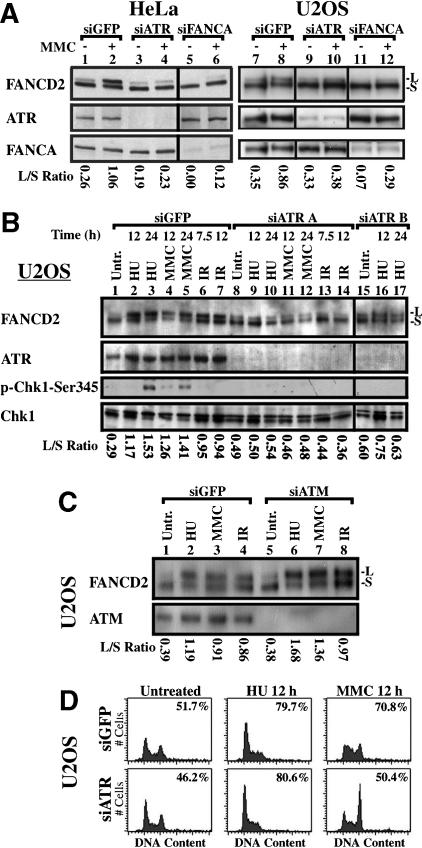
ATR is required for damage-inducible monoubiquitination of FANCD2. (A) ATR siRNA A strongly inhibits MMC-induced monoubiquitination of FANCD2 in HeLa and U2OS cells. Silencing of FANCA suppresses FANCD2 monoubiquitination either with or without exposure to MMC. (B) Silencing of ATR with siRNA in U2OS cells suppresses FANCD2 monoubiquitination following exposure to HU, MMC, or IR for the indicated times. Suppression of ATR activity is demonstrated by decreased phosphorylation of Chk1 on S345 in response to silencing. (C) Silencing of ATM does not decrease FANCD2 monoubiquitination. (D) Flow cytometric analysis demonstrates that inhibition of damage-induced monoubiquitination of FANCD2 by suppression of ATR is not due to an alteration of cell-cycle progression. Histograms of DNA content are displayed, with S-phase index measured by BrdU incorporation as described (Andreassen et al. 2001), inset in each histogram.
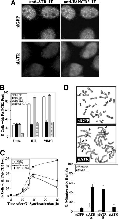
ATR is required for FANCD2 function following DNA damage. (A) ATR colocalizes with FANCD2 at DNA-damage foci formed by treatment of HeLa cells with 0.5 μM MMC for 24 h. Silencing of ATR with siATR A can be detected by immunofluorescence microscopy, and such cells are deficient for FANCD2 foci. (B) Silencing of ATR, but not ATM, leads to a strong decrease in the percentage of cells with FANCD2 foci following treatment with HU or MMC, relative to cells transfected with GFP siRNA. (C) HeLa cells treated with siGFP or siATR have similar levels of FANCD2 foci assembly during S phase in the absence of exogenous DNA damage. Cells were treated with 0.04 μg/mL nocodazole for 12 h, 4 d after transfection, and were synchronized in G1 by release in drug-free medium. MMC was added at 5 h release, where appropriate. Values represent three counts of 150 cells each. Standard deviation was <10% of the ordinate value. (D) Transformed human diploid fibroblasts (IMR90) deficient for ATR, following transfection with two different ATR siRNAs (A and B), have elevated formation of radial chromosomes in response to 48 h treatment with 0.032 μM MMC, relative to IMR90 cells transfected with GFP or ATM siRNA.
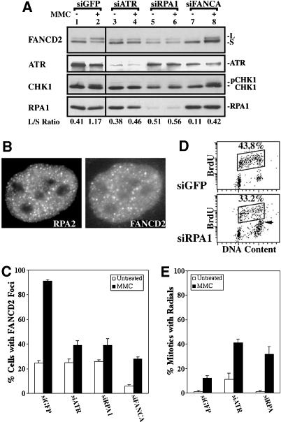
RPA1 is required for FANCD2 monoubiquitination following MMC treatment. (A) Immunoblots demonstrate that attenuation of RPA1 with siRNA strongly inhibits FANCD2 monoubiquitination in HeLa cells following treatment with 0.5 μM MMC for 24 h. (B) RPA2 colocalizes with FANCD2 in DNA-damage foci following 24 h treatment of HeLa cells with 0.5 μM MMC. (C) The formation of FANCD2 foci at 24 h treatment with 0.5 μM MMC is decreased strongly in HeLa cells in which either ATR or RPA1 had been attenuated with siRNA. (D) Dot plots of BrdU incorporation and DNA content demonstrate that siRPA does not suppress normal DNA replication during S phase (box). (E) Transformed human lung fibroblasts (IMR90) in which RPA1 has been silenced with siRNA form radial chromosomes in response to 48 h treatment with 0.032 μM MMC.
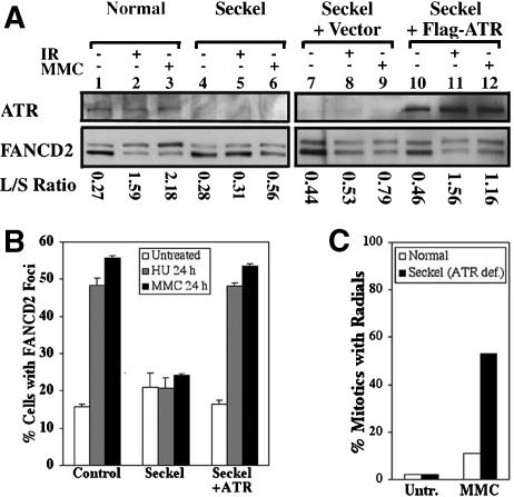
Cells derived from a Seckel syndrome patient are defective in the activation of the FA pathway. (A) Immunoblots with TERT immortalized fibroblasts, either wild type (PD846), ATR-deficient Seckel cells (FO2-98), Seckel cells with empty vector, or Seckel cells corrected by the expression of ATR were prepared from untreated specimens or cells exposed to 15 Gy IR for 12 h or 0.5 μM MMC for 24 h. (B) Damage-induced formation of FANCD2 foci occurs in control cells (PD846), but not in Seckel cells treated with 2 mM HU or 0.5 μM MMC for 24 h. (C) F02-98 Seckel cells, but not PD846 normal controls, show a highly elevated level of radial chromosomes in MMC.
References
-
- Abraham R.T. 2001. Cell cycle checkpoint signaling through the ATM and ATR kinases. Genes & Dev. 15: 2177-2196. - PubMed
-
- Bruun D., Folias, A., Akkari, Y., Cox, Y., Olson, S., and Moses, R. 2003. siRNA depletion of BRCA1, but not BRCA2, causes increased genome instability in Fanconi anemia cells. DNA Repair (Amst) 2: 1007-1013. - PubMed
-
- Casper A.M., Nghiem, P., Arlt, M.F., and Glover, T.W. 2002. ATR regulates fragile site stability. Cell 111: 779-789. - PubMed
-
- Chanan-Khan A., Holkova, B., Perle, M.A., Reich, E., Wu, C.D., Inghirami, G., and Takeshita, K. 2003. T-cell clonality and myelodysplasia without chromosomal fragility in a patient with features of Seckel syndrome. Haematologica 88: ECR14. - PubMed
Publication types
MeSH terms
Substances
Grants and funding
LinkOut - more resources
Full Text Sources
Other Literature Sources
Molecular Biology Databases
Research Materials
Miscellaneous