. 2004 Aug 15;18(16):1964-9.
doi: 10.1101/gad.305504.

Mosaic analyses using marked activation and deletion clones dissect Arabidopsis SCARECROW action in asymmetric cell division

Affiliations

Mosaic analyses using marked activation and deletion clones dissect Arabidopsis SCARECROW action in asymmetric cell division

Renze Heidstra et al. Genes Dev. .

Abstract

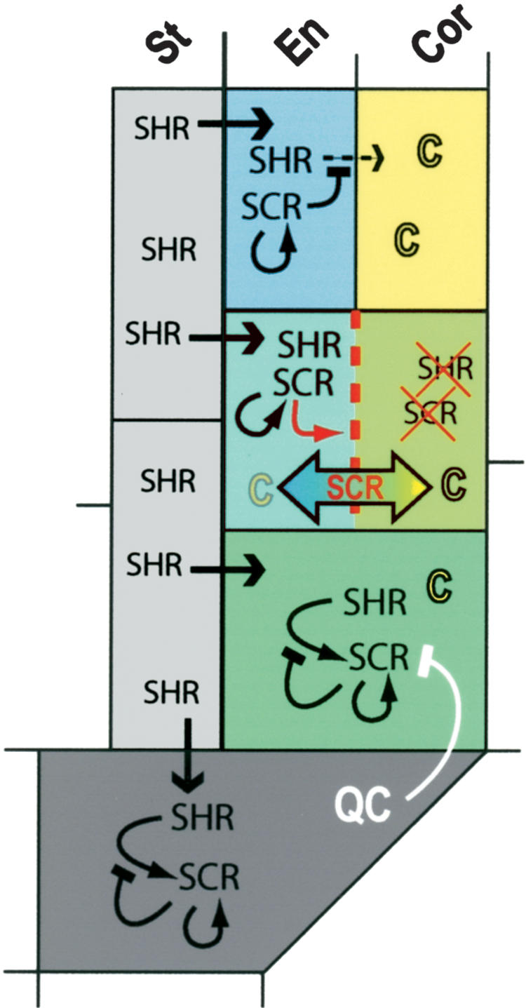
In the Arabidopsis root meristem, ground tissue stem cell daughters perform an asymmetric division to form endodermis and cortex. The putative transcription factors SCARECROW (SCR) and SHORTROOT (SHR) regulate this radial patterning event, and the mixed cell fate in scr mutants suggests an atypical role of the SCR gene in asymmetric cell division. Here we use a newly developed site-specific gene activation/deletion system in which induced clones are positively marked with green fluorescent protein (GFP). Using this system, we show that SCR acts cell-autonomously to control asymmetric cell division within the ground tissue. We provide evidence that SCR gene expression is under autoregulatory control, that SCR limits SHR movement, and that transient SCR action is sufficient to separate endodermis and cortex fates by asymmetric cell division.


Figures

Figure 1.
Figure 1.
Site-specific SCR gene activation and deletion to investigate alternative possibilities for SCR function in asymmetric division. (a) Schematic representation of cell types present in the Arabidopsis root meristem. Radially organized are the stele (St), endodermis (En), cortex (Cor), epidermis (Epi), and lateral root cap (Lrc). In the proximal-distal plane, quiescent center (QC) and columella (Col). (gtsc) Ground tissue stem cell; (scd) stem cell daughter. (b) Possibilities for SCR function in establishing asymmetry during periclinal ground tissue cell division. SCR is required for the execution of the periclinal division (1,2,3). (1) SCR and SHR are required to maintain endodermis fate; C determines cortex fate. (2) SCR plays no part in division asymmetry, only external factors determine endodermal and cortex fate. (3) During periclinal division, SCR separates endodermis and cortex fates. (c) Two possible signaling pathways allowing periclinal cell division in the ground tissue stem cell daughters. (1) Mature cells act as a patterning template and reinforce periclinal cell division. (2) QC signaling prevents differentiation and periclinal division in the stem cell. (d) Clonal SCR gene activation in scr-4 using vectors pCB1 and pG7HSCRE-USCR. Heat-shock activation of CRE recombinase mediates excision of the sequences between the direct repeat lox recombination sites. The 35S constitutive promoter then drives GAL4VP16 expression, resulting in transactivation of SCR expression and GFPER, thereby marking the cells with recombination events. (e) Clonal deletion of SCR using pCB1-SCR complemented scr-4 and pG7HSCRE. Heat-shock activation of CRE recombinase mediates excision of SCR, resulting in the 35S-driven GAL4VP16 and transactivation of GFPER expression, thereby marking the scr-4 mutant cells. Color-filled objects represent active promoters (green), transcribed genes (blue, green, orange, yellow), and terminators (red).
Figure 2.
Figure 2.
Induction of periclinal cell division by SCR activation clones. (a) Root tip showing GFPER-marked clones with SCR expression 16 has. (b) Close-up of section depicted in a, showing ground tissue clone. (c) Ground tissue clone from a and b has divided periclinally 24 has. (d) Additional periclinal ground tissue divisions (arrowhead) in scr-2 roots marked by SCR promoter-driven GFPER. (e) GFPER-marked SCR activation clone at the border of the meristem shows a periclinally divided ground tissue cell flanked by undivided proximal cells (arrowheads, 16 has). (f) At 24 has, the GFPER-marked cells have elongated and not divided again indicating their displacement from the meristem. (g,h) Vascular and epidermal clones do not induce periclinal divisions in the neighboring ground tissue. (i) SHR promoter-driven GFP:SHR expression in wild-type roots. GFP:SHR protein is localized to the nucleus in the endodermis, ground tissue stem cells, and QC. (j) Expression of pSCR::YFPH2B in endodermis, ground tissue stem cells, and QC in a wild-type root. (k) pSCR::YFPH2B is absent from the QC and low in scr-4 mutant ground tissue. (l,m) pSCR::YFPH2B is present in both ground tissue layers after periclinal divisions in scr-4 roots. (n) GFP:SHR is present in both ground tissue layers after periclinal divisions in scr-4 roots. Brackets indicate ground tissue. Internal tissues are to the left.
Figure 3.
Figure 3.
Activation clones reveal cell-autonomous SCR function in asymmetric division. (a-d) pSCR::YFPH2B is induced in a SCR activation clone and divided over the inner and outer ground tissue cells on periclinal division at 16 has (a,b). Later, pSCR::YFPH2B separates asymmetrically to the inner cells. Induced periclinal ground tissue divisions observed in cells flanking the clone (arrows) exhibit low levels of pSCR::YFPH2B that is divided equally over both cells (c,d; 40 has). (e,f) GFPER-marked ground tissue SCR activation clone (e; 16 has) induces periclinal division in a flanking GFPER- cell (f; arrow; 24 has). (g,h) GFPER- ground tissue cells flanking a periclinal GFPER-marked SCR activation G2-clone (g, arrows, 16 has) are nonautonomously induced to perform periclinal divisions (h; arrows; 24 has). (i-k) Periclinal division in GFPER- ground tissue cells flanking a SCR:GFP activation clone in the same (i) and adjacent (j) cell file 40 has. (k) Optical cross-section of the root in i and j, revealing two nonautonomously induced periclinal ground tissue divisions in cell files flanking the SCR:GFP activation clone (arrows). Nuclear SCR:GFP strictly colocalizes with the GFPER-marked activation clone. (l-o) pSCR::YFPH2B is divided over both ground tissue layers following periclinal division in a SCR activation clone (l,m; 16 has). At 24 has, functional SCR gene expression marked by GFPER expression throughout the cells localizes to the outer cells in the periclinal G2-clone (n; arrow), whereas nuclear pSCR::YFPH2B still separates asymmetrically to the inner cells (o). (a,c,l,n) GFPER and YFPH2B are visualized simultaneously in the GFP mode (see Materials and Methods). Brackets indicate ground tissue. Internal tissues are to the left.
Figure 4.
Figure 4.
Cell autonomy, autoregulation, and ground tissue determination revealed by SCR deletion clones. (a,b,e) GFPER-marked single-layered ground tissue SCR deletion clones (arrowheads) originating from a scr-/- stem cell are not induced to divide periclinally by neighboring cells expressing wild-type SCR levels. (e) Optical cross-section of a root containing a GFPER-marked SCR deletion clone circumpherentially flanked by wild-type ground tissue. (c,f) QC, ground tissue stem cells, and their daughters rapidly lose pSCR::YFPH2B when SCR expression is eliminated (arrowheads), whereas pSCR::YFPH2B is maintained in the already formed endodermis 40 has. (d,g) In a different root at 72 has, pSCR::YFPH2B is reduced to background levels in the already formed endodermis within a SCR deletion clone (open arrowheads). pSCR::YFPH2B remains absent in the single ground tissue layer originating from the mutant stem cell (arrowheads). (h) pCo2::YFPH2B marker expression in the cortex of wild-type roots is excluded from QC, ground tissue stem cells, and their single-layered daughters. (i) pCo2::YFPH2B expression in the single-layered ground tissue and QC (arrow) in scr-4 roots. (j,k) Massive GFPER marked the SCR deletion clone, including the ground tissue that contains a pile of single-layered cells (arrowheads, 48 has). pCo2::YFPH2B remains present in the cortex and is also present in the single-layered ground tissue (k, arrows). (l) pEn7::YFPH2B expression in the endodermis of wild-type roots is only excluded from the QC cells. (m) pEn7::YFPH2B expression in the single layered ground tissue in scr-4 roots. (n,o) Massive GFPER-marked SCR deletion clone, including the ground tissue that contains a pile of single layered cells (arrowheads, 48 has). (o) pEn7::YFPH2B remains present in the endodermis (arrows) and is also present in the single-layered ground tissue. Brackets indicate ground tissue. (e) Endodermis; (c) cortex. (a,b,h,i,l,m) Internal tissues are to the left.
Figure 5.
Figure 5.
SCR function in QC and ground tissue. SHR moves from the stele to the QC and ground tissue cells. In the QC and ground tissue stem cells, SCR expression is induced by SHR and maintained by autoregulation. The inductive capacity of SHR is lost on SCR expression. The QC prevents SCR induced periclinal cell division in the ground tissue stem cell. SCR induces the periclinal division in the ground tissue stem cell daughter. SCR mediates rapid endodermal (SHR) and cortical (C) fate separation and segregation of factors required for execution of the periclinal division. Residual SHR and SCR in the outer cell is degraded. In the endodermis, SCR prevents SHR movement to the neighboring cortex cell file, thereby preventing additional periclinal divisions.

Similar articles

Cited by

References

    1. Benfey P.N., Linstead, P.J., Roberts, K., Schiefelbein, J.W., Hauser, M.T., and Aeschbacher, R.A. 1993. Root development in Arabidopsis: Four mutants with dramatically altered root morphogenesis. Development 119: 57-70. - PubMed
    1. Di Laurenzio L., Wysockadiller, J., Malamy, J.E., Pysh, L., Helariutta, Y., Freshour, G., Hahn, M.G., Feldmann, K.A., and Benfey, P.N. 1996. The SCARECROW gene regulates an asymmetric cell division that is essential for generating the radial organization of the Arabidopsis root. Cell 86: 423-433. - PubMed
    1. Dolan L., Janmaat, K., Willemsen, V., Linstead, P., Poethig, S., Roberts, K., and Scheres, B. 1993. Cellular organization of the Arabidopsis root. Development 119: 71-84. - PubMed
    1. Helariutta Y., Fukaki, H., Wysocka-Diller, J., Nakajima, K., Jung, J., Sena, G., Hauser, M.T., and Benfey, P.N. 2000. The SHORT-ROOT gene controls radial patterning of the Arabidopsis root through radial signaling. Cell 101: 555-567. - PubMed
    1. Horvitz H.R., and Herskowitz, I. 1992. Mechanisms of asymmetric cell division: Two Bs or not two Bs, that is the question. Cell 68: 237-255. - PubMed