

Comparative Study
doi: 10.1101/gad.1219904.
Epub 2004 Aug 16.
Conjugation-specific small RNAs in Tetrahymena have predicted properties of scan (scn) RNAs involved in genome rearrangement
Affiliations
- PMID: 15314029
- PMCID: PMC515285
- DOI: 10.1101/gad.1219904
Item in Clipboard
Comparative Study
Conjugation-specific small RNAs in Tetrahymena have predicted properties of scan (scn) RNAs involved in genome rearrangement
Genes Dev.
.
Abstract
We proposed a scan-RNA model for genome rearrangement based on finding small RNAs that hybridized preferentially to micronuclear-specific sequences and on the properties of Twi1p, a PPD protein required for both sequence elimination and small RNA accumulation in Tetrahymena. Here we show that Twi1p interacts with the small RNAs in both the old and the developing macronucleus, and is required for their stability. We show that the specificity of the small RNAs for micronuclear-limited sequences increases during conjugation. These results indicate that the small RNAs observed in conjugating cells have the properties predicted for scan RNAs.
Figures

Construction and characterization of TWI1 germ-line knockout strains and Flag-TWI1 strains. (A) Genotyping of TWI1 germ-line knockout strains and Flag-TWI1 strains. The genotypes of the TWI1 loci in macro- and micronuclei of each cell type are illustrated at the top. TWI1 and its targeted loci are schematically drawn on the left. Arrows indicate the locations of primers used for genotyping PCR. (Lanes O1,O2) Some homozygous TWI1 knockout homokaryon strains had deletions in the neo3 cassette and gave both intact (4.3 kb) and deleted (∼2.5 kb) products. (Lanes O1′,O2′) Because macronuclear chromosomes are randomly segregated when macronuclei divide, intact targeted loci can be sorted out and knockout cells containing only deleted targeted loci can be obtained. (Lane F) The Flag-TWI1 strain was constructed in a homozygous TWI1 knockout homokaryon background and complete disruption of endogenous TWI1 loci in this strain was confirmed. Note that the region corresponding to the downstream primer was absent in pD5H8-Flag-TWI1, and thus the Flag-TWI1 gene could not be detected by PCR. (B) IES elimination assay. Double horizontal lines indicate DNA retained in the macronucleus and the filled box indicates an IES. Four primers (arrows on the horizontal lines) were used for nested PCR. The sizes of the processed (macronuclear form) and unprocessed (micronuclear form) of the M region are marked by arrowheads with “a” and “i”, respectively. (M) Molecular weight marker. (C) Expression of small RNA in wild-type and ΔTWI1 homozygous homokaryon cells. Total RNA was extracted from mating wild-type or ΔTWI1 homozygous homokaryon cells at 0, 4, 8, and 12 h postmixing and the RNA corresponding to 5 × 10⁴ cells was analyzed. The RNA was fractionated in 12% acrylamide-urea gels and stained with ethidium bromide. In vitro transcribed, 17-, 26-, and 51-nt RNAs were used as markers (M). The gel was partially destained and tRNAs amounts were observed to normalize the loading. The amount of the small RNAs in wild-type cells at 4 h postmixing was adjusted as 1.00 and the relative ratio of the small RNAs at other times is indicated at the bottom. Total RNA was used for Northern hybridization with a probe for PDD1 (631–943 bp, GenBank TTU66363) mRNA which is specifically expressed in conjugating cells.
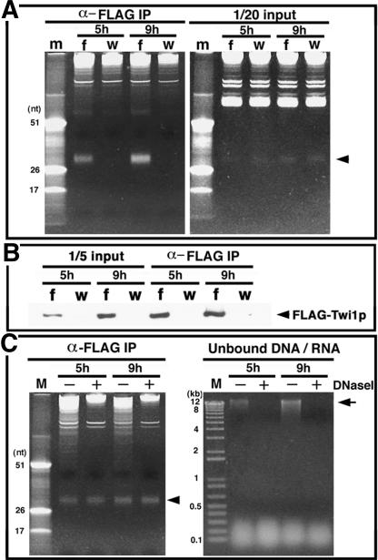
Coimmunoprecipitation of small RNAs with Flag-Twi1p. (A) Whole cell lysates were prepared from the mating of Flag-TWI1 × CU428 (f) or B2086 × CU428 (w) and the Flag-Twi1p-containing complex was immunoprecipitated using anti-Flag antibody. RNA extracted from one-twentieth of the lysates (right panel) and from the fraction eluted from the immunoprecipitate with Flag peptide (left panel) was analyzed by 12% acrylamide-urea gel electrophoresis followed by ethidium bromide staining. The marker (m) was the same as in Figure 1. The arrowhead shows the position of the small RNAs. (B) Flag-Twi1p in the elution (right) and the lysate (left, corresponding to one-fifth of total protein used for immunoprecipitation), was analyzed on Western blot using anti-Flag antibody. (C, Left) Immunoprecipitation was performed with (+) or without (-) DNase treatment and RNA was analyzed. The arrowhead shows the position of the small RNA. (Right) To confirm that genomic DNA in lysates had been digested, nucleic acids were extracted from the unbound fraction after immunoprecipitation with phenol/chloroform and fractionated in a 1% agarose gel followed by ethidium bromide staining. The arrow indicates the position of high-molecular-weight (genomic) DNA.

Hybridization of macro- and micronuclear DNA with labeled small RNAs. (A) Macronuclear (a) or micronuclear (i) DNA was isolated and digested with EcoRI. The DNA was separated in a 1% agarose gel and transferred to a membrane. To prepare the probe, total RNA from wild-type conjugating cells at 2, 4, 6, 8, 10, and 12 h postmixing was fractionated in a 12% acrylamide-urea gel and the small RNA was extracted from the gel. The purified small RNA was end-labeled with 32P and hybridized to macro- and micronuclear DNAs on a Southern blot. As a loading control, a probe for the HHO1 gene encoding the histone H1 was hybridized to the same blot. Note that approximately three times more DNA was loaded on lanes containing macronuclear DNA than on micronuclear DNA lanes. (B) The ratio of the small RNAs hybridized to micronuclear DNA versus macronuclear DNA from different time points is shown as a bar graph. The loading was normalized by using the signal obtained with the HHO1 probe.
References
-
- Godiska R., James, C., and Yao, M.C. 1993. A distant 10-bp sequence specifies the boundaries of a programmed DNA deletion in Tetrahymena. Genes & Dev. 7: 2357-2365. - PubMed
Publication types
MeSH terms
Substances
Grants and funding
LinkOut - more resources
Full Text Sources
Other Literature Sources