. 2004 Aug 16;166(4):455-63.
doi: 10.1083/jcb.200312048.

Dynamic targeting of the replication machinery to sites of DNA damage

Affiliations

Dynamic targeting of the replication machinery to sites of DNA damage

David A Solomon et al. J Cell Biol. .

Abstract

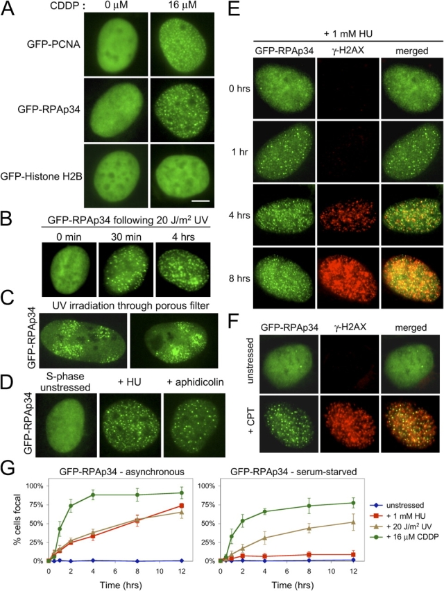
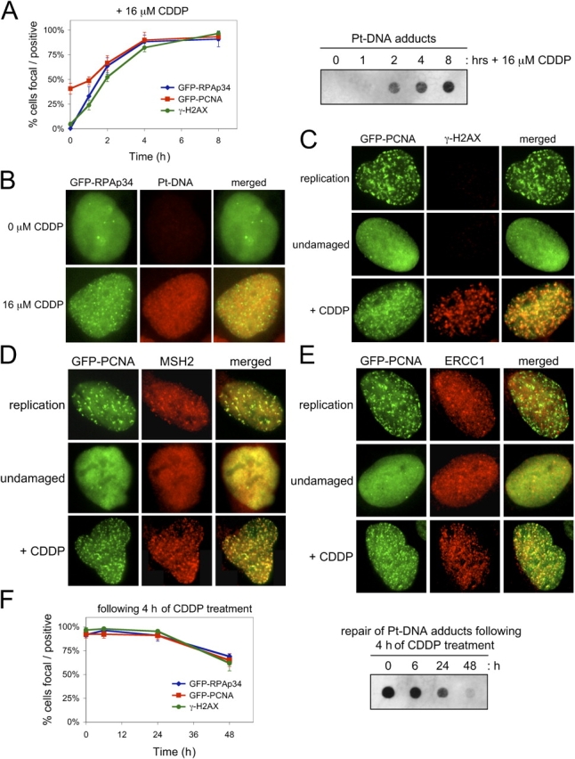
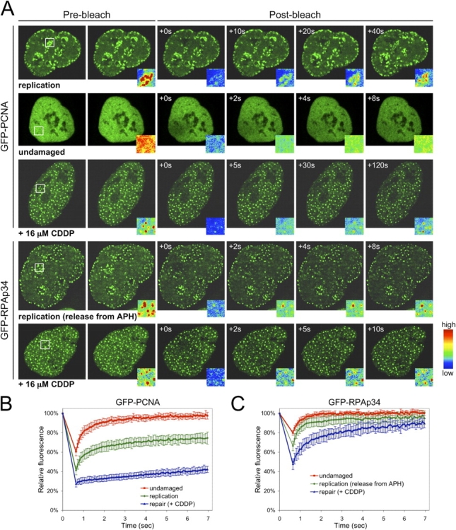
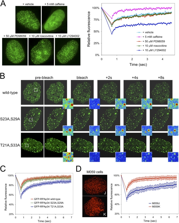
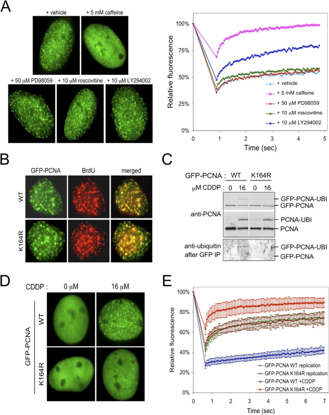
Components of the DNA replication machinery localize into discrete subnuclear foci after DNA damage, where they play requisite functions in repair processes. Here, we find that the replication factors proliferating cell nuclear antigen (PCNA) and RPAp34 dynamically exchange at these repair foci with discrete kinetics, and this behavior is distinct from kinetics during DNA replication. Posttranslational modification is hypothesized to target specific proteins for repair, and we find that accumulation and stability of PCNA at sites of damage requires monoubiquitination. Contrary to the popular notion that phosphorylation on the NH2 terminus of RPAp34 directs the protein for repair, we demonstrate that phosphorylation by DNA-dependent protein kinase enhances RPAp34 turnover at repair foci. Together, these findings support a dynamic exchange model in which multiple repair factors regulated by specific modifications have access to and rapidly turn over at sites of DNA damage.


Figures

Figure 1.
Figure 1.
Visualization of replication factors in the DNA damage response. (A) CDDP induces the relocalization of GFP-PCNA and GFP-RPAp34, but not GFP-histone H2B, into discrete foci within the nucleus of stably expressing Rat-1 cells. Bar, 5 μm. (B) RPAp34 rapidly accumulates into foci in response to UV irradiation. (C) Cells expressing GFP-RPAp34 were exposed to UV irradiation through a porous polycarbonate filter. GFP-RPAp34 only accumulates into foci within the irradiated microdomains 3 μm in diameter. (D) RPAp34 accumulates into foci in response to the stalling of DNA replication with HU or APH. (E) RPAp34 foci appear before the accumulation of H2AX phosphorylation after HU treatment. (F) Camptothecin (CPT) induces the formation of RPAp34 foci that colocalize with γ-H2AX foci. (G) Quantitation of the number of cells with punctate versus diffuse localization of GFP-RPAp34 in asynchronous (left) or serum-starved (right) cultures after various stresses over time. 200 cells were counted at each time point from each of three independent experiments. After 48 h serum-starvation, 4% of the unstressed cells labeled BrdU positive during the 12-h time course of the experiment.
Figure 2.
Figure 2.
Kinetic induction of repair foci by cisplatin. (A) Cells were treated with 16 μM CDDP, and total DNA was analyzed for Pt-DNA levels at the indicated times by dot blotting (right). Cells were concurrently scored for PCNA/RPAp34 foci and γ-H2AX reactivity (left). (B) Representative images of GFP-RPAp34 and Pt-DNA adducts after 4 h of CDDP damage. (C–E) Representative images of GFP-PCNA and γ-H2AX (C), MSH2 (D), or ERCC1 (E) localization in replicating, undamaged, and CDDP-treated cells. (F) Cells were treated with 16 μM CDDP for 4 h and then cultured in the absence of CDDP for the indicated times. DNA was analyzed for Pt-DNA levels by dot blotting (right), and cells were concurrently scored for PCNA/RPAp34 foci and γ-H2AX reactivity (left). The error bars are the SD in the cell, counting from three independent experiments.
Figure 3.
Figure 3.
Components of the replication machinery turn over at repair foci with distinct kinetics. (A) FRAP on undamaged, replicating, and CDDP-treated Rat-1 cells expressing GFP-PCNA and GFP-RPAp34. Pseudocolored insets display the intensity profile of the photobleached region boxed in white, 2.9 μm × 2.9 μm. (B and C) Quantitative FRAP analysis of GFP-PCNA (B) and GFP-RPAp34 (C) in undamaged, replicating, and CDDP-treated (4 h) Rat-1 cells. These replication proteins exhibit distinct dynamic properties and recover more slowly during repair than replication. The error bars are the SD in the FRAP measurements.
Figure 4.
Figure 4.
Phosphorylation by DNA-PK enhances turnover of RPAp34 at repair foci. (A) Localization and quantitative FRAP analysis of U2OS cells stably expressing GFP-RPAp34 after damage with 16 μM CDDP in combination with the indicated drug for 4 h. (B) FRAP on U2OS cells stably expressing GFP-RPAp34 wild type, a CDK phosphorylation site mutant (S23A and S29A), or a DNA-PK phosphorylation site mutant (T21A and S33A) after damage with 16 μM CDDP for 4 h. Pseudocolored insets display the intensity profile of the photobleached region boxed in white, 2.9 μm × 2.9 μm. (C) Quantitative FRAP analysis of cells in B. (D) Representative images of endogenous RPA (left) and quantitative FRAP analysis of GFP-RPAp34 (right) expressed in M059J and M059K cells after damage with 16 μM CDDP for 4 h. The error bars are the SD in the FRAP measurements.
Figure 5.
Figure 5.
Monoubiquitination of Lys-164 targets PCNA for DNA damage repair. (A) Localization and quantitative FRAP analysis of U2OS cells stably expressing GFP-PCNA after damage with 16 μM CDDP in combination with the indicated drug for 4 h. (B) Colocalization of GFP-PCNA wild type and K164R mutant with sites of active BrdU incorporation. (C) K164R mutation renders PCNA refractory to ubiquitination after genotoxic insult. Total cell lysate (top) and protein immunoprecipitated with GFP antibody (bottom) from U2OS cells stably expressing GFP-PCNA wild type or K164R mutant after treatment with vehicle or CDDP were resolved by SDS-PAGE, and the indicated protein species were detected by immunoblot. (D) Localization of PCNA wild type and K164R mutant after insult with CDDP for 4 h. (E) Quantitative FRAP analysis of GFP-PCNA wild-type and K164R mutant in U2OS cells during replication and after damage with 16 μM CDDP for 4 h. The error bars are the SD in the FRAP measurements.

Similar articles

Cited by

References

    1. Angus, S.P., D.A. Solomon, L. Kuschel, R.F. Hennigan, and E.S. Knudsen. 2003. Retinoblastoma tumor suppressor: analyses of dynamic behavior in living cells reveal multiple modes of regulation. Mol. Cell. Biol. 23:8172–8188. - PMC - PubMed
    1. Barr, S.M., C.G. Leung, E.E. Chang, and K.A. Cimprich. 2003. ATR kinase activity regulates the intranuclear translocation of ATR and RPA following ionizing radiation. Curr. Biol. 13:1047–1051. - PubMed
    1. Dutta, A., and B. Stillman. 1992. cdc2 family kinases phosphorylate a human cell DNA replication factor, RPA, and activate DNA replication. EMBO J. 11:2189–2199. - PMC - PubMed
    1. Garcia-Higuera, I., T. Taniguchi, S. Ganesan, M.S. Meyn, C. Timmers, J. Hejna, M. Grompe, and A.D. D'Andrea. 2001. Interaction of the Fanconi anemia proteins and BRCA1 in a common pathway. Mol. Cell. 7:249–262. - PubMed
    1. He, Z., L.A. Henricksen, M.S. Wold, and C.J. Ingles. 1995. RPA involvement in the damage-recognition and incision steps of nucleotide excision repair. Nature. 374:566–569. - PubMed

Publication types

MeSH terms