Comparative Study
. 2004 Aug 16;200(4):541-7.
doi: 10.1084/jem.20040061.

Statins inhibit HIV-1 infection by down-regulating Rho activity

Affiliations
Comparative Study

Statins inhibit HIV-1 infection by down-regulating Rho activity

Gustavo del Real et al. J Exp Med. .

Abstract

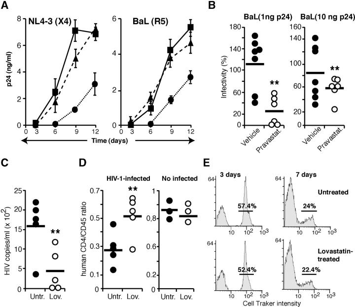
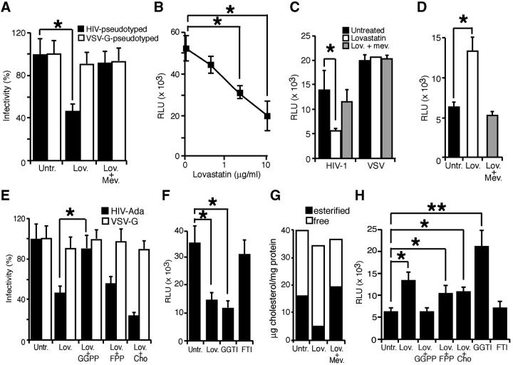
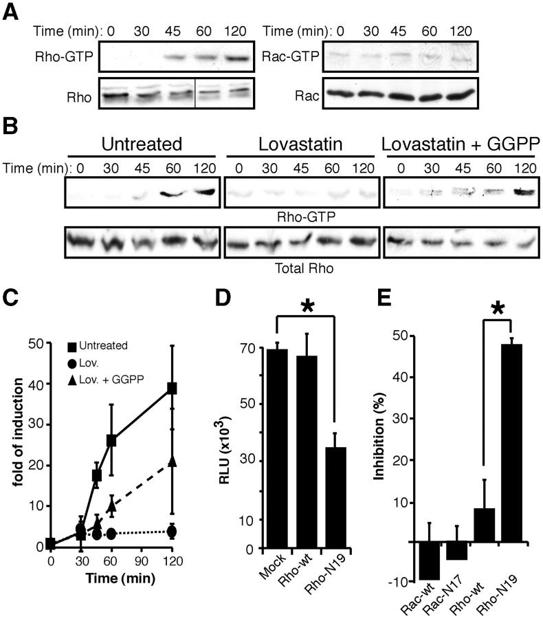
Human immunodeficiency virus (HIV)-1 infectivity requires actin-dependent clustering of host lipid raft-associated receptors, a process that might be linked to Rho guanosine triphosphatase (GTPase) activation. Rho GTPase activity can be negatively regulated by statins, a family of drugs used to treat hypercholesterolemia in man. Statins mediate inhibition of Rho GTPases by impeding prenylation of small G proteins through blockade of 3-hydroxy-3-methylglutaryl coenzyme A reductase. We show that statins decreased viral load and increased CD4+ cell counts in acute infection models and in chronically HIV-1-infected patients. Viral entry and exit was reduced in statin-treated cells, and inhibition was blocked by the addition of l-mevalonate or of geranylgeranylpyrophosphate, but not by cholesterol. Cell treatment with a geranylgeranyl transferase inhibitor, but not a farnesyl transferase inhibitor, specifically inhibited entry of HIV-1-pseudotyped viruses. Statins blocked Rho-A activation induced by HIV-1 binding to target cells, and expression of the dominant negative mutant RhoN19 inhibited HIV-1 envelope fusion with target cell membranes, reducing cell infection rates. We suggest that statins have direct anti-HIV-1 effects by targeting Rho.


Figures

Figure 1.
Figure 1.
Statins inhibit in vitro and in vivo HIV-1 infection of human PBMCs. (A) Infection of untreated (▪), Lov- (•), or Lov plus Mev–treated (▴) PHA-activated human PBMCs by X4 or R5 HIV-1 viral strains. Data are mean ± SD of triplicate points (n = 3). (B) PBMCs isolated from human volunteers, before and after vehicle or pravastatin treatment, were exposed to two doses of BaL HIV-1. Data are the ratio between post- and pretreatment p24 levels for PBMCs from each individual (**, P < 0.05). (C and D) Human PBMC-reconstituted SCID mice were Lov treated for 1 wk before HIV-1 infection. Viral load (C) and human CD4/CD45 ratio (D) was determined for each animal 1 wk after infection. One representative experiment out of two is shown (**, P < 0.05). The CD4+/CD45+ ratio was also determined in noninfected mice (D, right). (E) Lov-treated SCID mice were reconstituted with CellTracker-stained PBMCs and peritoneal cell labeling was examined at the indicated times. The numbers indicate the percentage of labeled cells.
Figure 2.
Figure 2.
Statins inhibit HIV-1 entry and exit. (A) Single-round infections were performed in untreated, Lov-, and Lov plus Mev–treated MT2-CCR5 cells using a replication-defective NL4-3 virus bearing the luciferase reporter pseudotyped with HIV-1Ada or VSV-G envelopes. Cell infection was normalized using untreated cells as 100%. (B) MT2-CCR5 cells were Lov treated (0.4, 2, or 10 μM), and then exposed to NL4-3 virus pseudotyped with HIV-1Ada envelope. The x axis is in log scale. (C) Virus production was measured by titration of viral stocks produced in untreated, Lov-, and Lov plus Mev–treated HEK-293T cells transfected with replication-defective NL4-3 virus. Relative luciferase units were calculated after normalization with luciferase activity from extracts of stock-producing cells. (D) LTR-driven gene expression was analyzed in untreated, Lov-, or Lov plus Mev–treated Jurkat cells transfected with pLTR-Luc, pcDNA-tat, and promoterless renilla for normalization. (E) Single-round infection experiments were performed using replication-defective NL4-3 virus in MT2-CCR5 cells treated with Lov or Lov plus the indicated compounds. Cell infection was normalized considering untreated cells as 100%. (F) Single-round infections performed with the HIV-1Ada–pseudotyped virus in MT2-CCR5 cells treated with Lov, GGTI, or FTI. (G) Free or esterified cholesterol levels in untreated, Lov-, or Lov plus Mev–treated MT2-CCR5 cells. One representative experiment out of two is shown. (H) LTR-driven gene expression in MT2-CCR5 cells treated with Lov, Lov plus the indicated compounds, or with GGTI-286 or FTI-277. (A–E, G, and H) Data are mean ± SD of duplicates (n = 3). Significant differences are indicated: *, P < 0.05; **, P < 0.01. Kruskal-Wallis test.
Figure 3.
Figure 3.
Statins inhibit HIV-1 infection by down-regulating Rho activation. (A) Serum-starved MT2-CCR5 cells were incubated with HIV-1 and cell lysates were assayed for active Rho or Rac. Total Rho or Rac was analyzed in parallel in crude cell extracts as a protein loading control. One experiment out of three is shown. Black line indicates that different sections of the same gel were juxtaposed. (B) Active Rho was determined in untreated, Lov-, or Lov plus GGPP–treated cells, as described above. Total Rho in crude cell extracts is shown as a loading control. One representative experiment out of three is shown. (C) Western blots from three independent experiments as in B were quantified by densitometry and values were normalized using Rho in crude cell extracts as a loading control. Data points are plotted relative to mean values of cells not exposed to virus (time 0) for each condition. (D) Single-round infections of MT2-CCR5 cells transfected with mock, wild-type Rho, or mutant Rho-N19 using an HIV-1Ada–pseudotyped, replication-defective virus. *, P < 0.05. Kruskal-Wallis test. (E) HeLa-CD4 cells transfected with wild-type Rac, wild-type Rho, Rac-N17, or Rho-N19 were mixed with HIV gp160–expressing BSC40 cells. Cell fusion events were measured and normalized relative to mock transfected cells. *, P < 0.05. Kruskal-Wallis test. (D and E) Data are mean ± SD of duplicate points (n = 3).

References

    1. Mañes, S., G. del Real, and C. Martínez-A. 2003. Pathogens: raft hijackers. Nat. Rev. Immunol. 3:557–568. - PubMed
    1. Mañes, S., G. del Real, R. Lacalle, P. Lucas, C. Gomez-Mouton, S. Sanchez-Palomino, R. Delgado, J. Alcami, E. Mira, and C. Martinez-A. 2000. Membrane raft microdomains mediate lateral assemblies required for HIV-1 infection. EMBO Rep. 1:190–196. - PMC - PubMed
    1. Nguyen, D., and J. Hildreth. 2000. Evidence for budding of human immunodeficiency virus type 1 selectively from glycolipid-enriched membrane lipid rafts. J. Virol. 74:3264–3272. - PMC - PubMed
    1. Ono, A., and E. Freed. 2001. Plasma membrane rafts play a critical role in HIV-1 assembly and release. Proc. Natl. Acad. Sci. USA. 98:13925–13930. - PMC - PubMed
    1. del Real, G., S. Jiménez-Baranda, R. Lacalle, E. Mira, P. Lucas, C. Gómez-Moutón, A. Carrera, C. Martínez-A., and S. Mañes. 2002. Blocking of HIV-1 infection by targeting CD4 to nonraft membrane domains. J. Exp. Med. 196:293–301. - PMC - PubMed

Publication types

MeSH terms