. 2004 Sep;24(17):7435-43.
doi: 10.1128/MCB.24.17.7435-7443.2004.

Disruption of the SH2-B gene causes age-dependent insulin resistance and glucose intolerance

Affiliations

Disruption of the SH2-B gene causes age-dependent insulin resistance and glucose intolerance

Chaojun Duan et al. Mol Cell Biol. 2004 Sep.

Abstract

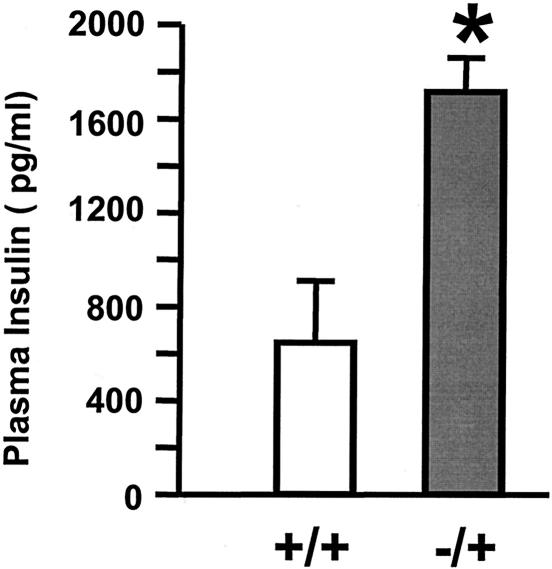
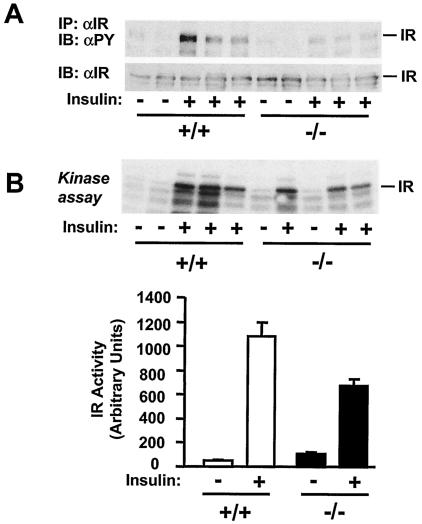
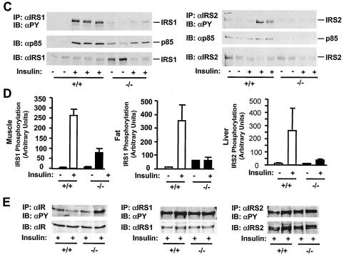
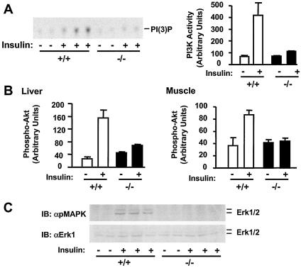
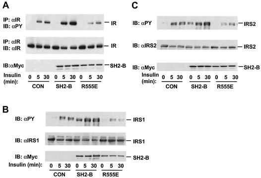
Insulin regulates glucose homeostasis by binding and activating the insulin receptor, and defects in insulin responses (insulin resistance) induce type 2 diabetes. SH2-B, an Src homology 2 (SH2) and pleckstrin homology domain-containing adaptor protein, binds via its SH2 domain to insulin receptor in response to insulin; however, its physiological role remains unclear. Here we show that SH2-B was expressed in the liver, skeletal muscle, and fat. Systemic deletion of SH2-B impaired insulin receptor activation and signaling in the liver, skeletal muscle, and fat, including tyrosine phosphorylation of insulin receptor substrate 1 (IRS1) and IRS2 and activation of the phosphatidylinositol 3-kinase/Akt and the Erk1/2 pathways. Consequently, SH2-B-/- knockout mice developed age-dependent hyperinsulinemia, hyperglycemia, and glucose intolerance. Moreover, SH2-B directly enhanced autophosphorylation of insulin receptor and tyrosine phosphorylation of IRS1 and IRS2 in an SH2 domain-dependent manner in cultured cells. Our data suggest that SH2-B is a physiological enhancer of insulin receptor activation and is required for maintaining normal insulin sensitivity and glucose homeostasis during aging.


Figures

FIG. 1.
FIG. 1.
Disruption of the SH2-B gene in mice. (A) Tissue distributions of SH2-B protein. Tissue extracts were prepared from a wild-type male (5 weeks old; 129/Sv and C57BL/6 mixed background), immunoprecipitated, and immunoblotted with anti-SH2-B. Proteins (2 mg for spleen, pancreas, and liver or 1 mg for muscle and fat) were used for immunoprecipitation. (B) Schematic representation of target vector and homologous recombination. The positions of the probe for Southern blotting (P) and BamHI sites (B) were marked. (C) Southern blots for the SH2-B gene. Genomic DNAs were digested with BamHI and subjected to Southern blot analysis. (D) Liver extracts were prepared from wild-type or SH2-B−/− males. SH2-B was immunoprecipitated (IP) and immunoblotted (IB) with anti-SH2-B (αSH2-B) (upper panel). Liver extracts were immunoblotted with anti-Erk1 (αERK1) (lower panel).
FIG. 2.
FIG. 2.
SH2-B−/− mice develop hyperglycemia and hyperinsulinemia. Wild-type (n = 12) and SH2-B−/− (n = 10) males were fed a standard mouse diet (9% fat content) for 6 to 7 months. (A) Blood glucose from fasting mice; (B) plasma insulin from fasting mice; (C) blood glucose from randomly fed mice; (D) plasma insulin from randomly fed mice. *, P < 0.05 compared with wild-type controls.
FIG. 3.
FIG. 3.
SH2-B−/− mice develop glucose intolerance and reduced response to exogenous insulin. Wild-type (n = 12) and SH2-B−/− (n = 10) males were a fed standard mouse diet (9% fat content) for 6 to 7 months. (A) GTT. Mice were injected intraperitoneally with d-glucose (2 mg/kg of body weight), and blood glucose was monitored at 0, 15, 30, 60, 90, 120, and 150 min after injection. (B) ITT. Mice were injected intraperitoneally with human insulin (1 IU/kg of body weight), and blood glucose was monitored at 0, 15, 30, and 60 min after injection.
FIG. 4.
FIG. 4.
Disruption of the SH2-B gene causes hyperplasia of islets. Pancreatic sections (5 μm thick) from wild-type or SH2-B−/− knockout males (6 months old) were stained with hematoxylin and eosin (H&E) or antibodies against insulin or glucagon as indicated. Bar, 2.5 μm.
FIG. 5.
FIG. 5.
Heterozygous deletion of SH2-B enhances high-fat-diet-induced insulin resistance. Wild-type or heterozygous SH2-B+/− mice (5-month-old males, n = 5) were fed a high-fat diet (45% fat, D12451; Research Diets, Inc.) for 4 weeks. Animals were subjected to fasting overnight (16 h), and plasma insulin was measured by using rat insulin enzyme-linked immunosorbent assay kits. *, P < 0.05 compared with wild-type controls.
FIG. 6.
FIG. 6.
Disruption of the SH2-B gene inhibits IR activation, tyrosine phosphorylation of IRS1 and IRS2, and interaction of IRS proteins with p85. Wild-type or SH2-B−/− males (7 months old) were subjected to fasting overnight (∼18 h) and administered human insulin (3 U per mouse) or PBS as a control via the inferior vena cava. The liver, gastrocnemius muscle, and epididymal fat were isolated 5 min after stimulation and homogenized. (A) Muscle extracts were immunoprecipitated (IP) with anti-IR (αIR) and immunoblotted (IB) with antiphosphotyrosine (αPY, top panel) or anti-IR (bottom panel). Each lane represents an individual mouse. (B) Immunopurified hepatic IR was subjected to in vitro kinase assays in the presence of [γ-32P]ATP with (+) and without (−) insulin. (C) IRS1 was immunoprecipitated with anti-IRS1 (αIRS1) from muscle extracts and separated by SDS-PAGE. The blot was cut into two pieces along the molecular standard of 117 kDa. The upper piece was immunoblotted with antiphosphotyrosine and subsequently reprobed with anti-IRS1. The bottom piece was immunoblotted with the anti-p85 regulatory subunit of PI 3-kinase (αp85). Similarly, hepatic IRS2 was immunoprecipitated with anti-IRS2 (αIRS2) and immunoblotted with anti-phosphotyrosine, anti-p85, or anti-IRS2 as indicated. (D) Tyrosine phosphorylation of IRS1 or IRS2 was quantified and normalized to total IRS1 or IRS2, respectively. Each bar represents the average of the results from two to three mice. (E) SH2-B−/− and wild-type littermates (7-week-old males) were subjected to fasting overnight (∼18 h) and treated with human insulin (3 U per mouse) for 5 min. Liver extracts were immunoprecipitated with anti-IR, anti-IRS1, or anti-IRS2 and immunoblotted with antiphosphotyrosine. The blots were reprobed with anti-IR, anti-IRS1, or anti-IRS2 as indicated.
FIG. 6.
FIG. 6.
Disruption of the SH2-B gene inhibits IR activation, tyrosine phosphorylation of IRS1 and IRS2, and interaction of IRS proteins with p85. Wild-type or SH2-B−/− males (7 months old) were subjected to fasting overnight (∼18 h) and administered human insulin (3 U per mouse) or PBS as a control via the inferior vena cava. The liver, gastrocnemius muscle, and epididymal fat were isolated 5 min after stimulation and homogenized. (A) Muscle extracts were immunoprecipitated (IP) with anti-IR (αIR) and immunoblotted (IB) with antiphosphotyrosine (αPY, top panel) or anti-IR (bottom panel). Each lane represents an individual mouse. (B) Immunopurified hepatic IR was subjected to in vitro kinase assays in the presence of [γ-32P]ATP with (+) and without (−) insulin. (C) IRS1 was immunoprecipitated with anti-IRS1 (αIRS1) from muscle extracts and separated by SDS-PAGE. The blot was cut into two pieces along the molecular standard of 117 kDa. The upper piece was immunoblotted with antiphosphotyrosine and subsequently reprobed with anti-IRS1. The bottom piece was immunoblotted with the anti-p85 regulatory subunit of PI 3-kinase (αp85). Similarly, hepatic IRS2 was immunoprecipitated with anti-IRS2 (αIRS2) and immunoblotted with anti-phosphotyrosine, anti-p85, or anti-IRS2 as indicated. (D) Tyrosine phosphorylation of IRS1 or IRS2 was quantified and normalized to total IRS1 or IRS2, respectively. Each bar represents the average of the results from two to three mice. (E) SH2-B−/− and wild-type littermates (7-week-old males) were subjected to fasting overnight (∼18 h) and treated with human insulin (3 U per mouse) for 5 min. Liver extracts were immunoprecipitated with anti-IR, anti-IRS1, or anti-IRS2 and immunoblotted with antiphosphotyrosine. The blots were reprobed with anti-IR, anti-IRS1, or anti-IRS2 as indicated.
FIG. 7.
FIG. 7.
Deletion of SH2-B impairs insulin-induced activation of the PI 3-kinase/Akt and the MAPK pathways. Wild-type or SH2-B−/− males (7 months old) were subjected to fasting overnight (∼18 h) and administered human insulin (3 U per mouse) (+) or PBS (−) as a control via the inferior vena cava. Tissues were isolated 5 min after stimulation and homogenized. (A) IRS1 proteins were immunoprecipitated from the gastrocnemius muscle with anti-IRS1, and IRS1-associated PI 3-kinase was subjected to in vitro kinase assays. (B) Tissue extracts were immunoblotted with anti-phospho-Akt that recognizes Akt phosphorylated specifically on Thr308. The same blots were stripped and reprobed with anti-Akt. Akt phosphorylation was normalized to the total amounts of Akt proteins. (C) Interscapular brown fat extracts were immunoblotted (IB) with anti-phospho-MAPK (αpMAPK) that specifically recognizes phosphorylated and activated Erk1/2. The same blots were stripped and reprobed with anti-Erk1 (αErk1) that recognizes both Erk1 and Erk2. Each lane represents the results from an individual mouse.
FIG. 8.
FIG. 8.
The SH2 domain is required for SH2-B stimulation of IR. (A) HEK293 cells were transiently cotransfected with plasmids encoding IR and Myc-tagged SH2-Bβ or SH2-B (R555E). IR was immunoprecipitated (IP) with anti-IR (αIR) and immunoblotted (IB) with antiphosphotyrosine (αPY). The same blots were reprobed with anti-IR. SH2-B in cell extracts was immunoblotted with anti-Myc (αMyc). (B) HEK293 cells were transiently cotransfected with plasmids encoding IRS1 and Myc-tagged SH2-Bβ or SH2-B (R555E) in the presence of IR. Proteins in cell extracts were immunoblotted with antiphosphotyrosine, anti-IRS1, or anti-Myc. (C) HEK293 cells were transiently cotransfected with plasmids encoding IRS2 and Myc-tagged SH2-Bβ or SH2-B (R555E) in the presence of IR. Proteins in cell extracts were immunoblotted with antiphosphotyrosine, anti-IRS2 (αIRS2), or anti-Myc. CON, control.

Similar articles

Cited by

References

    1. Aguirre, V., T. Uchida, L. Yenush, R. Davis, and M. F. White. 2000. The c-Jun NH(2)-terminal kinase promotes insulin resistance during association with insulin receptor substrate-1 and phosphorylation of Ser(307). J. Biol. Chem. 275:9047-9054. - PubMed
    1. Aguirre, V., E. D. Werner, J. Giraud, Y. H. Lee, S. E. Shoelson, and M. F. White. 2002. Phosphorylation of Ser307 in insulin receptor substrate-1 blocks interactions with the insulin receptor and inhibits insulin action. J. Biol. Chem. 277:1531-1537. - PubMed
    1. Ahmed, Z., and T. S. Pillay. 2003. Adapter protein with a pleckstrin homology (PH) and an Src homology 2 (SH2) domain (APS) and SH2-B enhance insulin-receptor autophosphorylation, extracellular-signal-regulated kinase and phosphoinositide 3-kinase-dependent signalling. Biochem. J. 371:405-412. - PMC - PubMed
    1. Ahmed, Z., B. J. Smith, K. Kotani, P. Wilden, and T. S. Pillay. 1999. APS, an adapter protein with a PH and SH2 domain, is a substrate for the insulin receptor kinase. Biochem. J. 341(Pt 3):665-668. - PMC - PubMed
    1. Ahmed, Z., B. J. Smith, and T. S. Pillay. 2000. The APS adapter protein couples the insulin receptor to the phosphorylation of c-Cbl and facilitates ligand-stimulated ubiquitination of the insulin receptor. FEBS Lett. 475:31-34. - PubMed

Publication types

MeSH terms

Substances