. 2004 Sep;24(17):7456-68.
doi: 10.1128/MCB.24.17.7456-7468.2004.

MUC20 suppresses the hepatocyte growth factor-induced Grb2-Ras pathway by binding to a multifunctional docking site of met

Affiliations

MUC20 suppresses the hepatocyte growth factor-induced Grb2-Ras pathway by binding to a multifunctional docking site of met

Toshio Higuchi et al. Mol Cell Biol. 2004 Sep.

Abstract

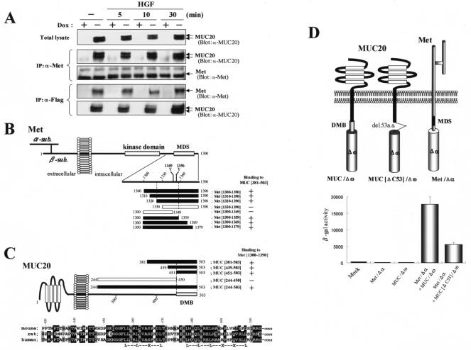
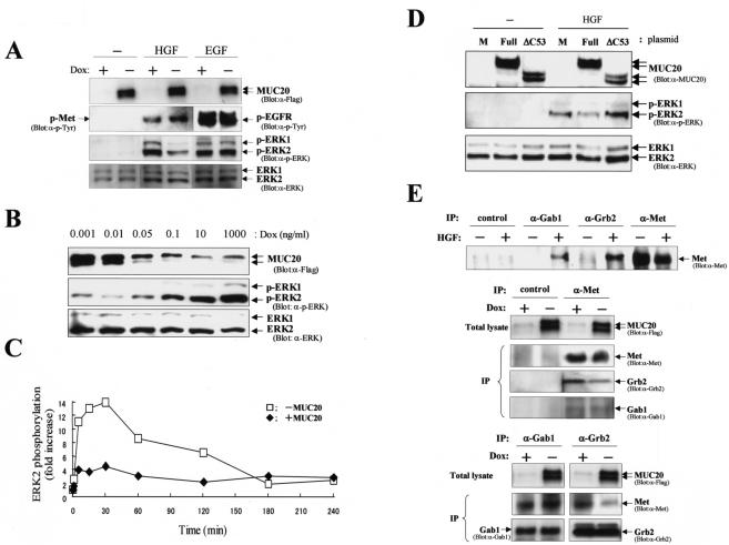
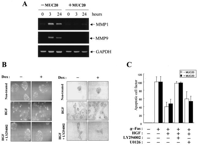
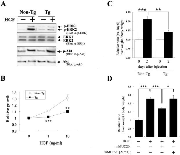
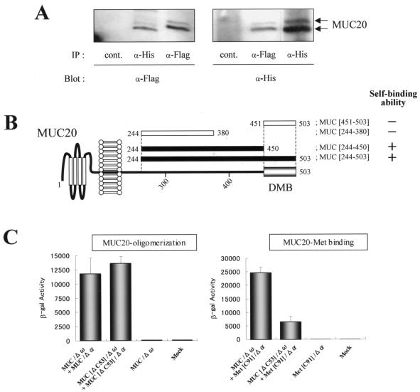
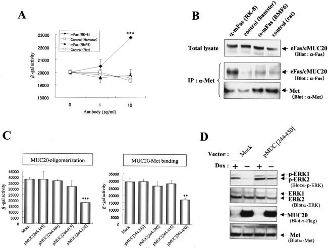
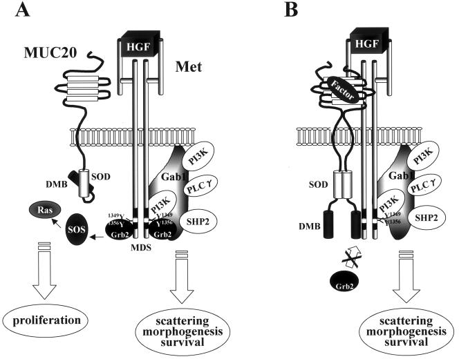
A cDNA encoding a novel mucin protein, MUC20, was isolated as a gene that is up-regulated in the renal tissues of patients with immunoglobulin A nephropathy. We demonstrate here that the C terminus of MUC20 associates with the multifunctional docking site of Met without ligand activation, preventing Grb2 recruitment to Met and thus attenuating hepatocyte growth factor (HGF)-induced transient extracellular signal-regulated kinase-1 and -2 activation. Production of MUC20 reduced HGF-induced matrix metalloproteinase expression and proliferation, which require the Grb2-Ras pathway, whereas cell scattering, branching morphogenesis, and survival via the Gab1/phosphatidylinositol 3-kinase (PI3K) pathways was not affected. Thus, MUC20 reduces HGF-induced activation of the Grb2-Ras pathway but not the Gab1/PI3K pathways. We further demonstrate that the cytoplasmic domain of MUC20 has the ability to oligomerize and that the oligomerization augments its affinity for Met. Taken together, these results suggest that MUC20 is a novel regulator of the Met signaling cascade which has a role in suppression of the Grb2-Ras pathway.


Figures

FIG. 1.
FIG. 1.
Interaction of MUC20 with Met. (A) Coimmunoprecipitation of MUC20 with Met. Lysates of cells transfected with a Flag-tagged MUC20 gene were subjected to immunoprecipitation (IP) with anti-Met (α-Met) or anti-Flag antibodies, and the precipitates were immunoblotted with the indicated antibodies. Two bands representing the MUC20 proteins were detected due to posttranslational modification (6). (B) MUC20 binding domain in Met. Peptides with progressive deletions from the N- or C-terminal end of Met[1300-1390] were used for prey. Positive or negative association of each peptide with the bait peptide, MUC[381-503], in the two-hybrid assay is indicated as + or −, respectively. The domain structure of Met is also shown (2). (C) Upper panel, Met binding domain in MUC20. Positive or negative association of each cytoplasmic peptide of MUC20 to Met[1300-1390] is shown as + or −, respectively. Lower panel, alignment of the C termini of human, rat, and mouse MUC20 proteins. Residues identical in human MUC20 and the other sequences are shown by reverse type, and conservative charges are shown by gray shading. Conserved leucine repeats are indicated at the bottom. (D) Evaluation of MUC20-Met binding by using the β-Gal mutant system. Upper panel, schematic representation of three chimeric constructs. Lower panel, β-Gal activities in the transfected CHO-K1 cells. Plasmids transfected into CHO-K1 cells are indicated below each bar. Data represent means ± standard deviations (n = 3). (E) Immunohistochemical analysis of MUC20 and Met in human kidney. Kidney sections were stained with anti-MUC20 (a), anti-Met (b), or anti-AQP1 (c) antibodies. G, glomerulus; DT, distal tubule; PT, proximal tubule.
FIG. 1.
FIG. 1.
Interaction of MUC20 with Met. (A) Coimmunoprecipitation of MUC20 with Met. Lysates of cells transfected with a Flag-tagged MUC20 gene were subjected to immunoprecipitation (IP) with anti-Met (α-Met) or anti-Flag antibodies, and the precipitates were immunoblotted with the indicated antibodies. Two bands representing the MUC20 proteins were detected due to posttranslational modification (6). (B) MUC20 binding domain in Met. Peptides with progressive deletions from the N- or C-terminal end of Met[1300-1390] were used for prey. Positive or negative association of each peptide with the bait peptide, MUC[381-503], in the two-hybrid assay is indicated as + or −, respectively. The domain structure of Met is also shown (2). (C) Upper panel, Met binding domain in MUC20. Positive or negative association of each cytoplasmic peptide of MUC20 to Met[1300-1390] is shown as + or −, respectively. Lower panel, alignment of the C termini of human, rat, and mouse MUC20 proteins. Residues identical in human MUC20 and the other sequences are shown by reverse type, and conservative charges are shown by gray shading. Conserved leucine repeats are indicated at the bottom. (D) Evaluation of MUC20-Met binding by using the β-Gal mutant system. Upper panel, schematic representation of three chimeric constructs. Lower panel, β-Gal activities in the transfected CHO-K1 cells. Plasmids transfected into CHO-K1 cells are indicated below each bar. Data represent means ± standard deviations (n = 3). (E) Immunohistochemical analysis of MUC20 and Met in human kidney. Kidney sections were stained with anti-MUC20 (a), anti-Met (b), or anti-AQP1 (c) antibodies. G, glomerulus; DT, distal tubule; PT, proximal tubule.
FIG. 2.
FIG. 2.
MUC20 attenuates HGF-induced ERK1/2 phosphorylation. (A) Attenuation of HGF-induced ERK1/2 phosphorylation by MUC20. HEK/tet-MUC20 cells cultured in the presence or absence of DOX were stimulated with HGF (20 ng/ml) or EGF (100 ng/ml). The lysates from those cells were used for immunoblotting with the indicated antibodies. p, phosphorylated; α-, anti-. (B) Dose-dependent effect of MUC20 on ERK1/2 phosphorylation. Cells cultured with various concentrations of DOX were stimulated with HGF. Equivalent amounts of the cell lysates were used for immunoblotting with the indicated antibodies. (C) Activation profile of ERK2 elicited by HGF with or without MUC20 production. Cell lysates were analyzed by immunoblotting with anti-ERK and anti-p-ERK antibodies. The signal intensity for the phospho-ERK2 bands was measured by densitometry, and the intensity of each signal was normalized against that of the protein band detected with anti-ERK antibody. (D) Involvement of the DMB in the suppressive effect of MUC20. HEK293 cells transfected with either phMUC20 (Full), phMUC20[ΔC53] (ΔC53), or pcDNA3 vector (M) were stimulated with HGF and then subjected to immunoblotting with the indicated antibodies. (E) Effects of MUC20 on Gab1 and Grb2 recruitment to activated Met. Immunoprecipitation (IP) with the indicated antibodies was carried out on lysates from HGF-stimulated HEK/tet-MUC20 cells with or without MUC20 production. The precipitates were used for immunoblotting with the indicated antibodies.
FIG. 3.
FIG. 3.
Effects of MUC20 on HGF-induced biological responses. (A) RT-PCR analysis of MMP-1, MMP-9, and GAPDH mRNAs in HEK/tet-MUC20 cells at various times after HGF stimulation, with or without MUC20 production. (B) Left panels, micrographs showing cell dissociation and scatter of MDCK/tet-MUC20 cells with or without MUC20 production. The cells were pretreated with or without LY294002 prior to HGF stimulation. Right panels, phase-contrast micrographs of MDCK/tet-MUC20 cells with or without MUC20 production, grown in a three-dimensional collagen matrix. Cultures were maintained in the presence of HGF alone, HGF plus LY294002, or neither. (C) The antiapoptosis effect of HGF does not depend on MUC20 dose. HEK/tet-MUC20 cells with or without MUC20 production were pretreated with LY294002 or U0126 prior to HGF stimulation and then treated with an agonistic anti-Fas (α-Fas) antibody. Apoptosis was quantified by enzyme-linked immunosorbent assay for free nucleosomes. Results are represented as the ratio to apoptosis in the agonistic anti-Fas antibody-treated cells without HGF stimulation. Data represent mean ± standard deviations (n = 3).
FIG. 4.
FIG. 4.
MUC20 impairs HGF-induced proliferation. (A) Reduction of HGF-induced ERK1/2 phosphorylation by MUC20. Primary renal tubular cells from MUC20-Tg and non-Tg mice were stimulated with HGF, and the lysates were subjected to immunoblotting with the indicated antibodies (α-). (B) Attenuation of HGF-induced proliferation in primary cells from MUC20-Tg mice. Proliferation was measured by a cell viability assay. Data represent means ± standard deviations (n = 3). Similar results were obtained with primary cells from another strain of MUC20-Tg mice (data not shown). **, P < 0.01; ***, P < 0.001 (versus non-Tg mice in a two-tailed Student t test). (C) Attenuation of exogenous hHGF-induced liver weight gain in MUC20-Tg mice. The liver and body weights of the naked phHGF-injected mice were recorded at days 0 and 2. Data are presented as means ± standard deviations (n = 6). **, P < 0.01; ***, P < 0.001 (two-tailed Student t test). No differences in the amount of plasma hHGF were observed between non-Tg and Tg mice (1.56 ± 0.31 and 2.20 ± 1.29 ng/ml [means ± standard deviations], respectively [n = 6]). (D) Involvement of the DMB in suppression of the hHGF-induced liver weight gain. Either pmMUC20 or pmMUC20[ΔC53] was injected along with phHGF into ICR mice. No differences in the amount of plasma hHGF were observed among mice injected with phHGF alone, phHGF plus pmMUC20, or phHGF plus pmMUC20[ΔC53] (1.94 ± 0.92, 2.10 ± 0.95, and 1.64 ± 1.04 ng/ml [means ± standard deviations], respectively [n = 6]). The liver and body weights of these mice were recorded at day 2 after injection. Data are presented as means ± standard deviations (n = 8). *, P < 0.05; ***, P < 0.001 (two-tailed Student t test).
FIG. 5.
FIG. 5.
MUC20 oligomerization domain. (A) Coimmunoprecipitation of tagged MUC20s. Lysates from HEK293 cells transfected with both phMUC20(Flag) and phMUC20(His) were subjected to immunoprecipitation (IP) and then to immunoblotting with the indicated antibodies (α-). (B) Determination of the region responsible for the oligomerization of MUC20. Self-binding and nonbinding abilities of each peptide in the two-hybrid assay are indicated as + and −, respectively. (C) β-Gal activities of CHO-K1 cells transfected with the chimeric constructs indicated below each bar. Data are represented as mean ± standard deviations (n = 3).
FIG. 6.
FIG. 6.
Oligomerization of MUC20 augments its affinity to Met. (A) Oligomerization of the chimeric molecules by an agonistic anti-Fas antibody. HEK293 cells cotransfected with eFas/cMUC20/Δω and eFas/cMUC20/Δα were treated with anti-Fas (RK-8 or RMF6) or normal immunoglobulin G (control) antibodies, and then β-Gal activities were measured. Data are represented as mean ± standard deviations (n = 3). ***, P < 0.001 versus control in a two-tailed Student t test. (B) Augmentation of Met binding by oligomerization of the chimeric proteins. Immunoprecipitations (IP) with anti-Met (α-Met) antibody were performed with lysates from HEK293 cells expressing eFas/cMUC20. Cells were treated with the indicated antibodies for 3 h prior to preparation of the lysates. The precipitates were immunoblotted with the indicated antibodies. (C) Reduction of MUC20-Met binding by interference with MUC20 oligomerization. Each plasmid, pMUC[244-345], pMUC[244-380], pMUC[244-415], and pMUC[244-450], was transfected into CHO-K1 cells along with MUC/Δω plus MUC/Δα or MUC/Δω plus Met[C91]/Δα, and β-Gal activities were measured. Data are represented as means ± standard deviations (n = 3). **, P < 0.01; ***, P < 0.001 (versus mock transfectants in a two-tailed Student t test). (D) Release from suppression of the Grb2-Ras pathway by reduction of MUC20 oligomerization. pMUC[244-450] or pcDNA3 (Mock) was transfected into HEK/tet-MUC20 cells cultured in the presence or absence of DOX. After stimulation with HGF, the cell lysates were used for immunoblotting with the indicated antibodies.
FIG. 7.
FIG. 7.
Model of MUC20 suppression. (A) A MUC20 monomer has less ability to associate with Met. In this condition, Grb2 is recruited to HGF-activated Met, followed by activation of signaling cascades leading to proliferation. (B) MUC20 oligomerization caused by either an overproduction of MUC20 or an unknown endogenous factor(s) leads to the association with Met, blocking Grb2 recruitment to Met and suppressing the Grb2-Ras pathway, but does not affect Gab1 recruitment. The oligomerizing domain in MUC20 is referred to as the self-oligomerizing domain (SOD). PLCγ, phospholipase C-γ.

Similar articles

Cited by

References

    1. Boccaccio, C., M. Ando', and P. M. Comoglio. 2002. A differentiation switch for genetically modified hepatocytes. FASEB J. 16:120-122. - PubMed
    1. Comoglio, P. M., and C. Boccaccio. 1996. The HGF receptor family: unconventional signal transducers for invasive cell growth. Genes Cells 1:347-354. - PubMed
    1. Di Renzo, M. F., R. P. Narsimhan, M. Olivero, S. Bretti, S. Giordano, E. Medico, P. Gaglia, P. Zara, and P. M. Comoglio. 1991. Expression of the Met/HGF receptor in normal and neoplastic human tissues. Oncogene 6:1997-2003. - PubMed
    1. Gandino, L., P. Longati, E. Medico, M. Prat, and P. M. Comoglio. 1994. Phosphorylation of serine 985 negatively regulates the hepatocyte growth factor receptor kinase. J. Biol. Chem. 269:1815-1820. - PubMed
    1. Grisendi, S., B. Chambraud, I. Gout, P. M. Comoglio, and T. Crepaldi. 2001. Ligand-regulated binding of FAP68 to the hepatocyte growth factor receptor. J. Biol. Chem. 276:46632-46638. - PubMed

MeSH terms