. 2004 Sep;24(17):7622-35.
doi: 10.1128/MCB.24.17.7622-7635.2004.

Internal ribosome entry site structural motifs conserved among mammalian fibroblast growth factor 1 alternatively spliced mRNAs

Affiliations

Internal ribosome entry site structural motifs conserved among mammalian fibroblast growth factor 1 alternatively spliced mRNAs

Yvan Martineau et al. Mol Cell Biol. 2004 Sep.

Abstract

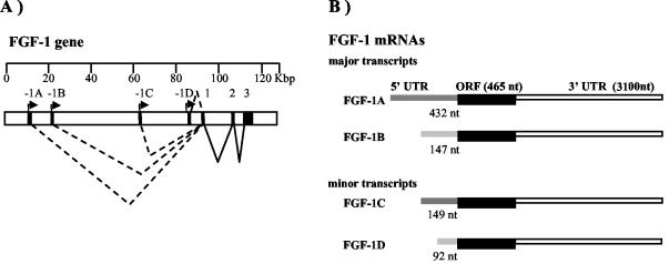
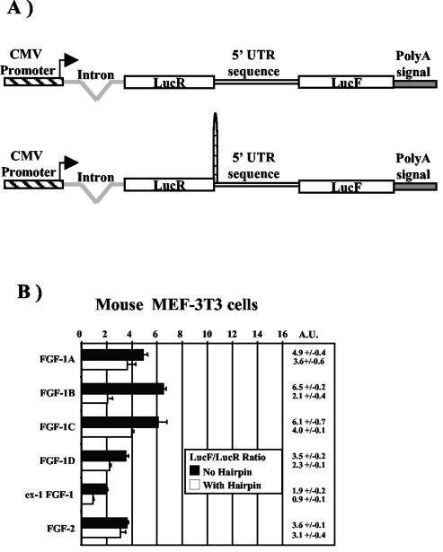
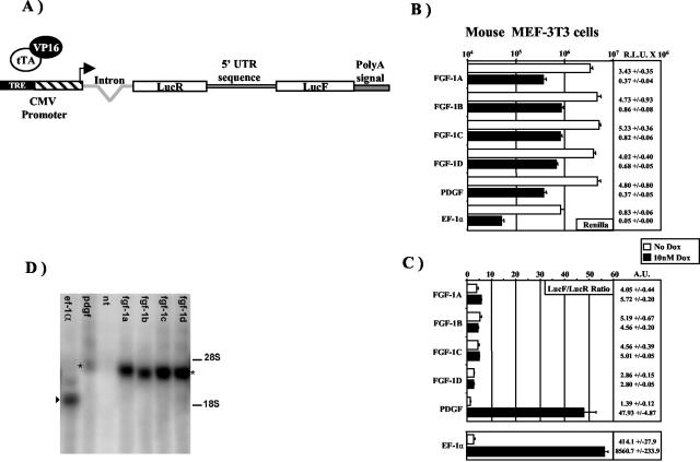
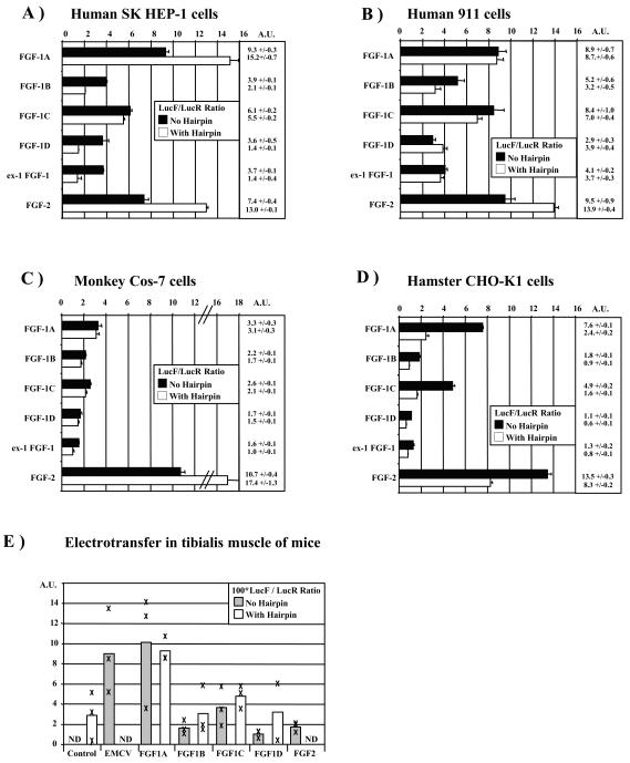
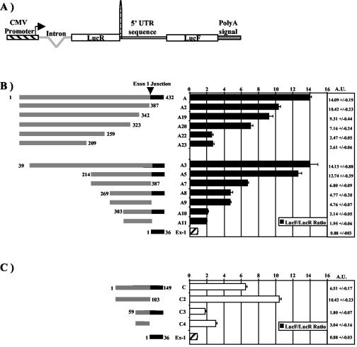
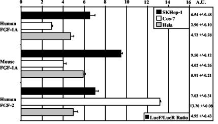
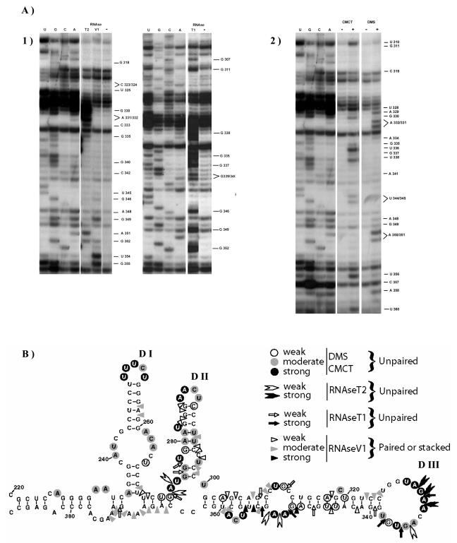
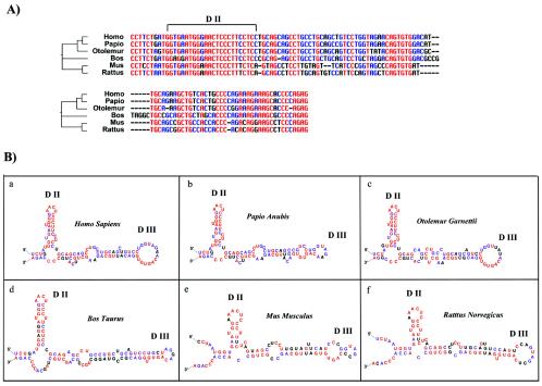
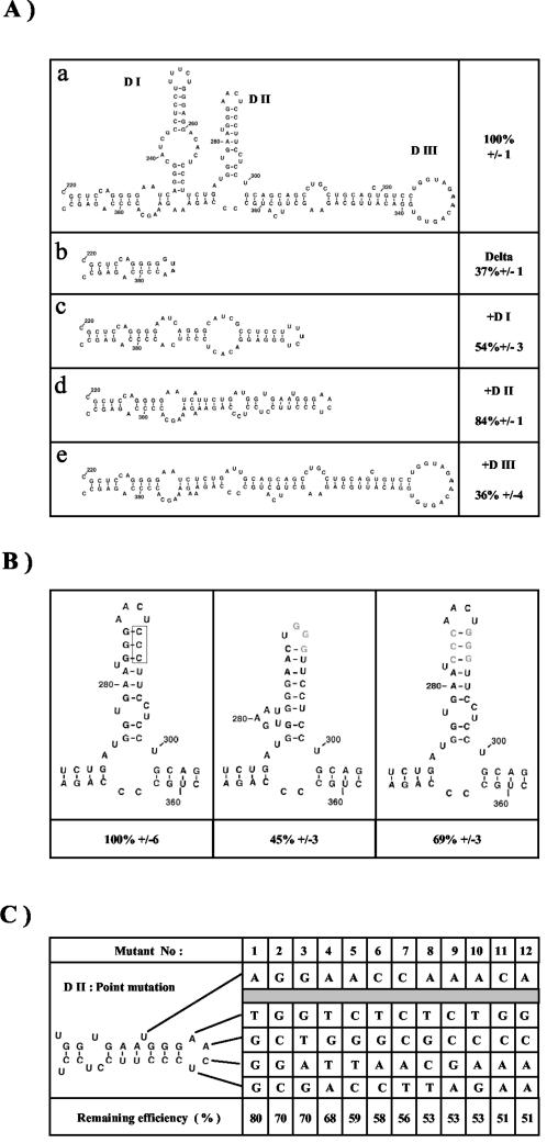
Fibroblast growth factor 1 (FGF-1) is a powerful angiogenic factor whose gene structure contains four promoters, giving rise to a process of alternative splicing resulting in four mRNAs with alternative 5' untranslated regions (5' UTRs). Here we have identified, by using double luciferase bicistronic vectors, the presence of internal ribosome entry sites (IRESs) in the human FGF-1 5' UTRs, particularly in leaders A and C, with distinct activities in mammalian cells. DNA electrotransfer in mouse muscle revealed that the IRES present in the FGF-1 leader A has a high activity in vivo. We have developed a new regulatable TET OFF bicistronic system, which allowed us to rule out the possibility of any cryptic promoter in the FGF-1 leaders. FGF-1 IRESs A and C, which were mapped in fragments of 118 and 103 nucleotides, respectively, are flexible in regard to the position of the initiation codon, making them interesting from a biotechnological point of view. Furthermore, we show that FGF-1 IRESs A of murine and human origins show similar IRES activity profiles. Enzymatic and chemical probing of the FGF-1 IRES A RNA revealed a structural domain conserved among mammals at both the nucleotide sequence and RNA structure levels. The functional role of this structural motif has been demonstrated by point mutagenesis, including compensatory mutations. These data favor an important role of IRESs in the control of FGF-1 expression and provide a new IRES structural motif that could help IRES prediction in 5' UTR databases.


Figures

FIG. 1.
FIG. 1.
Human FGF-1 gene and transcripts. (A) Organization of the human FGF-1 gene. Arrows represent the promoters 1A, 1B, 1C, and 1D. Alternative splicing of untranslated exons 1A, 1B, 1C, or 1D to exon 1 (depending on the promoter used) will generate mRNAs 1A, 1B, 1C, and 1D. (B) Structures of the FGF-1 mRNAs. mRNAs 1A and 1B are the most common and are expressed mainly in kidney brain and retina. mRNAs 1C and 1D are expressed at a very low level and can be induced by serum or transforming growth factor β (10).
FIG. 2.
FIG. 2.
Expression of FGF-1 leader-containing bicistronic vectors in transfected mammalian cell types. (A) Schematic representation of the bicistronic LucR-I-LucF vectors containing 1A, 1B, 1C, or 1D FGF-1 mRNA 5′ UTRs. Vector construction is described in Materials and Methods. Constructs contain the CMV promoter controlling the expression of a bicistronic LucR-I-LucF mRNA. A synthetic intron is present upstream from LucR, and a poly(A) site is present downstream from LucF (23). In the bottom construct, a hairpin has been introduced at the end of the luciferase renilla gene in order to prevent reinitiation and interaction between the FGF-1 5′ UTRs and the beginning of the bicistronic mRNA. (B) Transient transfection of murine embryo fibroblast MEF-3T3 cells with bicistronic LucR-I-LucF vectors containing either the 1A, 1B, 1C, or 1D FGF-1 5′ UTR in the intercistronic region, with or without a hairpin at the 3′ end of the LucR gene (see Materials and Methods). The bicistronic vectors pCRHL and pCRFL, containing either a hairpin or the FGF-2 IRES, were used as negative and positive controls, respectively (14). Cells were harvested 48 h after transfection, and luciferase activities present in cell extracts were measured. IRES activities were determined by calculating the LucF/LucR ratio. Data were calibrated by dividing the LucF/LucR ratio by the ratio of the pCRHL negative control, providing a relative IRES activity expressed in AU (23). The exact values are presented on the right. Experiments were repeated at least five times. Results represent means ± standard errors (calculated with Microsoft Excel software) from a representative experiment done in triplicate.
FIG. 3.
FIG. 3.
Development of a TET OFF regulatable system to check for the absence of cryptic promoters in the bicistronic Luc-I-Luc mRNAs. (A) Schematic representation of the bicistronic TET OFF LucR-I-LucF vector. We constructed a bicistronic vector with a promoter repressible by tetracycline, the TET OFF system. A tetracycline-responsive element (TRE) was placed in front of the CMV promoter (devoid of its enhancer). The principle is that in the absence of tetracycline, the chimeric tTA-VP16 protein is bound to the TRE element and thus the CMV promoter is active. In the presence of tetracycline or tetracycline analogs such as doxycycline, tTA will bind the antibiotic and will be unable to bind the TRE, thus preventing CMV promoter transactivation. Bicistronic TRE-CMV vectors were constructed with the different FGF-1 5′ UTRs or a hairpin in the intercistronic region. As positive promoter controls, the EF-1α promoter or the PDGFb 5′ UTR (containing a cryptic promoter) was also introduced in the intercistronic region. (B and C) Transfection of MEF-3T3 cells expressing tTA-VP16 with the TET regulatable bicistronic constructs described for panel A. (B) At 2 h prior to transfection, cells were treated or not with 10 nM doxycycline (Dox). Luciferase activities were measured as described for Fig. 2. LucR activities, expected to reflect the bicistronic mRNA levels, are represented for each construct. (C) The LucF/LucR ratio is expressed in AU measured as described for Fig. 2. A different scale has been used to present the values for the EF-1α-containing vector, whose LucF/LucR ratio is a higher order of magnitude. (D) Analysis of bicistronic mRNAs by Northern blotting. Northern blotting was performed with a LucF 32P-labeled riboprobe with total RNAs purified from MEF-3T3 cells transfected with the bicistronic vectors used for Fig. 2 C, containing either the FGF-1A, -1B, -1C, or -1D or PDGFb 5′ UTR or the EF-1α promoter (see Materials and Methods). For each transfection, the fragment present in the intercistronic region is indicated at the top of the lane. Migrations of 28S (4.7 kb) and 18S (1.2 kb) rRNAs are indicated. Migration of the monocistronic LucF mRNA is indicated by an arrowhead. Migration of the bicistronic mRNAs is indicated by an asterisk.
FIG. 4.
FIG. 4.
Analysis of the FGF-1 5′ UTR IRES activities in cells from several mammalian species and in the tibialis anterior muscle of mice. (A to D) Human SK-Hep-1 adenocarcinoma cells (A), human 911 retinoblasts (B), simian kidney COS-7 cells (C), and Chinese hamster ovary CHO-K1 cells (D) were transiently transfected with bicistronic LucR-I-LucF vectors devoid of an intercistronic hairpin, and IRES activities were measured as described for Fig. 2B. Experiments were repeated three to five times. Results represent means ± standard errors from a representative experiment done in triplicate. (E) Expression of FGF-1 leader-containing bicistronic vectors in electrotransferred mouse muscle in vivo. FGF-1 IRES activities were analyzed in vivo by DNA electrotransfer in mouse muscle. The bicistronic LucR-I-LucF vectors used for Fig. 2 were electrotransferred in mouse muscle as described in Materials and Methods. Muscles were extracted, and luciferase activities were measured in muscle lysates. The LucF/LucR ratio is indicated by a cross for each mouse, and the histogram represents the average for each construct. These results are representative of those from experiments that were repeated at least three times for each construct. The negative control corresponds to the construct with an intercistronic hairpin. ND, not determined.
FIG. 5.
FIG. 5.
Mapping of FGF-1 IRESs A and C. (A) Hairpin-containing vector used for mapping. Different portions of FGF-1A or -1C 5′ UTRs were introduced in the intercistronic region, downstream from the hairpin. (B and C) SK-Hep-1 cells were transfected with the different bicistronic constructs, and IRES activities were calculated from the LucF/LucR ratio and expressed in AU as described for Fig. 2. For each leader, nucleotide numbering is from the 5′ end. The different deletions in the A or C 5′ UTRs are shown and are named An or Cn, respectively. (B) Leader A and exon 1 (Ex-1); (C) leader C and exon 1. The precise IRES activity values are also indicated. The black boxes in the 3′ ends of the leaders correspond to the exon 1 36 nt shared by the four FGF-1 5′ UTRs.
FIG. 6.
FIG. 6.
Comparison of human and mouse FGF-1A 5′ UTRs for sequence homology and IRES activity in mammalian cell types. The entire mouse FGF-1A 5′ UTR was introduced in the Luc-R-I-LucF bicistronic vector described for Fig. 2A. SK-Hep-1, COS-7, and HeLa cells were transfected with the constructs containing the human or murine sequences, and IRES activities were determined as described for Fig. 2 by measurement of the LucF/LucR ratio calibrated to the negative control pCRHL devoid of IRES. Human FGF-2 was used as a positive control. Precise values are indicated on the right. Results represent means ± standard errors from a representative experiment done in triplicate.
FIG. 7.
FIG. 7.
RNA secondary structure of human FGF-1 IRES A. (A)Enzymatic (panel 1) and chemical (panel 2) probing of in vitro-transcribed A9 fragment. Cleavage and modification sites were detected by primer extension with the 5′-32P-end-labeled RTA9 oligonucleotide, which hybridizes at positions 363 to 385 of the FGF-1A RNA leader. The resulting cDNA was separated on an 8% polyacrylamide-8 M urea sequencing gel and analyzed by autoradiography. RNA sequencing reactions were run in parallel. The nature and positions of cleaved (panel 1) and modified (panel 2) bases are indicated at the right. RNase V1, RNase T1, RNase T2, or the two chemical agents (DMS or CMCT) were added (+) or not (−) before the reverse transcription step. Identical results were found for in vitro-transcribed A5 fragment (data not shown). (B) RNA secondary structure model of the fragment from nt 220 to 386, showing results from enzymatic cleavage and chemical modification experiments. The A9 region is from nt 269 to 387. Sites of RNase cleavage and chemical modification are indicated.
FIG. 8.
FIG. 8.
Alignment and folding of six RNA orthologous sequences corresponding to FGF-1 IRES A. (A) A phylogenic tree was obtained with 5′ UTR orthologous sequences from human (Homo sapiens), baboon (Papio anubis), small-eared galago monkey (Otolemur garnettii), cow (Bos taurus), mouse (Mus musculus), and rat (Rattus norvegicus) by using ClustalW and Phylip software. Nucleotide blocks corresponding to the human minimal IRES (A9 fragment, nt 269 to 387) were compared for the six mammalian species. Alignment was obtained by using Multalin software. Red, blue, and black nucleotides are 100% conserved, more than 50% conserved, and not conserved, respectively. The position of domain DII is indicated. (B) The region corresponding to the DII and DIII domains (nt 270 to 368) of the human FGF-1 IRES A was folded by using Zuker-derived folding software for the five other mammalian species shown in panel A. Domains DII and DIII, conserved in the six species, are indicated.
FIG. 9.
FIG. 9.
Domain and nucleotides mutations of human FGF-1 IRES A. The IRES activities of the different constructs, analyzed as described for Fig. 2 by using the double luciferase vector in SH-Hep-1-transfected cells, are indicated as percentages of the activity of the wild-type complete leader 1A IRES. (A) Domain deletion analysis of human FGF-1 IRES A. DI, DII, and DIII domains were deleted from the complete sequence (a) to create mutant Delta (b), and then domain DI (c), DII (d), or DIII (e) was added to Delta in order to estimate their role in IRES A function. (B) Point mutations affecting domain DII structural stability in the complete FGF-1 leader A. The original DII domain contains a three-G/C pairing stem (RNA structure on the left). The three Cs were mutated to three Gs (in gray, structure in the middle) to suppress interaction and to disrupt the structure. Finally, the three original Gs were mutated to three Cs (in gray, structure on the right) in order to restore the G/C pairing and to rebuild the starting structure. (C) Point mutations of the five most accessible and conserved nucleotides in domain DII (U bulge and AACU loop). Using the QuikChange method, nucleotides were randomly and simultaneously mutated in the context of the complete human FGF-1A leader.

Similar articles

Cited by

References

    1. Akiri, G., D. Nahari, Y. Finkelstein, S. Y. Le, O. Elroy-Stein, and B. Z. Levi. 1998. Regulation of vascular endothelial growth factor (VEGF) expression is mediated by internal initiation of translation and alternative initiation of transcription. Oncogene 17:227-236. - PubMed
    1. Arnaud, E., C. Touriol, C. Boutonnet, M. C. Gensac, S. Vagner, H. Prats, and A. C. Prats. 1999. A new 34-kilodalton isoform of human fibroblast growth factor 2 is cap dependently synthesized by using a non-AUG start codon and behaves as a survival factor. Mol. Cell. Biol. 19:505-514. - PMC - PubMed
    1. Bernstein, J., O. Sella, S. Y. Le, and O. Elroy-Stein. 1997. PDGF2/c-sis mRNA leader contains a differentiation-linked internal ribosomal entry site (D-IRES). J. Biol. Chem. 272:9356-9362. - PubMed
    1. Bonnal, S., C. Schaeffer, L. Creancier, S. Clamens, H. Moine, A. C. Prats, and S. Vagner. 2003. A single internal ribosome entry site containing a G quartet RNA structure drives fibroblast growth factor 2 gene expression at four alternative translation initiation codons. J. Biol. Chem. 278:39330-39336. - PMC - PubMed
    1. Brunel, C., and P. Romby. 2000. Probing RNA structure and RNA-ligand complexes with chemical probes. Methods Enzymol. 318:3-21. - PubMed

Publication types

MeSH terms

LinkOut - more resources