. 2004 Sep;24(17):7643-53.
doi: 10.1128/MCB.24.17.7643-7653.2004.

Role of atypical protein kinase C in estradiol-triggered G1/S progression of MCF-7 cells

Affiliations

Role of atypical protein kinase C in estradiol-triggered G1/S progression of MCF-7 cells

Gabriella Castoria et al. Mol Cell Biol. 2004 Sep.

Abstract

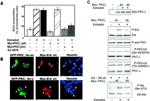
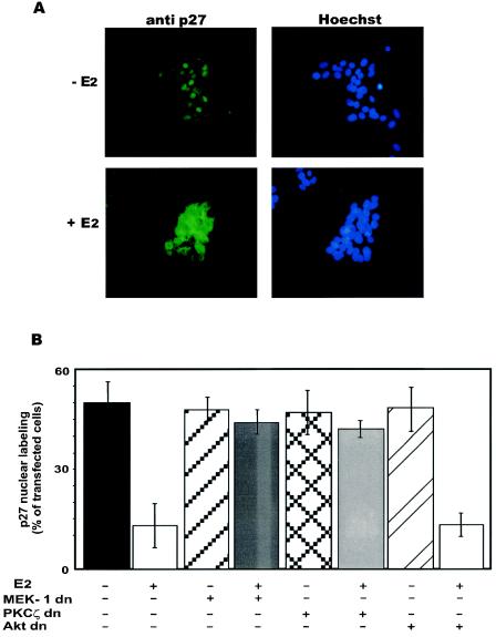
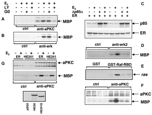
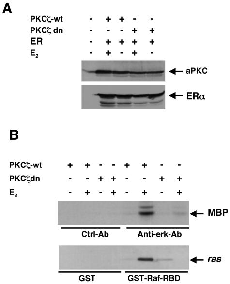
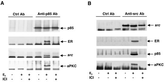
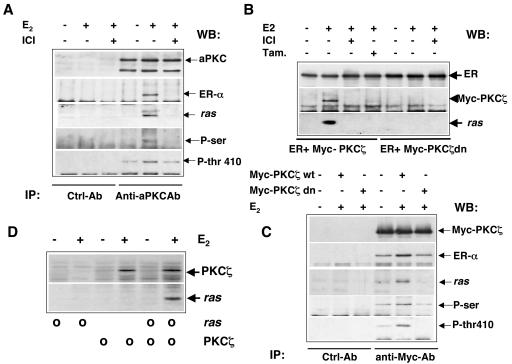
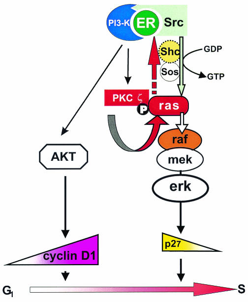
Expression of a dominant negative atypical protein kinase C (aPKC), PKCzeta, prevents nuclear translocation of extracellular regulated kinase 2 (ERK-2), p27 nuclear reduction, and DNA synthesis induced by estradiol in human mammary cancer-derived MCF-7 cells. aPKC action upstream of these events has been analyzed. In hormone-stimulated NIH 3T3 and Cos cells ectopically expressing human estrogen receptor alpha (hERalpha), aPKC is activated by phosphatidylinositol 3-kinase (PI 3-kinase) and, in turn, controls the Ras/MEK-1/ERK cascade. In MCF-7 and Cos cells stimulated by hormone, PI 3-kinase activates PKCzeta by Thr410 phosphorylation. Serine phosphorylation of PKCzeta is simultaneously induced. PKCzeta activation leads to recruitment of Ras to a multimolecular complex that also includes hERalpha, Src, PI 3-kinase, and aPKC. We propose that PKCzeta pushes Ras and the signaling complex close together in such a way that it facilitates the Src-dependent Ras activation. This activation is crucial for the interplay between estradiol-triggered signaling and cell cycle machinery.


Figures

FIG. 1.
FIG. 1.
Involvement of aPKC in S-phase entry and ERK-2 nuclear translocation induced by estradiol in MCF-7 cells. Quiescent MCF-7 cells were seeded on coverslips. (A) Cells were untransfected or transfected with the indicated plasmids and left unstimulated or stimulated with 10 nM estradiol. Untransfected cells were also stimulated with estradiol in the presence of 1 μM GO6976 (Calbiochem). After BrdU pulsing, coverslips were analyzed for BrdU incorporation. In transfected cells, this was calculated as the percentage of BrdU-positive cells = [(number of transfected BrdU-positive cells/number of transfected cells)] × 100 and compared to BrdU incorporation of untransfected cells from the same coverslips. For each plasmid, data are derived from scoring of at least 200 cells. Results of more than two independent experiments have been averaged; values shown are means and standard errors of the mean. A construct expressing the Myc-tagged pSG5 empty plasmid was transfected into quiescent MCF-7 cells as a control. The cells were left unstimulated or stimulated with estradiol. BrdU was added, and after 24 h the coverslips were analyzed for DNA synthesis. A total of 62% of the Myc-tag-expressing cells entered S phase, whereas the percentage of BrdU incorporation in unstimulated cells was less than 13%. (B) Cells were transfected with the Myc-tagged ERK-2 wild type along with either GFP-antisense dominant negative PKCζ (GFP-PKCζ dn as) or GFP-sense dominant negative PKCζ (GFP-PKCζ dn s) (left panels). The middle panels show the staining of the Myc-tagged ERK-2 wild type. The results of corresponding nuclear staining (Hoechst) are shown (right panels). The micrographs represent the results of two independent experiments. (C) Quiescent MCF-7 cells were transfected with either wild-type or dominant negative Myc-tagged PKCζ. Expression of PKCζ was revealed by the anti-Myc-tagged antibody (top). In the results shown in the upper panels, the cells were left unstimulated or stimulated for 3 min with 10 nM estradiol, and lysates were immunoblotted with the antibodies against the indicated proteins. In the results shown in the lower panels, the cells were cotransfected with the HA-tagged Akt wild type. They were then left unstimulated or stimulated for 3 min with 10 nM estradiol. Lysates were immunoprecipitated with anti-HA-tagged antibody. Immunoprecipitated proteins were hybridized with the antibodies against the indicated proteins. wt, wild type; dn, dominant negative.
FIG. 2.
FIG. 2.
Involvement of aPKC in the p27 nuclear localization modulated by estradiol in MCF-7 cells. Quiescent MCF-7 cells were seeded on coverslips. (A) MCF-7 cells were left untreated (top) or treated with 10 nM estradiol for 8 h (bottom). p27 localization was analyzed by fluorescence microscopy with anti-p27 MAb (left panels). The corresponding nuclear staining (Hoechst) is shown (right panels). (B) Cells were untransfected or transfected with the indicated plasmids and then left unstimulated or stimulated with 10 nM estradiol for 8 h. The p27 staining of cells was then analyzed by fluorescence microscopy. p27 staining of transfected cells was compared with that of nontransfected cells from the same coverslips. Data are derived from scoring of at least 200 cells for each coverslip, and results are expressed as the percentage of cells showing predominantly p27 nuclear staining. Results of more than three independent experiments have been averaged; the values shown are the means and standard errors of the mean. The MCF-7 cells used were also analyzed for BrdU incorporation. Incorporation was observed in 12% of total nuclei of unstimulated cells. This value shifted to 68% after cells were treated with estradiol. Overexpression of either Myc-tagged dominant negative PKCζ, A221-MEK-1 or Myc-His-tagged (dn) Akt inhibited the estradiol-stimulated BrdU incorporation by 68, 70, and 82%, respectively. E2, estradiol.
FIG. 3.
FIG. 3.
Estradiol (E2) triggers PI 3-kinase-dependent aPKC activity in MCF-7 cells and transfected NIH 3T3 cells. Quiescent MCF-7 cells were preincubated for 10 min with and without LY294002 (10 μM) (LY) or GO6976 (1 μM) (GO). Cells were then left untreated or treated with 10 nM estradiol for 3 min. Lysates were immunoprecipitated with either anti-aPKC antibodies (A) or ERK-2 antibodies (B). Parallel samples were immunoprecipitated with control antibodies (ctrl). Kinase activities were assayed using MBP as a substrate. NIH 3T3 fibroblasts were transfected with hERα-expressing plasmid alone or with Δp85α-expressing plasmid. Quiescent cells were left untreated or treated for 3 min with 10 nM estradiol. (C) Lysates were analyzed for either p85 or ERα expression by immunoblotting with the appropriate antibodies. (D) Lysates were immunoprecipitated with either control (ctrl) or anti-ERK-2 (anti-erk2) antibodies, and the kinase activity was assayed using MBP as a substrate. (E) Lysates were incubated with either GST or GST-Raf-RBD. Eluted proteins were blotted with anti-pan Ras antibody. (F) Lysates were immunoprecipitated with either control (ctrl) or anti-aPKC (anti-aPKC) antibodies, and the kinase assay was performed with MBP as a substrate. (G) NIH 3T3 fibroblasts were transfected with either the wild-type ERα (ER) or the transcriptionally inactive Δ250-303 ERα (HE241)-expressing plasmid. Quiescent cells were left untreated or treated for 3 min with 10 nM estradiol, and lysates were immunoprecipitated with the anti-aPKC antibody (Gibco-BRL). (Top) Immunoprecipitates were hybridized with aPKC antibodies; (bottom) the immunoprecipitates were assayed for kinase activity with MBP as a substrate. No aPKC or kinase activity was detected in control immunoprecipitates (ctrl). The bottom panel shows the expression of ERα (HEG0) or its mutant (HE241G) revealed by immunoblotting of lysates with H222 rat anti-ER MAb. Lysates from NIH 3T3 fibroblasts transfected with the pSG5 empty plasmid were also immunoblotted as a control (pSG5).
FIG. 4.
FIG. 4.
Estradiol-induced Ras and ERK-2 activation depends on aPKC activity in hERα-expressing Cos cells. Cos cells were transfected with either the pSG5 empty plasmid or hERα-expressing plasmid, alone or with a wild-type or dominant negative PKCζ-expressing plasmid. Quiescent cells were left untreated or treated for 3 min with 10 nM estradiol. (A) Lysates were analyzed for expression of either PKCζ or hERα by immunoblotting with the appropriate antibodies. (B) Lysates were immunoprecipitated with either control (Ctrl-Ab) or anti-ERK-2 (Anti-erk-Ab) antibodies. The kinase activity was assayed with MBP as a substrate (top). Lysates were submitted to either GST or GST-Raf-RBD. Eluted proteins were immunoblotted with anti-pan Ras antibodies (bottom). ER, hERα; E2, estradiol; wt, wild type; dn, dominant negative.
FIG. 5.
FIG. 5.
Estradiol triggers association of ERα, Src, p85, and aPKC in MCF-7 cells. Quiescent MCF-7 cells were left untreated or treated for 3 min with 10 nM estradiol in the absence or presence of the antiestrogen ICI 182,780. Lysates were immunoprecipitated with either anti-p85 antibody (A) or anti-Src antibody (B). Parallel lysate samples were incubated with control antibodies (Ctrl Ab). Immunoprecipitates were blotted with antibodies against the indicated proteins. ICI, ICI 182,780; E2, estradiol.
FIG. 6.
FIG. 6.
The estradiol-stimulated ERα/PKCζ/Ras complex assembly depends on PKCζ activation and is associated with serine phosphorylation of aPKC. (A) Quiescent MCF-7 cells were left untreated or treated for 3 min with 10 nM estradiol in the absence or presence of the antiestrogen ICI 182,780. Cell lysates were immunoprecipitated (IP) with anti-aPKC antibody (Santa Cruz). Lysate samples were incubated in parallel with control antibodies (Ctrl-Ab). Immunoprecipitates were immunoblotted with antibodies against the indicated proteins. (B) Cos cells were cotransfected with hERα (ER) and wild-type (wt) or dominant negative (dn) Myc-tagged PKCζ. Quiescent cells were left untreated or treated for 3 min with 10 nM estradiol (E2) in the absence or presence of the antiestrogens ICI 182,780 (ICI) or OH-tamoxifen (Tam). Lysates were immunoprecipitated with anti-ER antibody (ER), and immunoprecipitates were blotted with antibodies against the indicated proteins. (C) Cos cells were transfected with hERα alone or together with the wild-type or dominant negative Myc-tagged PKCζ. Quiescent cells were left untreated or treated for 3 min with 10 nM estradiol. Lysates were immunoprecipitated with anti-Myc-tagged antibody, and immunoprecipitates were blotted with antibodies against the indicated proteins. (D) GST-HEG14 was incubated with the 35S-labeled proteins (circles) in the absence (−) or presence (+) of 10 nM estradiol. Eluted proteins were resolved by sodium dodecyl sulfate-polyacrylamide gel electrophoresis and revealed by fluorography. WB, Western blot.
FIG. 7.
FIG. 7.
Model of the role of aPKC in the estradiol-induced signaling complex. PKCζ, activated by the p85-associated PI 3-kinase, induces recruitment of Ras to the ERα/Src/p85/PKCζ complex and allows Ras stimulation by the Src/Shc-SOS pathway. Activation of PI 3-kinase- and Ras-dependent kinase cascades leads to increased cyclin D1 transcription and decreased p27 nuclear localization, respectively. These events are required for the G1/S transition of MCF-7 cells.

Similar articles

Cited by

References

    1. Aktas, H., H. Cai, and G. M. Cooper. 1997. Ras links growth factor signaling to the cell cycle machinery via regulation of cyclin D1 and the Cdk inhibitor p27KIP1. Mol. Cell. Biol. 17:3850-3857. - PMC - PubMed
    1. Berra, E., M. T. Diaz-Meco, J. Lozano, S. Frutos, M. M. Municio, P. Sanchez, L. Sanz, and J. Moscat. 1995. Evidence for a role of MEK and MAPK during signal transduction by protein kinase C zeta. EMBO J. 14:6157-6163. - PMC - PubMed
    1. Cariou, S., J. C. Donovan, W. M. Flanagan, A. Milic, N. Bhattacharya, and J. M. Slingerland. 2000. Down-regulation of p21WAF1/CIP1 or p27Kip1 abrogates antiestrogen-mediated cell cycle arrest in human breast cancer cells. Proc. Natl. Acad. Sci. USA 97:9042-9046. - PMC - PubMed
    1. Castoria, G., M. Lombardi, M. V. Barone, A. Bilancio, M. Di Domenico, D. Bottero, F. Vitale, A. Migliaccio, and F. Auricchio. 2003. Androgen-stimulated DNA synthesis and cytoskeletal changes in fibroblasts by a nontranscriptional receptor action. J. Cell Biol. 161:547-556. - PMC - PubMed
    1. Castoria, G., M. V. Barone, M. Di Domenico, A. Bilancio, D. Ametrano, A. Migliaccio, and F. Auricchio 1999. Non-transcriptional action of estrogen and progestin triggers DNA synthesis. EMBO J. 18:2500-2510. - PMC - PubMed

Publication types

MeSH terms