. 2004 Sep;24(17):7681-94.
doi: 10.1128/MCB.24.17.7681-7694.2004.

Single-chain estrogen receptors (ERs) reveal that the ERalpha/beta heterodimer emulates functions of the ERalpha dimer in genomic estrogen signaling pathways

Affiliations

Single-chain estrogen receptors (ERs) reveal that the ERalpha/beta heterodimer emulates functions of the ERalpha dimer in genomic estrogen signaling pathways

Xiaodong Li et al. Mol Cell Biol. 2004 Sep.

Abstract

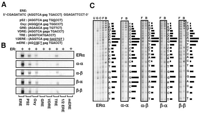
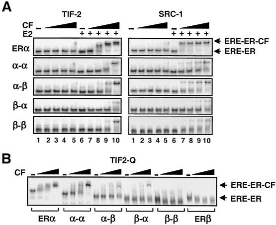
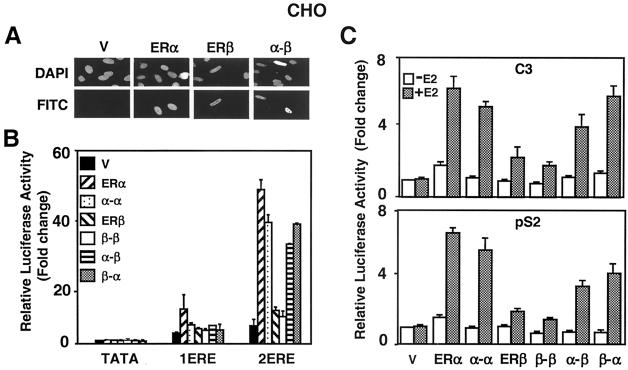
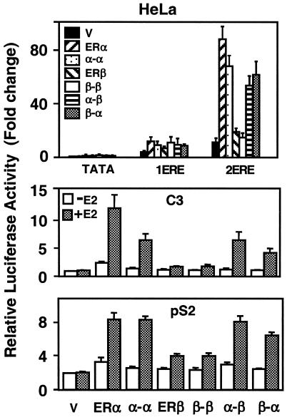
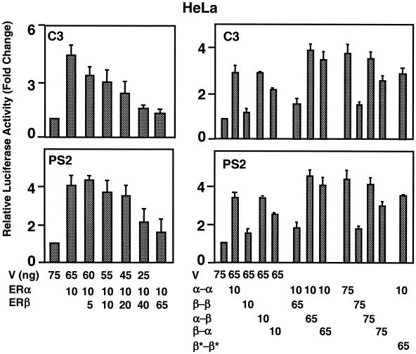
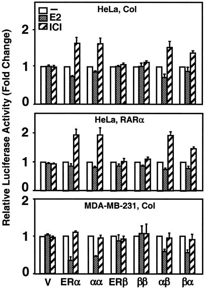
The effects of estrogens, particularly 17beta-estradiol (E2), are mediated by estrogen receptor alpha (ERalpha) and ERbeta. Upon binding to E2, ERs homo- and heterodimerize when coexpressed. The ER dimer then regulates the transcription of target genes through estrogen responsive element (ERE)-dependent and -independent pathways that constitute genomic estrogen signaling. Although ERalpha and ERbeta have similar ERE and E2 binding properties, they display different transregulatory capacities in both ERE-dependent and -independent signaling pathways. It is therefore likely that the heterodimerization provides novel functions to ERs by combining distinct properties of the contributing partners. The elucidation of the role of the ER heterodimer is critical for the understanding of physiology and pathophysiology of E2 signaling. However, differentially determining target gene responses during cosynthesis of ER subtypes is difficult, since dimers formed are a heterogeneous population of homo- and heterodimers. To circumvent the pivotal dimerization step in ER action and hence produce a homogeneous ER heterodimer population, we utilized a genetic fusion strategy. We joined the cDNAs of ERalpha and/or ERbeta to produce single-chain ERs to simulate the ER homo- and heterodimers. The fusion ERs interacted with ERE and E2 in a manner similar to that observed with the ER dimers. The homofusion receptors mimicked the functions of the parent ER dimers in the ERE-dependent and -independent pathways in transfected mammalian cells, whereas heterofusion receptors emulated the transregulatory properties of the ERalpha dimer. These results suggest that ERalpha is the functionally dominant partner in the ERalpha/beta heterodimer.


Figures

FIG. 1.
FIG. 1.
In vitro and in situ synthesis of ERα and ERβ. (A) For in vitro synthesis of ERs, 1.5 μg of linearized pBS-KS bearing no cDNA or a cDNA for an ER was transcribed by T3 polymerase and translated by rabbit reticulocyte lysate in the presence of 4 μl of [3H]methionine in a total reaction volume of 50 μl. Equal amounts (5 μl) of reaction mixtures were subjected to SDS-7.5% PAGE, visualized by fluorography (3H) or by Western blotting with an antibody directed to the carboxyl terminus of ERα (HC), of ERβ (PA1) or the amino-terminal Flag epitope (Flag) of both ERs. For in situ synthesis of ERs, CHO cells were transiently transfected with 3 μg of expression vector bearing none (V) or a cDNA for an ER. After 24 h, cells were lysed, and equal amounts (10 μg) of total cellular proteins were resolved by SDS-7.5% PAGE, followed by Western blotting with Flag, HC, or PA1 antibody. Endogenously expressed ERα in T47D cells (T47D) and ERβ in PA-1 cells (PA-1) were also analyzed by Western blotting with HC and PA1 antibodies, respectively. NS, nonspecific protein. (B) In vitro (InV, 10 μl) and in situ (CHO, 10 μg) synthesized ERβ were analyzed by Western blotting with Flag antibody. The ERE binding of ERβ synthesized in vitro (InV) or in CHO cells (CHO) was assessed by EMSA. Briefly, 1, 2, 4, 7, and 10 μl of transcription-translation mixture and 1, 2, 4, 7, and 10 μg of total protein from cell extract were incubated with 62.5 pmol of 32P-labeled ERE-containing DNA fragment for 30 min at 4°C and then subjected to 5% nondenaturing PAGE. The gel is dried and exposed to a PhosphorImager. Unbound ERE (ERE) and ERE-ERβ complexes are indicated. (C) The binding of ERα (lanes 2, 3, and 4) and ERβ (lanes 5, 6, and 7) synthesized in vitro (In Vitro) or in transiently transfected CHO cells (CHO) to the consensus ERE was assessed by EMSA. In this assay, 10 μl of in vitro-synthesized ERβ and 2 μl of ERα were used to obtain similar amounts of ERE-bound receptors. Reactions were incubated in the absence or presence (+) of the Flag (lanes 4 and 7), HC (lane 3), or PA1 (lane 6) antibody. ER-ERE complexes representing truncated-ERα homodimer (complex 1; the least prominent ER-ERE complex), truncated-ERα-WT-ERα heterodimer (complex 2), and WT-ERα homodimer (complex 3) are indicated. Lane 1 indicates reactions with the parent expression vector.
FIG. 2.
FIG. 2.
Coexpression of ERα and ERβ in vitro and in situ leads to formation of ERα/β heterodimer. For Western blotting of in vitro-synthesized ERs, 0.3 μg of pBS-KS vector bearing the Flag ERα cDNA was coexpressed with 0.3, 0.6, 1.2, or 1.8 μg of vector bearing the Flag-ERβ cDNA that correspond to 1-, 2-, 4-, or 6-fold-greater amounts, respectively, of the expression vector. Equal amounts (5 μl) of reaction mixtures were subjected to SDS-7.5% PAGE, followed by Western blotting with the Flag antibody. Migration of ERα and ERβ are indicated. The dimerization of ERα and ERβ was assessed by EMSA with equal amounts of transcription-translation mixture (5 μl). For Western blotting of in situ-synthesized ERs, 0.5 μg of the mammalian expression vector bearing the Flag-ERα cDNA was cotransfected with 0.5, 1.0, 2.0, or 3.0 μg (corresponding to 1, 2, 4, or 6, respectively) of the expression vector carrying the Flag-ERβ cDNA in CHO cells. Equal amounts of cell extracts (10 μg of total protein) were subjected to SDS-7.5% PAGE, followed by Western blotting with the Flag antibody. For EMSA, the cosynthesis of ERs was accomplished as described above, except that the cDNA for ERα does not contain sequences for the Flag epitope to distinguish ER species by the use of the antibody. Equal amounts of total protein (10 μg) of CHO cell extracts were subjected to EMSA. ERE-ER complexes were analyzed by using the HC or Flag antibody. ERE-bound ERα and ERβ homodimers and the ERα/β heterodimer are indicated. Unbound ERE is not shown.
FIG. 3.
FIG. 3.
Construction and synthesis of fusion ERs. (A) Schematics of ER fusion receptor cDNAs. A PCR-generated NdeI restriction enzyme site at the 5′ or 3′ end of an ER was used to genetically fuse two ER cDNAs in tandem. (B) Synthesis of fusion ERs in vitro with the parent vector bearing no cDNA (V) or a cDNA for an ER was accomplished as described for Fig. 1. Equal amounts of in vitro reaction mixtures (5 μl) or of (10 μg of total protein) of cell extracts from transfected CHO cells (Flag-CHO) were subjected to SDS-7.5% PAGE. The in vitro samples were visualized by fluorography (3H). The same samples were also probed with the HC, the PA1, or the Flag antibody (Flag-InV). (C) ERE binding of in vitro or in situ (CHO)-synthesized fusion receptors. Equal amounts (2 μl) of reaction mixtures, with the exception of the mixture containing ERβ (10 μl), were incubated with radiolabeled ERE in the absence or presence of the Flag antibody. Reaction mixtures were electrophoresed by 5% nondenaturing PAGE. (D) Equal amounts of CHO cell extracts (10 μg) were subjected to EMSA. The results from a representative experiment of two independent experiments are shown.
FIG. 4.
FIG. 4.
(A) The fusion ERs bind to ERE as monomers. The expression plasmids bearing ERα and the same (1:1) or a twofold-greater (1:2) concentration of the expression vector bearing the heterofusion Flag-α-β were cosynthesized in the presence of [3H]methionine in vitro. Equal amounts (5 μl) of reaction mixtures were subjected to SDS-7.5% PAGE or EMSA in the absence or presence (+) of the Flag antibody. (B) Intact DBDs are required for binding to ERE. Radiolabeled WT or DNA-binding defective (✽) ERα, the homofusion α-α, or the heterofusion α-β were subjected to SDS-PAGE and EMSA in the absence or presence (+) of Flag antibody. (C) Effect of dimerization surfaces in the LBDs on the ability of fusion receptor to bind to ERE. Equal amounts (5 μl) of WT or variant, with mutations in dimerization interface (indicated by the “dd” subscript), ERα, α-α, or α-β synthesized in vitro were subjected to SDS-PAGE. Then, 100 pM concentrations of each construct were electrophoresed on 5% nondenaturing PAGE (EMSA) in the absence or presence (+) of Flag antibody. (D) Dimerization interfaces in LBDs are required for an efficient binding of the fusion receptors to ERE. Equal molar concentrations (0, 6, 12, 25, and 50 pM [lanes 1, 2, 3, 4, and 5, respectively]) of WT and mutant (subscript dd) α-α or α-β were subjected to EMSA. For all experimental series, a representative experiment from at least two independent experiments is shown.
FIG. 5.
FIG. 5.
(A) Upper strand of the sequence containing the consensus ERE (in brackets), pS2, and Oxy ERE, GRE, VDRE, and TRE responsive elements and 1/2 ERE and mERE with the underlined five- and three-nucleotide changes from the ERE, respectively. The central base spacer is shown in lowercase. (B) Fusion ERs bind to the same spectrum of response elements. Equal molar concentrations of in vitro-synthesized ERα dimer or a fusion receptor were subjected to EMSA. A representative phosphorimage of two independent experiments is shown. (C) Fusion receptors bind to ERE in a manner similar to ERα. Critical nucleosides for ER-ERE interaction were identified by missing nucleoside hydroxyl radical assay. End-labeled DNA fragment containing consensus ERE was randomly cleaved by hydroxyl radical, incubated with 50 μl of transcription-translation mixtures, and subjected to 5% nondenaturing PAGE. Radioactive bands containing bound and free DNA were excised, eluted, and subjected to 15% sequencing gel electrophoresis. The intensities of individual DNA bands from sequencing gels were quantified by using a PhosphorImager. The ratio of free (F) to bound (B) DNA at each base was plotted as horizontal bars, the length of which approximates the strength of nucleoside contact with the protein. Uncut (U), Maxam-Gilbert G reaction-cut (G), and randomly cut (C) DNA fragments were also electrophoresed. A representative phosphorimage from at least two independent experiments is shown.
FIG. 6.
FIG. 6.
(A) Interactions of cofactors (CF) with fusion ERs as assessed by EMSA. Equal molar concentrations of in vitro-synthesized receptors were preincubated in the absence (−, lanes 1 to 5) or presence (+, lanes 6 to 10) of 10−7 M E2 (E2), followed by the addition of the end-labeled consensus ERE. The reactions were then incubated with 0 (−),1.56, 6.25, 25, or 100 ng (lanes 1 to 5 and 6 to 10, respectively) of GST fusion TIF-2623-986 (TIF-2) or 0, 0.125, 0.25, 0.5, and 1 μg (lanes 1 to 5 and 6 to 10, respectively) of SRC-1219-399 (SRC-1). Reactions were resolved on 5% nondenaturing PAGE. A representative phosphorimage of two independent experiments is shown. Unbound ERE is not shown. (B) Interaction of increasing concentrations (0.3, 0.6, and 1 μg) of TIF21125-1325 (TIF2-Q)-GST fusion protein with ERE-bound ERs. A representative phosphorimage of two independent experiments is presented. Free DNA is not shown.
FIG. 7.
FIG. 7.
(A) Intracellular localization of fusion ERs was examined by immunocytochemistry. The expression vector without (V) or with a cDNA for Flag ERα, ERβ, or fusion α-β was transiently transfected into CHO cells. The proteins were probed with the Flag antibody and visualized by using fluorescein-conjugated secondary antibody (FITC). DAPI (4′,6′-diamidino-2-phenylindole) staining indicates the nucleus. There was no protein detectable in cells transfected with the parent vector. The absence or presence (data not shown) of 10−9 M E2 did not affect the intracellular localization of the ERs. (B) CHO cells were transiently transfected with 75 ng of expression vector bearing a cDNA for an ER, together with 125 ng of reporter plasmid bearing no (TATA), one (1ERE), or two copies of the consensus ERE (2ERE). The TATA box promoter drives the expression of the firefly luciferase cDNA. The transfection efficiency was monitored by determining the coexpression of 2 ng of reporter plasmid bearing the Renilla luciferase cDNA. Cells were treated without (data not shown) or with 10−9 M E2 (E2) for 24 h. The data represent the means ± the standard errors of the mean (SEM) of three independent experiments performed in duplicate. (C) CHO cells transiently transfected as described above by using a reporter plasmid bearing the C3 or pS2 promoter that drive the expression of the firefly luciferase enzyme cDNA. Cells were treated in the absence (−E2) or presence (+E2) of 10−9 M E2 for 24 h. The results from three independent experiments in duplicate are represented as the means ± the SEM.
FIG. 8.
FIG. 8.
HeLa cells were transiently transfected as described for Fig. 7. The data represent the means ± the SEM of three experiments performed in duplicate.
FIG. 9.
FIG. 9.
Effects of coexpression of ERs and fusion receptor on transcriptional responses. (Left) HeLa cells were transiently transfected with a constant amount (in nanograms) of ERα expression vector and increasing amounts of the ERβ expression vector. Cells were cotransfected with a reporter plasmid bearing the C3 or pS2 promoter that drives the expression of the firefly luciferase enzyme cDNA. Cells were treated without (data not shown for simplicity) or with 10−9 M E2 for 24 h. (Right) HeLa cells were transiently transfected with an expression vector bearing a fusion ER cDNA alone or together with another fusion ER cDNA. Cells were also transfected with the C3 or pS2 reporter plasmid. After transfection, cells were incubated in the absence (data not shown) or presence of 10−9 M E2 for 24 h. V, parent expression vector. The data are the means ± the SEM of three independent experiments performed in duplicate.
FIG. 10.
FIG. 10.
Transcriptional responses to fusion ERs from the ERE-independent signaling pathway. HeLa or MDA-MB-231 cells were transiently transfected with 125 ng of expression vector bearing no cDNA (V) or a cDNA for an ER, together with 125 ng of reporter vector that contained the Col or the RARα promoter driving the expression of firefly luciferase enzyme cDNA. Cells were treated without (−) or with 10−9 M E2 (+E2) or 10−7 M ICI (ICI) for 40 h. The means ± the SEM of four independent experiments are shown.

Similar articles

Cited by

References

    1. Anzick, S. L., J. Kononen, R. L. Walker, D. O. Azorsa, M. M. Tanner, X. Y. Guan, G. Sauter, O. P. Kallioniemi, J. M. Trent, and P. S. Meltzer. 1997. AIB1, a steroid receptor coactivator amplified in breast and ovarian cancer. Science 277:965-968. - PubMed
    1. Benecke, A., P. Chambon, and H. Gronemeyer. 2000. Synergy between estrogen receptor alpha activation functions AF1 and AF2 mediated by transcription intermediary factor TIF2. EMBO Rep. 1:151-157. - PMC - PubMed
    1. Brzozowski, A. M., A. C. Pike, Z. Dauter, R. E. Hubbard, T. Bonn, O. Engstrom, L. Ohman, G. L. Greene, J. A. Gustafsson, and M. Carlquist. 1997. Molecular basis of agonism and antagonism in the estrogen receptor. Nature 389:753-758. - PubMed
    1. Cowley, S. M., S. Hoare, S. Mosselman, and M. G. Parker. 1997. Estrogen receptors α and β form heterodimers on DNA. J. Biol. Chem. 272:19858-19862. - PubMed
    1. Cowley, S. M., and M. G. Parker. 1999. A comparison of transcriptional activation by ER α and ERβ. J. Steroid Biochem. Mol. Biol. 69:165-175. - PubMed

Publication types

MeSH terms

LinkOut - more resources