. 2004 Sep;24(17):7779-94.
doi: 10.1128/MCB.24.17.7779-7794.2004.

Jun blockade of erythropoiesis: role for repression of GATA-1 by HERP2

Affiliations

Jun blockade of erythropoiesis: role for repression of GATA-1 by HERP2

Kamaleldin E Elagib et al. Mol Cell Biol. 2004 Sep.

Erratum in

Abstract

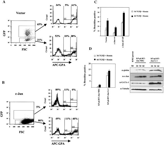
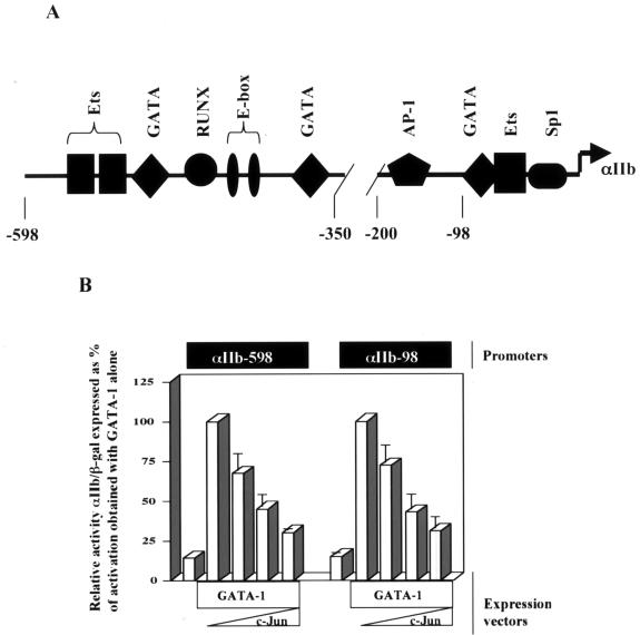
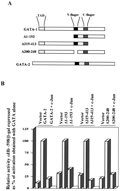
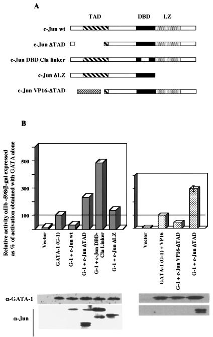
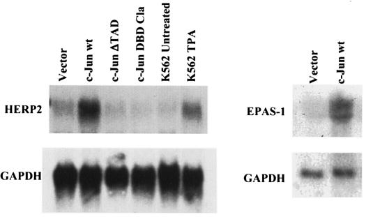
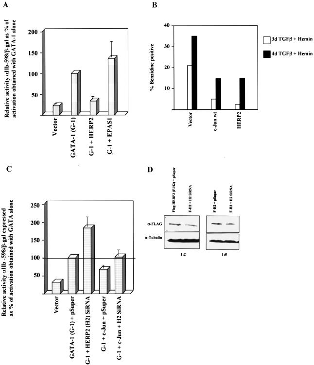
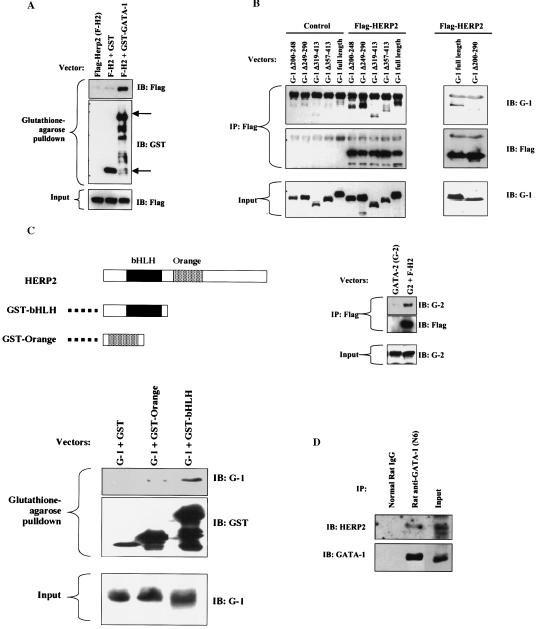
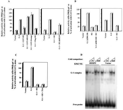
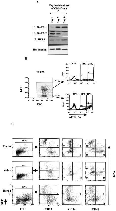
Although Jun upregulation and activation have been established as critical to oncogenesis, the relevant downstream pathways remain incompletely characterized. In this study, we found that c-Jun blocks erythroid differentiation in primary human hematopoietic progenitors and, correspondingly, that Jun factors block transcriptional activation by GATA-1, the central regulator of erythroid differentiation. Mutagenesis of c-Jun suggested that its repression of GATA-1 occurs through a transcriptional mechanism involving activation of downstream genes. We identified the hairy-enhancer-of-split-related factor HERP2 as a novel gene upregulated by c-Jun. HERP2 showed physical interaction with GATA-1 and repressed GATA-1 transcriptional activation. Furthermore, transduction of HERP2 into primary human hematopoietic progenitors inhibited erythroid differentiation. These results thus define a novel regulatory pathway linking the transcription factors c-Jun, HERP2, and GATA-1. Furthermore, these results establish a connection between the Notch signaling pathway, of which the HERP factors are a critical component, and the GATA family, which participates in programming of cellular differentiation.


Figures

FIG. 1.
FIG. 1.
Expression of c-Jun inhibits erythroid differentiation of primary human CD34+ hematopoietic progenitors and of K562 cells. (A) Purified human CD34+ hematopoietic progenitor cells transduced with the control vector (MSCV-IRES-GFP) were cultured in erythroid medium for 7 days, followed by flow cytometric analysis for the erythroid marker glycophorin A (GPA). As shown, GFP-positive (transduced) and GFP-negative (nontransduced) populations were separately gated for analysis. FSC, forward angle light scatter. (B) CD34+ cells transduced with the c-Jun-encoding retrovirus (MSCV-c-Jun-IRES-GFP) were cultured in erythroid medium and analyzed for GPA expression as in A. (C) K562 cells transduced with the indicated retroviral constructs were subjected to erythroid induction as indicated, followed by benzidine staining for hemoglobin. Results represent the mean of three determinations ± standard error of the mean. (D) Overexpression of GATA-1 reverses c-Jun inhibition of erythroid differentiation. K562 cells stably overexpressing c-Jun were superinfected with either the control vector (K5-pLRT-Jun MIG) or a GATA-1-encoding retroviral vector (K5-pLRT-Jun G-1). Erythroid differentiation was induced with transforming growth factor β (TGFβ) plus hemin for the indicated durations, followed by either benzidine staining or immunoblot analysis of whole-cell lysates.
FIG. 2.
FIG. 2.
c-Jun inhibits transcriptional activation by GATA-1. (A) Map of the GATA-responsive αIIb promoter, showing positions of binding sites based on use of the TESS and TFSearch programs as previously described (14). (B) GATA-1 repression by c-Jun does not depend on cis-acting AP-1 sites. The potency of Jun-mediated repression of GATA-1 transcriptional activation was compared using two GATA-responsive reporter constructs, one possessing an AP-1 binding site (αIIb-598) and one lacking an AP-1 binding site (αIIb-98), as depicted in the diagram in A; 2 μg of the GATA-1 expression construct (pEF-GATA-1) was cotransfected with 0.2, 0.5, or 1.0 μg of the c-Jun expression construct (RSV-c-Jun). Results ± standard error of the mean of three experiments are shown as relative reporter activity compared with activation by GATA-1 alone, normalized to β-galactosidase expression.
FIG. 3.
FIG. 3.
c-Jun-mediated transcriptional repression operates through a FOG-independent mechanism. (A) Diagrams of GATA-1, GATA-2, and GATA-1 deletion mutants. (B) Analysis of transcriptional activation by the indicated GATA factors with and without c-Jun. Results ± standard error of the mean of three experiments are shown as relative reporter activity compared with activation by GATA-1 alone, normalized by β- galactosidase expression. For each GATA factor, the activation obtained with that factor alone was set at 100%.
FIG. 4.
FIG. 4.
Structural requirements for Jun-mediated inhibition of GATA-1; dominant negative c-Jun mutants potentiate GATA-1 function. (A) Diagrams of c-Jun mutants. TAD, transcription activation domain; DBD, DNA binding domain; LZ, leucine zipper. The VP16 ΔTAD mutant contains the herpes simplex virus VP16 TAD in place of the c-Jun TAD. (B) Analysis of transcriptional activation by GATA-1 with and without the indicated c-Jun mutants. Results ± standard error of the mean of three experiments are shown as relative reporter activity compared with activation by GATA-1 alone, normalized by β-galactosidase expression. Whole-cell lysates from a duplicate transfection were immunoblotted for GATA-1 or Jun factors.
FIG. 5.
FIG. 5.
Expression of wild-type c-Jun is associated with upregulation of the hairy-enhancer-of-split-related bHLH factor HERP2. HERP2 mRNA levels in transduced cell lines expressing wild-type (wt) or mutant c-Jun as indicated were analyzed by Northern blot analysis, probing with HERP2 cDNA. Also analyzed were parental K562 with and without tetradecanoyl phorbol acetate (TPA) treatment (25 nM, 48 h). The membrane was stripped and reprobed with labeled glyceraldehhyde-3-phosphate dehydrogenase (GAPDH) cDNA. The right panel shows EPAS-1 and GAPDH mRNA levels in transduced cell lines, as indicated.
FIG. 6.
FIG. 6.
HERP2 inhibits transcriptional activation by GATA-1 and erythroid differentiation of K562 cells, and HERP2 siRNA interferes with c-Jun-mediated inhibition and augments GATA-1 function. (A) Analysis of transcriptional activation by GATA-1 with and without either HERP2 or EPAS-1. Reporter assays were carried out as described for Fig. 2. (B) K562 cells transduced with the indicated retroviral constructs were subjected to erythroid induction followed by benzidine staining for hemoglobin. (C) Analysis of transcriptional activation by GATA-1 with and without siRNA for HERP2 and with and without c-Jun; 2 × 10⁶ K562 cells were transfected with 2 μg of pEF-GATA-1, 3 μg of pSUPER or pSUPER-siHERP2.2, 1 μg of pCMV-c-Jun, 1.5 μg of αIIb-598-Luc, and 0.5 μg of pCMV-βgal, as indicated. After 72 h of incubation, cells were harvested for standard luciferase and β-galactosidase assays. Results shown represent three independent experiments ± standard error of the mean. (D) Demonstration of Flag-HERP2 knockdown by the HERP2 siRNA construct. K562 cells cotransfected with pcDNA3.1(−)-Flag-HERP2 plus either the pSUPER vector or pSUPER-siHERP2.2 were analyzed by immunoblotting (IB) for Flag or tubulin. Transfections were carried out with 5 μg of DNA per 2 × 10⁶ cells. Ratios below the panels indicate the relative amounts (in micrograms) of the Flag-HERP2 and siRNA vectors, respectively.
FIG. 7.
FIG. 7.
HERP2 interacts physically with GATA factors. (A) Physical interaction of HERP2 and GATA-1. 293T cells cotransfected with the indicated expression vectors were subjected to glutathione-agarose pulldown followed by immunoblotting with anti-Flag to detect Flag-HERP2 or with anti-GST to detect GST and GST-GATA-1. The upper arrow indicates the position of GST-GATA-1, and the lower arrow indicates the position of GST. (B) Physical interaction of HERP2 and GATA factors. 293T cells cotransfected with the indicated expression vectors were subjected to immunoprecipitation with anti-Flag antibody followed by immunoblotting with antibodies to GATA-1 (IB: G-1), GATA-2 (IB: G-2), or Flag, as indicated. The GATA mutants consist of the following deletions: N-finger (Δ200-248), C-finger (Δ249-290), carboxy-terminal regions (Δ319-413 and Δ357-413), and both fingers (Δ200-290). (C) Physical interaction of GATA-1 with isolated HERP2 domains. A diagram of HERP2 and of GST fusion proteins is shown. Cotransfections, pulldowns, and immunoblotting were performed as indicated. (D) Physical association of endogenous GATA-1 and HERP2. Parental K562 cellular extracts were subjected to immunoprecipitation with 10 μg of either normal rat immunoglobulin G or rat anti-GATA-1 (N6) monoclonal antibody. Immunoprecipitates were immunoblotted with rabbit anti-HERP2 followed by stripping and reprobing with rabbit anti-GATA-1. The input consisted of 20 μg of cellular extracts.
FIG. 8.
FIG. 8.
Characterization of the HERP2 repressive function. (A) Effects of various HERP factors on GATA-1 function. GST fusions contained full-length HERP2 (GST-H2) or isolated bHLH or orange domains. Also shown are results with Flag-HERP1 (Flag-H1) and a HERP2 truncation mutant lacking the bHLH domain (H2 ΔbHLH). Reporter assays were carried out as in Fig. 2. (B) HERP2 repression of GATA-1 lacking the N-finger (G-1 Δ200-248) and of GATA-2 (G-2). Reporter assays were carried out as in Fig. 2. (C) Histone deacetylase-independent repression of GATA-1 by HERP2. Transfectants were incubated with either vehicle control (0.05% dimethyl sulfoxide) or 500 nM trichostatin A (TSA). Reporter assays were carried out as in Fig. 2. (D) Lack of HERP2 influence on GATA-1 DNA binding. The electrophoretic mobility shift assay employed nuclear extracts (NE) from K562 cells transduced with either HERP2-encoding retrovirus or parent vector. Cells were treated for 48 h with 0.5 ng of transforming growth factor β per ml plus 60 μM hemin prior to harvest.
FIG.9.
FIG.9.
Expression and function of HERP2 in erythroid differentiation of primary human CD34+ hematopoietic progenitor cells. (A) Expression of HERP2, GATA-1, and GATA-2 proteins in CD34+ cells cultured in erythroid medium for the indicated durations. Whole-cell lysates were analyzed by immunoblotting with the indicated antibodies. (B) HERP2-transduced CD34+ cells show impaired erythroid differentiation. Cells transduced with MSCV-HERP2-IRES-GFP were cultured in erythroid medium for 7 days, followed by flow cytometric analysis for the erythroid marker glycophorin A (GPA), as in Fig. 1. Results from parallel transduction with the control vector are shown in Fig. 1A. Similar results were obtained from two independent experiments. (C) c-Jun and HERP2 both interfere with erythroid maturation. CD34+ cells transduced with constructs encoding the vector, c-Jun, or HERP2 were cultured in erythroid medium for 7 days, followed by flow cytometric analysis with costaining for GPA plus CD13 (granulocytic lineage marker), GPA plus CD34 (immaturity marker), or GPA plus CD41 (megakaryocyte-associated antigen).

Similar articles

Cited by

References

    1. Alitalo, R., J. Partanen, L. Pertovaara, E. Holtta, L. Sistonen, L. Andersson, and K. Alitalo. 1990. Increased erythroid potentiating activity/tissue inhibitor of metalloproteinases and jun/fos transcription factor complex characterize tumor promoter-induced megakaryoblastic differentiation of K562 leukemia cells. Blood 75:1974-1982. - PubMed
    1. Andrews, N. C., and D. C. Faller. 1991. A rapid micropreparation technique for extraction of DNA-binding proteins from limiting numbers of mammalian cells. Nucleic Acids Res. 19:2499. - PMC - PubMed
    1. Bamberger, A. M., K. Milde-Langosch, E. Rossing, C. Goemann, and T. Loning. 2001. Expression pattern of the AP-1 family in endometrial cancer: correlations with cell cycle regulators. J. Cancer Res. Clin. Oncol. 127:545-550. - PubMed
    1. Bauknecht, T., P. Angel, M. Kohler, F. Kommoss, G. Birmelin, A. Pfleiderer, and E. Wagner. 1993. Gene structure and expression analysis of the epidermal growth factor receptor, transforming growth factor-alpha, myc, jun, and metallothionein in human ovarian carcinomas. Classification of malignant phenotypes. Cancer 71:419-429. - PubMed
    1. Blobel, G. A., T. Nakajima, R. Eckner, M. Montminy, and S. H. Orkin. 1998. CREB-binding protein cooperates with the transcription factor GATA-1 and is required for erythroid differentiation. Proc. Natl. Acad. Sci. USA 95:2061-2066. - PMC - PubMed

Publication types

MeSH terms