. 2004 Sep;24(17):7795-805.
doi: 10.1128/MCB.24.17.7795-7805.2004.

Spliced-leader RNA trans splicing in a chordate, Oikopleura dioica, with a compact genome

Affiliations

Spliced-leader RNA trans splicing in a chordate, Oikopleura dioica, with a compact genome

Philippe Ganot et al. Mol Cell Biol. 2004 Sep.

Abstract

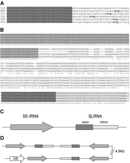
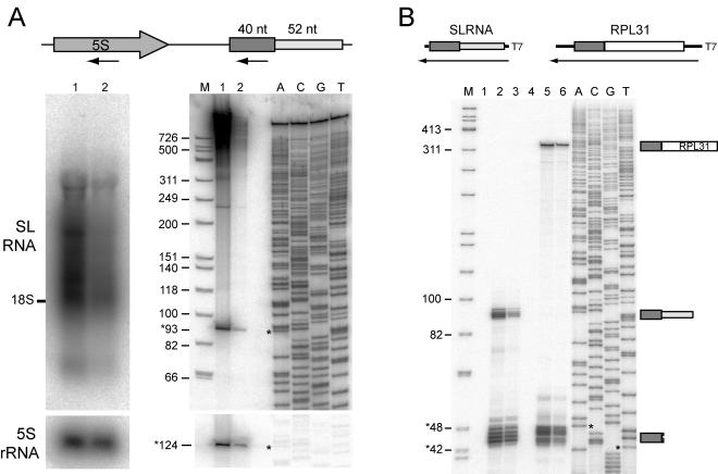
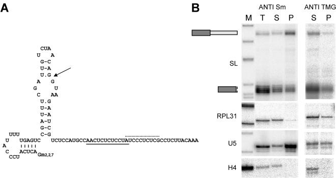
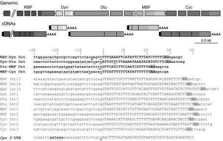
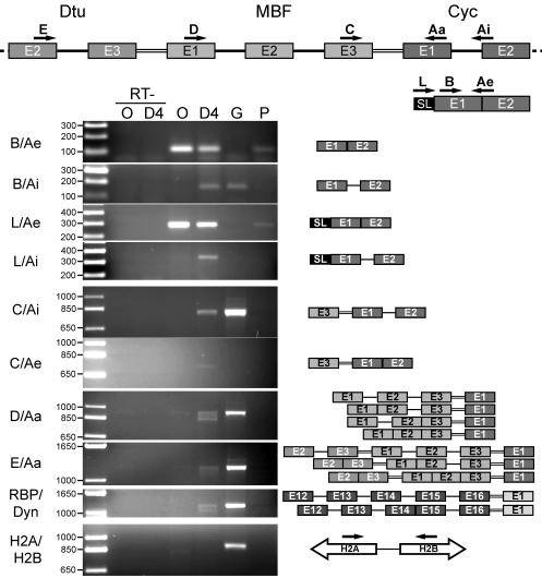
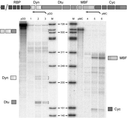
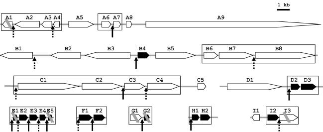
trans splicing of a spliced-leader RNA (SL RNA) to the 5' ends of mRNAs has been shown to have a limited and sporadic distribution among eukaryotes. Within metazoans, only nematodes are known to process polycistronic pre-mRNAs, produced from operon units of transcription, into mature monocistronic mRNAs via an SL RNA trans-splicing mechanism. Here we demonstrate that a chordate with a highly compact genome, Oikopleura dioica, now joins Caenorhabditis elegans in coupling trans splicing with processing of polycistronic transcipts. We identified a single SL RNA which associates with Sm proteins and has a trimethyl guanosine cap structure reminiscent of spliceosomal snRNPs. The same SL RNA, estimated to be trans-spliced to at least 25% of O. dioica mRNAs, is used for the processing of both isolated or first cistrons and downstream cistrons in a polycistronic precursor. Remarkably, intercistronic regions in O. dioica are far more reduced than those in either nematodes or kinetoplastids, implying minimal cis-regulatory elements for coupling of 3'-end formation and trans splicing.


Figures

FIG. 1.
FIG. 1.
O. dioica SL RNA genes. (A) Alignment of 5′ ends of cDNAs for the O. dioica cyclin D3-like homologue (Cyc), MBF, delta-tubulin (Dtu), dynein light chain (Dyn), RBP, and RPL31 (RPL). The common 40-nt leader sequence is capitalized and highlighted in grey, and the deduced methionine initiation codon is boldfaced. The deduced initiation codon for the cyclin cDNA is further downstream. (B) Alignment of SL RNA genomic loci. The 4 most divergent sequences out of 19 independent loci are shown. The 5′ region highlighted in grey is the 5S rRNA coding sequence. Dark and light grey backgrounds in the 3′ region indicate SL RNA exon and intron sequences, respectively. Asterisks indicate conserved nucleotides. (C) Schematic representation of the 5S rRNA-SL RNA locus assembled from contigs in the genomic shotgun database. (D) 5S rRNA-SL RNA head-to-tail repeats in a BAC sequence. Elements are represented as in panel C. The U6 snRNA gene is upstream of one of the 5S rRNA genes.
FIG. 2.
FIG. 2.
SL RNA expression. (A) Northern blot, with arrows in the schema at the top indicating regions used as probes. A 1% denaturing agarose gel (left panels) and a 6% polyacrylamide denaturing gel (right panels) were probed with oligonucleotides specific to the SL RNA exon (upper gels) and 5S rRNA (lower gels). Lanes: 1, oocyte RNA; 2, day 4 RNA; M, molecular size marker (with sizes given in thousands on the left); ACGT, dideoxy sequencing reaction. Sizes of bands indicated by asterisks are given. Intense smearing in the upper portion of lane 1 in the right panel may be partially due to nonspecific binding of polysaccharides in addition to the specific detection of trans-spliced RNAs seen in lane 2 or the left panel. (B) RNase protection, with schema at the top showing the antisense probes used for full-length SL RNA (lanes 1, 2, and 3) and the 5′ end of RPL31 RNA (lanes 4, 5, and 6). Lanes: 1 and 4, yeast tRNA control; 2 and 5, oocyte total RNA; 3 and 6, day 4 total RNA; M and ACGT, as explained for panel A. Diagrams to the right of the autoradiograph indicate protected fragments corresponding to visualized bands. Minor variation in band lengths observed for full-length SL RNA likely results from slight sequence differences in the 3′-terminal region, yielding alteration over a few base pairs in the length of the protected fragments.
FIG. 3.
FIG. 3.
O. dioica SL-RNP. (A) Model of SL RNA secondary structure. Arrow points to the exon-intron boundary; dotted and solid lines, possible Sm binding sites within the intron. Gm2,2,7, TMG cap. (B) Immunoprecipitation with anti-Sm (left) and anti-TMG (right) antibodies evaluated by RNase protection with probes specific for SL RNA, RPL31 mRNA, U5 snRNA, and histone H4 mRNA. Lanes: M, molecular size marker; T, total input RNA; S, unbound RNA fraction; P, antigen-bound RNA fraction.
FIG. 4.
FIG. 4.
Intergenic sequences in a cluster of trans-spliced genes are very short and lack a consensus polyadenylation signal. (Top) Genomic organization of the cluster and its processed mRNAs, with gene nomenclature as explained for Fig. 1. Rectangles, gaps, and lines represent exons, introns, and intercistronic regions, respectively. Rectangles of the same greyscale represent exons of the same gene, with 3′ UTRs in a lighter shade. On mature mRNAs, black rectangles indicate the trans-spliced leader and AAAA indicates the poly(A) tail. Schema is drawn to scale except for the 4,446-bp RBP16 gene, which has been truncated (parallel diagonal lines). A putative exon is present immediately upstream of the RBP16 gene, suggesting that the compact gene cluster continues further upstream. (Bottom) Sequence alignments of intercistronic regions (boldfaced) and introns. Flanking residues 30 nt upstream and 6 nt downstream of the splice sites are included. A portion of the sequence of the 3′ UTR of the cyclin gene is also shown. Capital letters, intronic and intergenic sequences. AG (highlighted in grey), 3′ cis or trans acceptor splice site. For each gene, sequencing of several cDNAs revealed alternative poly(A) cleavage sites. The various mapped cleavage sites are underlined, with the statistically most frequent occurring at the exon-intercistronic region boundary. Position +1 corresponds to the first nucleotide of the intercistronic region or intron. The consensus polyadenylation signal of the cyclin gene is boldfaced with a dotted underline, and sequences most closely approximating a polyadenylation signal in the 3′ regions of the upstream genes are indicated with a dotted underline. Polyadenylation cleavage sites were assigned as the first C, G, or U upstream of the sequenced poly(A) tail.
FIG. 5.
FIG. 5.
The gene cluster containing the cyclin D3-like gene is transcribed as a polycistronic transcript. Schema at the top shows the positions and orientations of primers used for RT-PCR analysis of RNA products from portions of the cyclin D3-like gene cluster. Rectangles, exons; single lines, introns; double lines, intercistronic regions. Primers were exon specific except for Ai and Ae. Ai amplifies cDNAs produced from mRNAs containing the first intron of the cyclin D gene, and Ae amplifies the spliced product between exons 1 and 2 of the same gene. Gels show PCR products resulting from use of the primer pairs given at the left. Leftmost lanes, molecular weight ladders, with sizes (in thousands) to the left. Template DNAs: O, oocyte cDNA; D4, cDNA from day 4 animals; G, genomic DNA; P, phage cDNA library from day 4 animals. RT− lanes, controls run in the absence of reverse transcriptase. The bottom two panels show RT-PCR products amplified between exon 12 of the RBP gene and exon 1 of the Dyn gene (RBP/Dyn) and the divergently transcribed histone H2A and H2B genes (H2A/H2B), respectively. Schema to the right of the gels depicts results from sequencing of RT-PCR products, showing various cis- and trans-spliced RNA intermediates. Schema for H2A and H2B shows genomic organization of the histone genes targeted for amplification.
FIG. 6.
FIG. 6.
Expression of genes in the cyclin D3-like gene cluster. A schematic representation of probes used for the RNase protection assay is shown at the top. pDD encompasses exon 1 (partially), intron 1, and exon 2 of the dynein gene, the intergenic region between the dynein and delta-tubulin genes, and part of exon 1 of the delta-tubulin gene. pMC encompasses part of exon 3 of the MBF gene, the intergenic region, and part of exon 1 of the cyclin gene. Drawings beside the autoradiographs represent identities of protected fragments corresponding to visualized bands (indicated by brackets). Lanes: 1 and 4, yeast tRNA control; 2 and 5, oocyte total RNA; 3 and 6, day 4 total RNA; pDD and pMC, 1/1,000 dilution of the probe alone; M, molecular size marker.
FIG. 7.
FIG. 7.
Putative operons in the O. dioica genome. Shown is the genomic organization of nine regions containing genes that are trans spliced (acceptor site [arrows]) or possibly trans spliced (candidate acceptor site localized next to the start codon [dotted arrows]). Solid arrows, ribosomal protein genes. Candidate operons (distance between translated regions, <300 bp) are boxed. Double parallel diagonal bars indicate regions of discontinuity in sequence information. A1, phospholipid-transporting ATPase VA; A2, cisplatin resistance-related protein CRR9P; A3, LMP7-like protein; A4, CG9166 protein; A5, speckle-type POZ protein; A6, prefoldin subunit 2; A7, dUTP nucleotidohydrolase; A8, prediction; A9, basement membrane-specific heparan sulfate proteoglycan core protein precursor; B1, myeloblast KIAA0230; B2, prediction; B3, CG14213 protein; B4, ribosomal protein S2; B5, prediction; B6, prediction; B7, adenosine deaminase; B8, autoantigen; C1, mediator subunit SUR2; C2, Trp4-associated protein TAP1; C3, retinoblastoma binding proteins 4 and 7; C4, protein phosphatase 2, regulatory subunit A (PR 65); C5, similar to SMAD; D1, nucleoporin 155; D2, ribosomal protein L17; D3, ribosomal protein L8; E1, ribosomal protein S20; E2, ribosomal protein L26; E3, ribosomal protein L11; E4, ribosomal protein S13; E5, ribosomal protein S5; F1, ribosomal protein S16; F2, ribosomal protein L6; G1, related to guanine nucleotide-binding protein; G2, ribosomal protein L10; H1, ribosomal protein L23; H2, ubiquitin A 52-residue ribosomal protein fusion product 1 (UBA52) (includes ribosomal protein L40); I1, related to thioredoxin; I2, ribosomal protein L24; I3, DNA replication licensing factor mcm2.

Similar articles

Cited by

References

    1. Blumenthal, T. 1995. trans-splicing and polycistronic transcription in Caenorhabditis elegans. Trends Genet. 11:132-136. - PubMed
    1. Campbell, D. A., N. R. Sturm, and M. C. Yu. 2000. Transcription of the kinetoplastid spliced leader RNA gene. Parasitol. Today 16:78-82. - PubMed
    1. Chioda, M., R. Eskeland, and E. M. Thompson. 2002. Histone gene complement, variant expression, and mRNA processing in a urochordate Oikopleura dioica that undergoes extensive polyploidization. Mol. Biol. Evol. 19:2247-2260. - PubMed
    1. Clayton, C. E. 2002. Life without transcriptional control? From fly to man and back again. EMBO J. 21:1881-1888. - PMC - PubMed
    1. Colgan, D. F., and J. L. Manley. 1997. Mechanism and regulation of mRNA polyadenylation. Genes Dev. 11:2755-2766. - PubMed

Publication types