. 2004 Aug 16;32(14):4358-67.
doi: 10.1093/nar/gkh744. Print 2004.

Triplex DNA-mediated downregulation of Ets2 expression results in growth inhibition and apoptosis in human prostate cancer cells

Affiliations

Triplex DNA-mediated downregulation of Ets2 expression results in growth inhibition and apoptosis in human prostate cancer cells

Giuseppina M Carbone et al. Nucleic Acids Res. .

Abstract

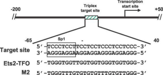
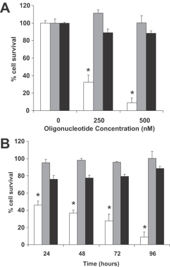
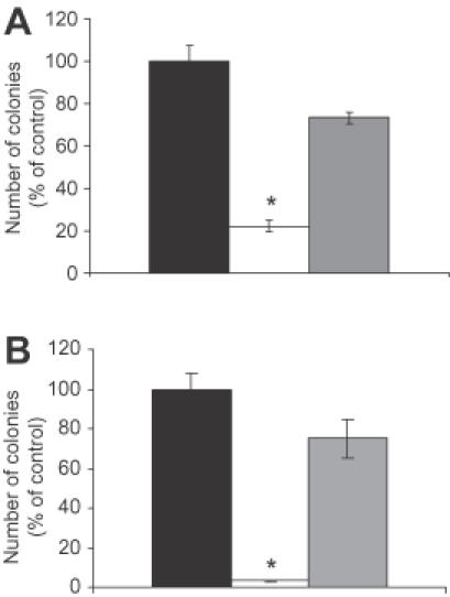
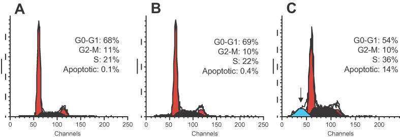
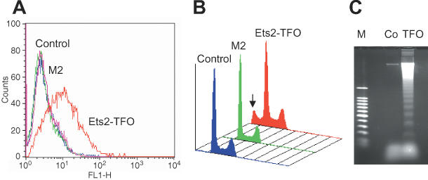
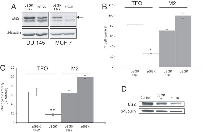
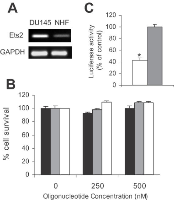
Ets2 is a member of the Ets family of transcription factors that in humans comprise 25 distinct members. Various Ets-domain transcription factors have been implicated in cancer development. Ets2 is expressed in prostate and breast cancer cells and is thought to have a role in promoting growth and survival in these cell types. However, a definitive role and the mechanisms whereby Ets2 acts in cancer cells are still unclear. Structural and functional similarities as well as overlapping DNA binding specificities complicate the identification of the specific roles of the various Ets factors. In this study, we used a triplex-forming oligonucleotide (TFO) to selectively inhibit Ets2 transcription in prostate cancer cells. We had previously shown that the Ets2-targeting TFO, which was directed to a unique purine-rich sequence critical for Ets2 promoter activity, acted with a high degree of sequence-specificity and target selectivity. TFO-mediated downregulation of Ets2 in prostate cancer cells induced important phenotypic changes, including inhibition of anchorage-dependent and anchorage -independent growth, cell cycle alterations and induction of apoptotic cell death. Expression of Ets2 under the control of a heterologous promoter abolished the anti-proliferative effects of the TFO in both short- and long-term assays, suggesting that these effects were a direct result of downregulation of Ets2 transcription and confirming target selectivity of the TFO. Furthermore, normal human fibroblasts, which expressed low levels of Ets2, were not affected by the Ets2-targeting TFO. Downregulation of Ets2 in prostate cancer cells was associated with reduced levels of the anti-apoptotic protein bcl-x(L) and growth regulatory factors cyclin D1 and c-myc. These data revealed a specific role of this transcription factor in promoting growth and survival of prostate cancer cells. Furthermore, the activity and selectivity of the Ets2-targeting TFO suggest that it might represent a valid approach to prostate cancer therapy.


Figures

Figure 1
Figure 1
Sequences of the Ets2-TFO, M2 control oligonucleotide and target site in the Ets2 promoter. The positions of the triplex target site and transcription start site in the Ets2 promoter are indicated. Numbers are relative to the transcription start site.
Figure 2
Figure 2
TFO-mediated inhibition of prostate cancer cell growth. DU145 cells were transfected for 4 h with Ets2-TFO or M2 control oligonucleotide in the presence of DOTAP or DOTAP alone. Viable cell number was determined using the MTT assay. Data are presented as percentage of viable cell number compared to untreated control samples. Data are mean ± SD of triplicate samples from a representative experiment. Similar results were obtained in three separate experiments. (A) Fraction of surviving cells was determined at 96 h post-transfection with the indicated concentrations of oligonucleotides. Final concentrations of DOTAP in culture medium were 0, 10 and 20 μg/ml, respectively. (B) Fraction of surviving cells was measured at 24 h intervals after transfection with 500 nM TFO and M2 control oligonucleotide or incubation with 20 μg/ml of DOTAP alone. Empty bars, Ets2-TFO; gray bars, M2 control oligonucleotide; black bars, DOTAP alone. Asterisk indicates p < 0.001 compared to DOTAP- and M2-treated cells.
Figure 3
Figure 3
Inhibition of colony forming ability of prostate cancer cells by the Ets2-targeting TFO. DU145 cells were transfected for 4 h with 500 nM Ets2-TFO or M2 control oligonucleotide in the presence of DOTAP or DOTAP alone. Cells were counted and plated to determine colony foming ability in anchorage-depedent and anchorage-independent growth conditions. (A) Cells (1 × 10³ per well) were plated in 35 mm tissue culture dishes and colonies were counted after 8–10 days. (B) Cells (2 × 10³ per well) were plated in 0.3% soft agar as described in Materials and Methods. Colonies were counted after 21–24 days. Data are mean ± SD of triplicate samples from a representative experiment. Black bars, DOTAP alone, empty bars, Ets2-TFO; gray bars, M2 control oligonucleotide. Asterisk indicates p < 0.001 compared to DOTAP- and M2-treated cells.
Figure 4
Figure 4
Cell cycle effects of Ets2 downregulation in prostate cancer cells. DU145 cells were left untreated or transfected with 1 μM Ets2-TFO or M2 control oligonucleotide. At 48 h post-transfection, cells were collected, fixed, stained with propidium iodide and analyzed for DNA content by flow cytometry. Percentages of cells in different phase of cell cycle or showing sub-G1 DNA content (arrow) are indicated for each panel. (A) untreated control; (B) Ets2-TFO; (C) M2 control oligonucleotide.
Figure 5
Figure 5
Apoptosis and DNA fragmentation in Ets2-TFO-treated prostate cancer cells. DU145 cells were incubated with 500 nM Ets2-TFO or M2 control oligonucleotide or left untreated (control). After 72 h, cells were harvested and analyzed. (A) Apoptotic cells were detected by terminal deoxynucleotidyl-transferase-mediated nick end labeling (TUNEL assay) and flow cytometry. (B) Percentage of cells with sub-G1 DNA content (arrow) was determined by flow cytometry analysis of propidium iodide stained cells. (C) Soluble DNA was isolated and analyzed on a 1.2% agarose gel to determine the presence of internucleosomal DNA fragmentation. Co, untreated control; M, 100 bp DNA ladder.
Figure 6
Figure 6
Forced expression of Ets2 rescues cells from growth inhibition induced by the Ets2-TFO. (A) Expression of Ets2 in transiently transfected DU145 and MCF-7 cells was determined by western blot at 48 h post-transfection. (B) DU145 were co-transfected with the Ets2 expression vector (pSGK-Ets2) or an empty vector (pSGK) along with 250 nM of Ets2-TFO or M2 control oligonucleotide for 4 h. Percentage of surviving cells was determined with MTS assay after 72 h. *P < 0.001 compared to cells transfected with pSGK-Ets2 and Ets2-TFO. (C) Cells were transfected with Ets2-TFO or M2 oligonucleotide with or without pSGK-Ets2 and empty vector (pSGK) as described above and plated in 35 mm tissue culture dishes. Colonies were counted after 8–10 days. Data are mean ± SD of triplicate samples from a representative experiment. **P < 0.01 compared to cells transfected with pSGK-Ets2 and Ets2-TFO. (D) Western blot analysis of DU145 cells left untreated (control) or transfected with Ets2-TFO along with pSGK-Ets2 or pSGK for 4 h as described above and then harvested after 72 h.
Figure 7
Figure 7
Insensitivity of normal human fibroblasts to the growth inhibitory effects of the Ets2-TFO. (A) Expression of Ets2 in normal human fibroblasts (NHF) and DU145 cells was determined by RT–PCR. GAPDH was used as control. (B) Human fibroblasts were transfected for 4 h with the indicated concentrations of Ets2-TFO or M2 control oligonucleotide in the presence of DOTAP or DOTAP alone. Viable cell number was determined after 96 h as indicated in the legend to Figure 2. (C) Human fibroblasts were transfected with pGL3-Ets2 luciferase reporter construct along with 500 nM Ets2-TFO or M2 oligonucleotide. Luciferase assays were performed after 24 h post-transfection. Data are mean ± SD of triplicate samples. P < 0.01 compared to M2-treated cells.
Figure 8
Figure 8
Expression of Ets2 downstream target genes in Ets2-TFO-treated prostate cancer cells. DU145 cells were transfected with 500 nM Ets2-TFO or M2 control oligonucleotide for 4 h. After 48 h, western blots were performed on cell lysates from Ets2-TFO- and M2-treated cells using antibodies against the indicated proteins. β-actin and α-tubulin were used as loading controls.

Similar articles

Cited by

References

    1. Gewirtz A.M., Sokol,D.L. and Ratajczak,M.Z. (1998) Nucleic acid therapeutics: state of the art and future prospects. Blood, 92, 712–736. - PubMed
    1. Dorsett Y. and Tuschl,T. (2004) siRNAs: applications in functional genomics and potential as therapeutics. Nature Rev. Drug Discov., 3, 318–329. - PubMed
    1. Chan P.P. and Glazer,P.M. (1997) Triplex DNA: fundamentals, advances, and potential applications for gene therapy. J. Mol. Med., 75, 267–282. - PubMed
    1. Praseuth D., Guieysse,A.L. and Helene,C. (1999) Triple helix formation and the antigene strategy for sequence-specific control of gene expression. Biochim. Biophys. Acta, 1489, 181–206. - PubMed
    1. Seidman M.M. and Glazer,P.M. (2003) The potential for gene repair via triple helix formation. J. Clin. Invest., 112, 487–494. - PMC - PubMed

Publication types

MeSH terms