. 2004 Aug 16;32(14):4368-76.
doi: 10.1093/nar/gkh754. Print 2004.

Sequential dimerization of human zipcode-binding protein IMP1 on RNA: a cooperative mechanism providing RNP stability

Affiliations

Sequential dimerization of human zipcode-binding protein IMP1 on RNA: a cooperative mechanism providing RNP stability

Jacob Nielsen et al. Nucleic Acids Res. .

Abstract

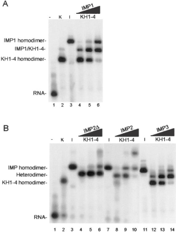
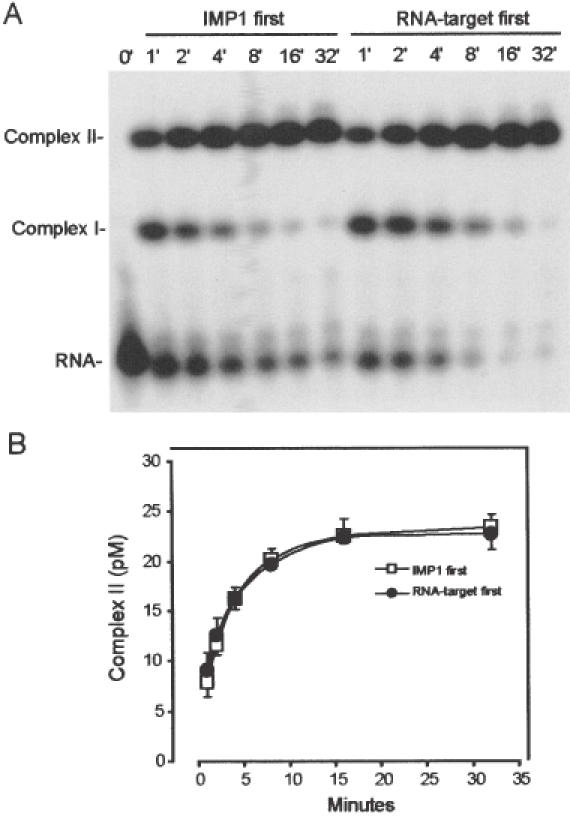
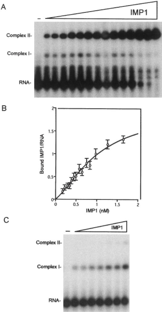
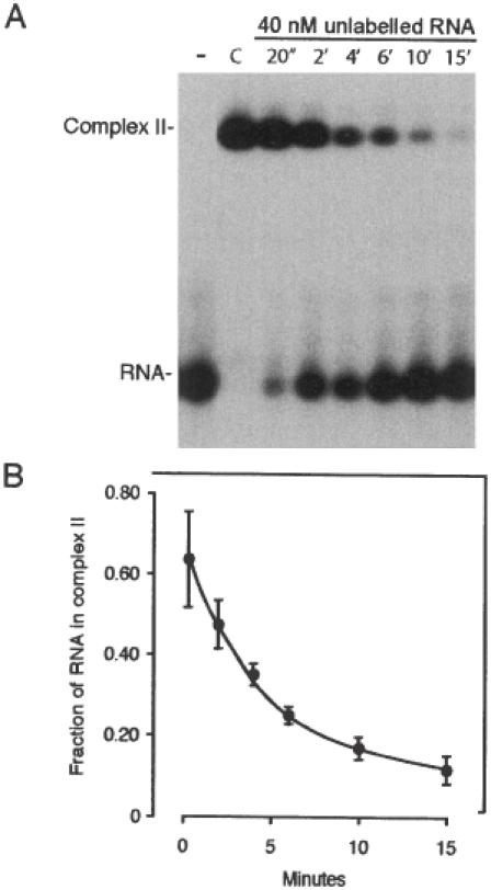
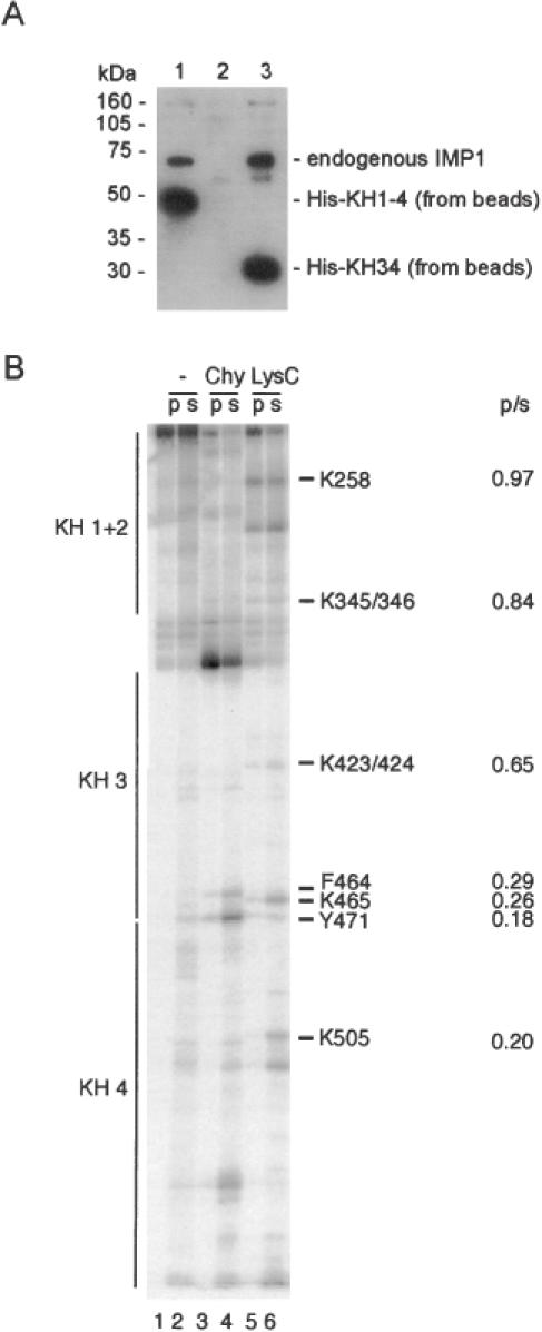
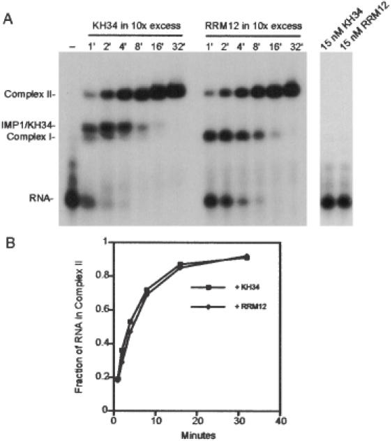
Active cytoplasmic RNA localization depends on the attachment of RNA-binding proteins that dictate the destination of the RNA molecule. In this study, we used an electrophoretic mobility-shift assay in combination with equilibrium and kinetic analyses to characterize the assembly of the human zipcode-binding protein IMP1 on targets in the 3'-UTR from Igf-II mRNA and in H19 RNA. In both cases, two molecules of IMP1 bound to RNA by a sequential, cooperative mechanism, characterized by an initial fast step, followed by a slow second step. The first step created an obligatory assembly intermediate of low stability, whereas the second step was the discriminatory event that converted a putative RNA target into a 'locked' stable RNP. The ability to dimerize was also observed between members of the IMP family of zipcode-binding proteins, providing a multitude of further interaction possibilities within RNP granules and with the localization apparatus.


Figures

Figure 1
Figure 1
Modular architecture of IMP1 and the constructs used in the present study.
Figure 2
Figure 2
Dimerization of IMP isoforms examined by gel mobility-shift analysis. (A) Track 1, radiolabelled RNA target (28 pM) on its own; track 2, in the presence of 1.5 nM KH1-4; track 3, in the presence of 1.5 nM IMP1; tracks 4–6, in the presence of both 1.5 nM KH1-4 and 0.5, 1.5 or 4.5 nM IMP1, respectively. (B) Similar to (A) but IMP1 is substituted with IMP2Δ in tracks 3–6, with IMP2 in tracks 7–10 and IMP3 in tracks 11–14. The slow migrating species in track 2 in (A) and in tracks 6 and 10 in (B) are oligomers of KH1-4, IMP2Δ and IMP2, respectively.
Figure 3
Figure 3
Sequential attachment of IMP1 to the radiolabelled RNA target. (A) Autoradiograph of the time course of the binding of 1.0 nM IMP1, pre-incubated in the absence of 28 pM RNA (left part), or diluted directly into the RNA-containing solution (right part). Complex I contains one IMP1 molecule and complex II contains two protein molecules. (B) InstantImager analysis of the formation of complex II as a function of time derived from three independent experiments.
Figure 4
Figure 4
Cooperative binding of IMP1 to the radiolabelled RNA target. (A) Twenty-eight picomolar RNA was incubated for 30 min with IMP1 concentrations in the 0.17–1.64 nM range and subsequently analysed by gel mobility-shift analysis. Complex I contains one, and Complex II contains two IMP1 molecules. (B) InstantImager data from seven independent experiments plotted as the number of bound IMP1 molecules versus the protein concentration, and the curve is a fit to the equation shown in Materials and Methods. (C) Twenty-eight picomolar RNA was incubated for 2 min with 1.3-fold increments of IMP1 in the 0.17–0.83 nM range and subsequently analysed by gel mobility-shift analysis.
Figure 5
Figure 5
Dissociation kinetics of Complex II. (A) Autoradiograph of the time course of dissociation of Complex II following the addition of 40 nM unlabelled RNA target. Complex II was formed initially by incubating 28 pM RNA with 1.5 nM IMP1 for 30 min under standard conditions (track C), and the unlabelled RNA was added at time 0. (B) InstantImager analysis of data from five independent dissociation experiments plotted as the fraction of RNA in Complex II versus time of incubation with unlabelled RNA.
Figure 6
Figure 6
The dimerization motif is located in the KH34 didomain. (A) Western analysis of the ability of endogenous IMP1 from an RNAse T1-treated cytoplasmic extract of RD-cells to associate with His–KH1-4 (track 1), His–KH12 (track 2) or His–KH34 (track 3) on nickel beads. The anti-IMP1 antibody is raised towards the C-terminal 10 amino acids (6), thus being unable to detect His–KH12 in track 2. (B) C-terminally labelled KH1-4 was partially cleaved with either chymotrypsin or LysC and then bound to immobilized GST–KH1-4. The pellet (p) or the supernatant (s) was analysed by polyacrylamide–SDS–tricin gel electrophoresis. The positions of the four KH domains are shown at the left, and amino acid positions and p/s ratios at the right of the autoradiograph.
Figure 7
Figure 7
Formation of Complex II in the presence of a 10-fold molar excess of the dimerization motif. (A) Left panel, autoradiograph of the time course of the formation of Complex II from 28 pM radiolabelled RNA and 1.5 nM IMP1 in the presence of either 15 nM KH34 or 15 nM RRM12. Complex I contains one, and Complex II contains two IMP1 molecules. IMP1–KH34 is a dimer between one molecule of full-length IMP1 and one molecule of the KH34 didomain from IMP1. (A) Right panel, autoradiograph showing that neither 15 nM KH34 nor 15 nM RRM12 is able to bind the radiolabelled RNA after 32 min. (B) InstantImager analysis of the formation of Complex II as a function of time derived from the data in (A).

References

    1. Chartrand P., Singer,R.H. and Long,R.M. (2001) RNP localization and transport in yeast. Annu. Rev. Cell Dev. Biol., 17, 297–310. - PubMed
    1. Tekotte H. and Davis,I. (2002) Intracellular mRNA localization: motors move messages. Trends Genet., 18, 636–642. - PubMed
    1. Yaniv K. and Yisraeli,J.K. (2002) The involvement of a conserved family of RNA binding proteins in embryonic development and carcinogenesis. Gene, 287, 49–54. - PubMed
    1. Havin L., Git,A., Elisha,Z., Oberman,F., Yaniv,K., Schwartz,S.P., Standart,N. and Yisraeli,J.K. (1998) RNA-binding protein conserved in both microtubule- and microfilament-based RNA localization. Genes Dev., 12, 1593–1598. - PMC - PubMed
    1. Deshler J.O., Highett,M.I., Abramson,T. and Schnapp,B.J. (1998) A highly conserved RNA-binding protein for cytoplasmic mRNA localization in vertebrates. Curr. Biol., 8, 489–496. - PubMed

Publication types