

Developmentally regulated mannose 6-phosphate receptor-mediated transport of a lysosomal enzyme across the blood-brain barrier
- PMID: 15314220
- PMCID: PMC515112
- DOI: 10.1073/pnas.0405042101
Developmentally regulated mannose 6-phosphate receptor-mediated transport of a lysosomal enzyme across the blood-brain barrier
Abstract
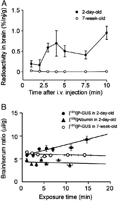
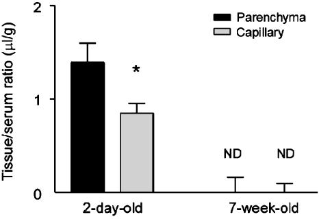
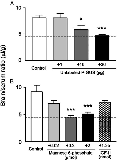
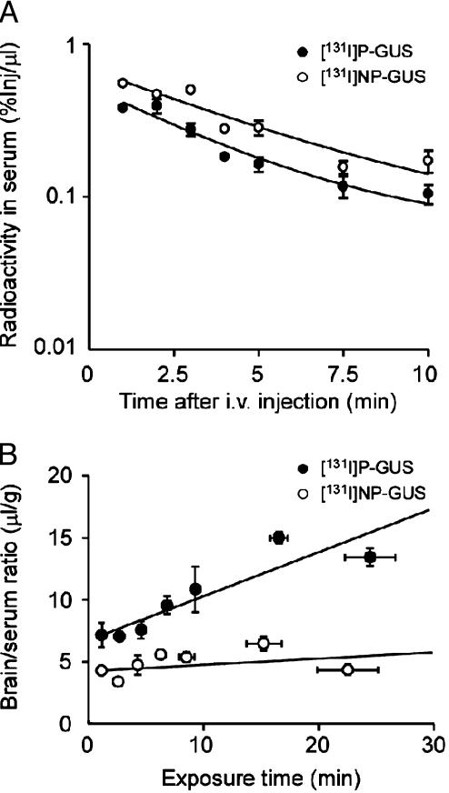
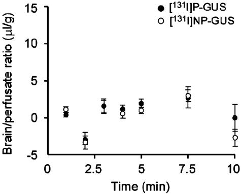
Mucopolysaccharidosis type VII is a lysosomal storage disorder resulting from inherited deficiency of beta-glucuronidase (GUS). Mucopolysaccharidosis type VII is characterized by glycosaminoglycan storage in most tissues, including brain. In these disorders, enzyme delivery across the blood-brain barrier (BBB) is the main obstacle to correction of lysosomal storage in the CNS. Prior studies suggested mouse brain is accessible to GUS in the first 2 weeks of life but not later. To explore a possible role for the mannose 6-phosphate/insulin-like growth factor II receptor in GUS transport across the BBB in neonatal mice, we compared brain uptake of phosphorylated GUS (P-GUS) and nonphosphorylated GUS (NP-GUS) in newborn and adult mice. (131)I-P-GUS was transported across the BBB after i.v. injection in 2-day-old mice. The brain influx rate (K(in)) of (131)I-P-GUS in 2-day-old mice was 0.21 microl/g.min and decreased with age. By 7 weeks of age, transport of (131)I-P-GUS was not significant. Capillary depletion revealed that 62% of the (131)I-P-GUS in brain was in brain parenchyma in 2-day-old mice. In addition, uptake of (131)I-P-GUS into brain was significantly reduced by coinjection of unlabeled P-GUS or M6P in a dose-dependent manner. In contrast, the K(in) of (131)I-NP-GUS (0.04 microl/g.min) was significantly lower than (131)I-P-GUS in 2-day-old mice. Transcardiac brain perfusion confirmed that neither (131)I-P-GUS nor (131)I-NP-GUS crossed the BBB in adult mice. These results indicate that (131)I-P-GUS transport into brain parenchyma in early postnatal life is mediated by the mannose 6-phosphate/insulin-like growth factor II receptor. This receptor-mediated transport is not observed in adult mice.
Figures


Similar articles
-
Epinephrine enhances lysosomal enzyme delivery across the blood brain barrier by up-regulation of the mannose 6-phosphate receptor.Proc Natl Acad Sci U S A. 2007 Jul 31;104(31):12873-8. doi: 10.1073/pnas.0705611104. Epub 2007 Jul 23. Proc Natl Acad Sci U S A. 2007. PMID: 17646643 Free PMC article.
-
Mannose 6-phosphate receptor-mediated transport of sulfamidase across the blood-brain barrier in the newborn mouse.Mol Ther. 2008 Jul;16(7):1261-6. doi: 10.1038/mt.2008.84. Epub 2008 Apr 29. Mol Ther. 2008. PMID: 18443601 Free PMC article.
-
Chemically modified beta-glucuronidase crosses blood-brain barrier and clears neuronal storage in murine mucopolysaccharidosis VII.Proc Natl Acad Sci U S A. 2008 Feb 19;105(7):2616-21. doi: 10.1073/pnas.0712147105. Epub 2008 Feb 11. Proc Natl Acad Sci U S A. 2008. PMID: 18268347 Free PMC article.
-
New strategies for enzyme replacement therapy for lysosomal storage diseases.Rejuvenation Res. 2010 Apr-Jun;13(2-3):229-36. doi: 10.1089/rej.2009.0920. Rejuvenation Res. 2010. PMID: 20345279 Free PMC article. Review.
-
Lysosomal storage diseases and the blood-brain barrier.Curr Pharm Des. 2008;14(16):1566-80. doi: 10.2174/138161208784705504. Curr Pharm Des. 2008. PMID: 18673198 Review.
Cited by
-
A Hitchhiker's guide to the blood-brain barrier: in trans delivery of a therapeutic enzyme.Mol Ther. 2014 Mar;22(3):483-484. doi: 10.1038/mt.2014.12. Mol Ther. 2014. PMID: 24584077 Free PMC article. No abstract available.
-
Inflammation-associated extracellular β-glucuronidase alters cellular responses to the chemical carcinogen benzo[a]pyrene.Arch Toxicol. 2016 Sep;90(9):2261-2273. doi: 10.1007/s00204-015-1593-7. Epub 2015 Oct 5. Arch Toxicol. 2016. PMID: 26438400 Free PMC article.
-
Cell replacement therapy in neurological disease.Philos Trans R Soc Lond B Biol Sci. 2006 Sep 29;361(1473):1463-75. doi: 10.1098/rstb.2006.1886. Philos Trans R Soc Lond B Biol Sci. 2006. PMID: 16939969 Free PMC article. Review.
-
Agile delivery of protein therapeutics to CNS.J Control Release. 2014 Sep 28;190:637-63. doi: 10.1016/j.jconrel.2014.06.017. Epub 2014 Jun 21. J Control Release. 2014. PMID: 24956489 Free PMC article. Review.
-
Insulin-Like Growth Factor-II/Cation-Independent Mannose 6-Phosphate Receptor in Neurodegenerative Diseases.Mol Neurobiol. 2017 May;54(4):2636-2658. doi: 10.1007/s12035-016-9849-7. Epub 2016 Mar 19. Mol Neurobiol. 2017. PMID: 26993302 Free PMC article. Review.
References
-
- Musa, B. U., Doe, R. P. & Seal, U. S. (1965) J. Biol. Chem. 240, 2811-2816. - PubMed
-
- Paigen, K. (1989) Prog. Nucleic Acid Res. Mol. Biol. 37, 155-205. - PubMed
-
- Sly, W. S., Quinton, B. A., McAlister, W. H. & Rimoin, D. L. (1973) J. Pediatr. 82, 249-257. - PubMed
-
- Vogler, C., Levy, B., Kyle, J. W., Sly, W. S., Williamson, J. & Whyte, M. P. (1994) Mod. Pathol. 7, 132-137. - PubMed
Publication types
MeSH terms
Substances
Grants and funding
LinkOut - more resources
Full Text Sources
Other Literature Sources
Miscellaneous
