. 2004 Aug;2(8):E219.
doi: 10.1371/journal.pbio.0020219. Epub 2004 Aug 17.

A focused and efficient genetic screening strategy in the mouse: identification of mutations that disrupt cortical development

Affiliations

A focused and efficient genetic screening strategy in the mouse: identification of mutations that disrupt cortical development

Konstantinos Zarbalis et al. PLoS Biol. 2004 Aug.

Abstract

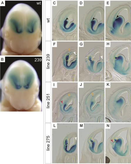
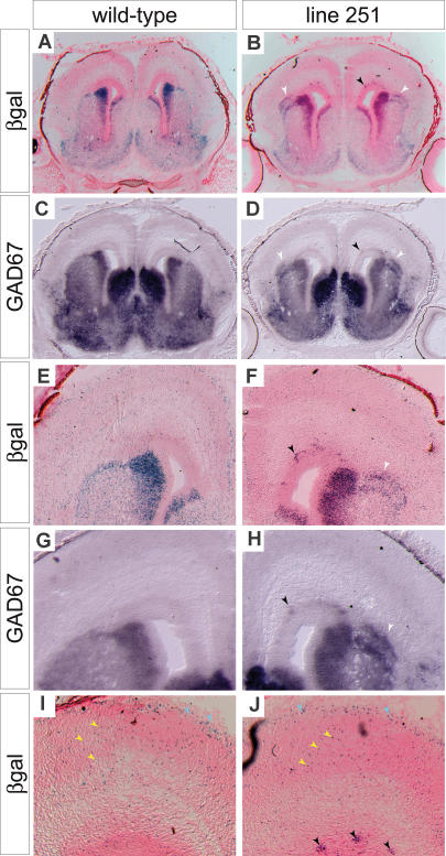
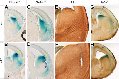
Although the mechanisms that regulate development of the cerebral cortex have begun to emerge, in large part through the analysis of mutant mice (Boncinelli et al. 2000; Molnar and Hannan 2000; Walsh and Goffinet 2000), many questions remain unanswered. To provide resources for further dissecting cortical development, we have carried out a focused screen for recessive mutations that disrupt cortical development. One aim of the screen was to identify mutants that disrupt the tangential migration of interneurons into the cortex. At the same time, we also screened for mutations that altered the growth or morphology of the cerebral cortex. We report here the identification of thirteen mutants with defects in aspects of cortical development ranging from the establishment of epithelial polarity to the invasion of thalamocortical axons. Among the collection are three novel alleles of genes for which mutant alleles had already been used to explore forebrain development, and four mutants with defects in interneuron migration. The mutants that we describe here will aid in deciphering the molecules and mechanisms that regulate cortical development. Our results also highlight the utility of focused screens in the mouse, in addition to the large-scale and broadly targeted screens that are being carried out at mutagenesis centers.


Conflict of interest statement

The authors have declared that no conflicts of interest exist.

Figures

Figure 1
Figure 1. The Strategy for Isolation of Recessive Mutations Disrupting Cortical Development
(A) Migrating interneuron precursors appear to migrate tangentially (yellow arrows) through both the MZ and the IZ/SVZ. Precursors also migrate radially (pink arrows) to reach the developing cortical plate (CP). (B) Migrating interneuron precursors expressing β-galactosidase can be seen in a whole-mount preparation of an E14.5 Dlx-LacZ mouse as diffuse cortical staining. (C) In coronal section, the densely labeled SVZ of the LGE can be seen, as can the streams of migrating interneuron precursors in the cortex. (D) To identify recessive mutations, male C57BL6/J mice were treated with ENU and then crossed to FVBN/J females that were homozygous for the Dlx-LacZ transgene. Male offspring of this cross were backcrossed to produce female offspring that were then backcrossed to their fathers. Embryonic litters from these backcrosses were stained and examined for defects at E13.5 or E14.5.
Figure 2
Figure 2. Novel Alleles of scribble, megalin, and Rfx4
(A and B) Comparison of E12.5 WT (A) and mutant (B) embryos showing the severe craniorachischisis of line 90 embryos. (C) Sequencing chromatograms show the thymine in the WT sequence (top) that is an adenine in line 90 (bottom). This thymine-to-adenine substitution changes an isoleucine codon to a lysine codon within a conserved LRR domain of SCRIBBLE. (D and E) Ribbon diagrams of an LRR fold, illustrating the predicted position (red arrowheads) of the line 90 amino acid substitution. The substitution is in a linker region between two stretches of β-sheet. An LRR fold has a β-sheet on the inside, and linker regions on the outside, of a broad half-circular curve. (F) The predicted domain structure of SCRIBBLE indicating where the line 90 and the Circletail alleles alter the protein relative to the three LRR and four PDZ domains. (G and H) Dorsal views of the cortex of E17.5 WT (G) and line 267 mutant (H) embryos stained to reveal the expression of the Dlx-LacZ transgene. The red arrowheads point to the choroid plexus of the third ventricle, which is stained because of its endogenous β-galactosidase expression, and which is greatly enlarged in the mutant. The cortex of the mutant is longer along the rostrocaudal axis and is altered in shape in the caudal portion (yellow arrows). The olfactory bulbs (blue arrowheads) are also altered in shape. (I) The choroid plexus persists in its hypertrophic state after birth and can be seen as a pinkish lump on the head of this postnatal day 14 (P14) pup. (J) A coronal section through the dorsal portion of the diencephalon of a P0 pup. (K) Sequencing chromatograms show the thymine in the WT sequence (top) that is an adenine in line 267 (bottom). (L) The predicted domain structure of MEGALIN indicating where the new stop codon is introduced by the line 267 mutation. (M and N) Dorsal views of the cerebral cortex of E14.5 WT (M) and line 269 mutant (N) embryos stained to reveal the expression of the Dlx-LacZ transgene. (O and P) Coronal sections of E14.5 WT (O) and line 269 mutant (P) forebrains. Red arrowheads indicate the apparent span over which dorsal midline structures are lost in the mutant. (Q) Sequence chromatograms showing the thymine in the WT sequence (top) that is a cytosine in line 269 (bottom). (R) The predicted domain structure of RFX4 indicating where the line 269 substitution alters the protein relative to the DNA binding and the extended (B, C, and Dim) dimerization domains.
Figure 3
Figure 3. Severe Disruption of Interneuron Migration in Line 154 Mutants
(A) Disseminated immature interneurons are seen as diffuse cortical (Cx) staining in WT embryos. Subcortical expression in the SVZ of the LGE can be seen as a darkly stained, inverted crescent. (B) Embryos homozygous for the line 154 mutation have little or no cortical staining, and the subcortical staining has aberrant streaks (pink arrowheads) and spots (white arrowhead), particularly in the frontal cortex. (C, E, and G) In this rostral-to-caudal series of coronal sections from WT embryos, the normal MZ and IZ/SVZ migratory streams are diffusely labeled (red arrowheads), and a sharp cortical-subcortical boundary (black arrowheads) is marked by the abrupt transition between the densely stained SVZ of the LGE and the diffuse staining of the migrating interneuron precursors. (D, F, and H) A rostral-to-caudal series of coronal sections from a line 154 mutant embryo shows the rostral spots (white arrowheads) visible in whole mount to be aggregates of cells in the cortex, and the streaky subcortical staining to be radially directed linear aggregates (pink arrowheads). The SVZ of the LGE is also noticeably thinner, and there is not a well-defined cortical-subcortical transition in the staining pattern. (I and J) Rostral views of Gad67 whole-mount in situ hybridization show the pattern of migration abnormalities in the cortex and significant defects in population of the olfactory bulb by GABAergic neurons.
Figure 4
Figure 4. Limb Patterning Defects in Tangential Migration Mutants
Anterior is to the right for all limbs. (A–C) Left hindlimbs are shown from E14.5 WT (A), line 251 (B), and line 239 (C). Yellow arrowheads point to extra digits on the anterior (thumb) side of the limb in mutants. Line 275 mutants also have anterior polydactyly. The mutants have a slight developmental delay that causes some differences in appearance of the limb buds at E14.5, when the limbs are growing rapidly. (D) This diagram illustrates components of the Shh/Fgf feedback loop that maintains Fgf4 expression in the AER. (E–J) In situ hybridization on E11.5 limb buds. Unlike in WT (E), expression of the posterior patterning gene Hoxd13 in left forelimb buds extends ectopically into an anterior domain (yellow arrowheads) in 251 (F) and 239 (G) mutants. Fgf4 expression in left hindlimb buds, restricted to the posterior AER in WT (H), has an ectopic expression domain at the far anterior edge of the AER (yellow arrowheads) in 251 (I) and 239 (J) mutants.
Figure 5
Figure 5. Line 239, 251, and 275 Mutants Have Similar Migration Defects
(A) Frontal view of an E14.5 WT embryo. (B) Frontal view of an E14.5 embryo homozygous for the line 239 mutation. The stream of cells leaving the SVZ of the LGE is streaky and aggregated in the rostral cortex of the mutant. (C–E) A rostral-to-caudal series of coronal sections from an E14.5 WT embryo. WT embryos have diffuse cortical staining and a sharp boundary (black arrowheads) between the subcortical and the cortical telencephalon. (F–H) Coronal sections from E14.5 line 239 mutant forebrains. Yellow circles indicate the area of the cortical-subcortical boundary where a large excess of migrating cells can be seen in the IZ/SVZ area. Orange arrowhead indicates aggregated cells in the IZ/SVZ of the lateral wall of the cortex. White arrowheads indicate aggregates in the medial wall. The staining in the MZ appears normal. Defects become less apparent in the more caudal sections. (I–K) Sections from an E14.5 line 251 mutant embryo. Orange arrowheads indicate aggregates in the lateral wall of the cortex. In (I), a linear aggregate of stained cells can be seen extending from the IZ/SVZ to the MZ (red arrowhead). The cortical-subcortical boundary is well defined in line 251 mutants. (L–N) Sections from an E14.5 line 275 mutant embryo. Yellow circles indicate aberrant staining in the cortical-subcortical boundary region. The white arrowhead indicates aggregates in the medial wall of the cortex, and the red arrowhead points to a radially directed aggregate of cells extending from the IZ/SVZ to the MZ.
Figure 6
Figure 6. Dlx-LacZ and GAD67 Expression Show that Interneuron Precursors Persist in the IZ/SVZ of 251 Mutant Cortices
(A) Coronal section through the forebrain of an E18.5 WT embryo stained for β-galactosidase and counterstained with nuclear fast red. (B) A similar section from a line 251 mutant embryo. Unusual accumulations of stained cells can be seen in the cortex just dorsal to the striatum (white arrowheads). Aggregates of cells in the IZ/SVZ can also be seen (black arrowhead). (C and D) Sections adjacent to those in (A) and (B) hybridized with a probe for Gad67 mRNA. Arrowheads in (D) point to the same features that are seen in (B). (E–H) Higher-magnification views of the dorsal portions of the sections in (A–D). (I and J) Higher-magnification view of cortex. In the WT cortex (I), cells can be seen dispersed through the cortical plate (yellow arrowheads) and scattered through the MZ (blue arrowheads). Similar distributions of labeled cells can be seen in the mutant cortex (J). In contrast to the WT, however, aggregates of cells are found in IZ/SVZ (black arrowheads).
Figure 7
Figure 7. Corticostriatal Delamination and Lack of the Thalamocortical Projection in Line 412 Mutants
(A–D) Coronal hemisections of E15.5 WT (A and C) and line 412 (B and D) mutant embryos stained for the Dlx-LacZ transgene. The cortex is thinner in 412 mutants, as can be seen in both the rostral (B) and the caudal (D) sections. Delamination of the corticostriatal boundary can be seen in the region between the red arrowheads in (D). (E and F) Immunostaining for L1 antigen labels the thalamocortical fibers in coronal hemisections from E14.5 WT (E) and mutant (F) embryos. The striatum and corticostriatal areas are shown. In the WT section, the thalamocortical fibers can be seen traversing the striatum through the internal capsule and coursing into the cortex. In the mutant section, a few fibers enter the striatum but do not traverse it to reach the cortex. Corticostriatal delamination can be seen as a hole in the right side of the section. (G and H) Immunostaining for TAG1 antigen (blue arrowheads) reveals corticofugal fibers.

Similar articles

Cited by

References

    1. Anderson SA, Eisenstat DD, Shi L, Rubenstein JL. Interneuron migration from basal forebrain to neocortex: Dependence on Dlx genes. Science. 1997;278:474–476. - PubMed
    1. Anderson SA, Marin O, Horn C, Jennings K, Rubenstein JL. Distinct cortical migrations from the medial and lateral ganglionic eminences. Development. 2001;128:353–363. - PubMed
    1. Ang ES, Haydar TF, Gluncic V, Rakic P. Four-dimensional migratory coordinates of GABAergic interneurons in the developing mouse cortex. J Neurosci. 2003;23:5805–5815. - PMC - PubMed
    1. Bilder D, Perrimon N. Localization of apical epithelial determinants by the basolateral PDZ protein Scribble. Nature. 2000;403:676–680. - PubMed
    1. Bilder D, Li M, Perrimon N. Cooperative regulation of cell polarity and growth by Drosophila tumor suppressors. Science. 2000;289:113–116. - PubMed

Publication types

MeSH terms