. 2004 Aug;2(8):E225.
doi: 10.1371/journal.pbio.0020225. Epub 2004 Aug 17.

Forgetting, reminding, and remembering: the retrieval of lost spatial memory

Affiliations

Forgetting, reminding, and remembering: the retrieval of lost spatial memory

Livia de Hoz et al. PLoS Biol. 2004 Aug.

Abstract

Retrograde amnesia can occur after brain damage because this disrupts sites of storage, interrupts memory consolidation, or interferes with memory retrieval. While the retrieval failure account has been considered in several animal studies, recent work has focused mainly on memory consolidation, and the neural mechanisms responsible for reactivating memory from stored traces remain poorly understood. We now describe a new retrieval phenomenon in which rats' memory for a spatial location in a watermaze was first weakened by partial lesions of the hippocampus to a level at which it could not be detected. The animals were then reminded by the provision of incomplete and potentially misleading information-an escape platform in a novel location. Paradoxically, both incorrect and correct place information reactivated dormant memory traces equally, such that the previously trained spatial memory was now expressed. It was also established that the reminding procedure could not itself generate new learning in either the original environment, or in a new training situation. The key finding is the development of a protocol that definitively distinguishes reminding from new place learning and thereby reveals that a failure of memory during watermaze testing can arise, at least in part, from a disruption of memory retrieval.


Conflict of interest statement

The authors have declared that no conflicts of interest exist.

Figures

Figure 1
Figure 1. Experimental Design
Outline of the different phases of testing. The platform position used during training is indicated by a red circle in the NE quadrant of the pool (large blue circle), although in practice platform locations were counterbalanced between NE and SW locations. The novel location, to which a subset of rats was exposed during reminding, is indicated by a black circle in the SW quadrant. This position was always opposite to that used during training. PT1 and PT2: probe test 1 and 2. The hatched areas represent the original training quadrant irrespective of the position of the platform (i.e., original or novel) during retention testing. PTn1 and PTn2: PTs during new context learning in the second pool.
Figure 2
Figure 2. Training
Mean latencies to escape from the water and climb onto the hidden platform during task acquisition. Data are averaged in blocks of five trials and grouped according to the lesion made at the end of training; note that all animals were unoperated during acquisition. Only rats that reached criterion (mean escape latency less than 15 s over the last ten trials) and whose lesions were considered acceptable (see Results: Surgery) are presented. Animals rapidly learned to locate the escape platform, and prospective lesion groups did not differ.
Figure 3
Figure 3. Lesion Analysis
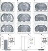
Representative photomicrographs of cresyl-violet-stained coronal brain sections taken from subjects belonging to each of the three lesion groups—partial hippocampal lesion (A), sham lesion (B), and complete hippocampal lesion (C). In each case, sections corresponding to anterior, middle, and posterior levels of the hippocampus are displayed. The mean area of spared hippocampal tissue in each group (see Materials and Methods for calculation) is plotted below in (D). Note that the volumes of spared tissue in the septal and temporal halves of the hippocampus are plotted separately, but these values are still expressed as percentages of the entire hippocampal volume—hence the value of 50% per half in shams. The cartoon hippocampi accompanying the graph indicate lesioned tissue in dark grey, and spared tissue in light cream. As intended, partially lesioned rats exhibited substantial sparing only in the septal (dorsal) half of the hippocampus, and rats with complete hippocampal lesions exhibited minimal sparing (less than 5% at either pole).
Figure 4
Figure 4. Retention Testing: Quadrant Analysis
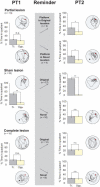
Percentage time during PT1 and PT2 spent in the training and opposite quadrants of the pool (left and right lanes) and the reminder treatment (grey central lane). The training location is represented as a red circle in the NE quadrant, and the novel location (novel subgroups only) as a black circle in the SW quadrant. In practice, NE and SW quadrants were counterbalanced. Rats with partial hippocampal lesions were unable to remember the platform location on PT1 but could be reminded of the training location by exposure, at the end of PT1, to a platform in the original or a novel location. (Note that the ‘reminder' lane simply refers to this exposure to a platform—PT1 is itself the ‘reminder trial.') The key finding is that the improvement in PT2 occurred irrespective of the platform location during reminding. In contrast, sham-lesioned animals exhibited some reversal learning upon exposure to the platform in a novel location. Complete-lesioned rats did not remember the platform location during either PT1 or PT2. *p < 0.05; **p < 0.01; n.s. = nonsignificant; comparisons with chance = 50%; one-sample t-tests. Representative swim paths are included.
Figure 5
Figure 5. Retention Testing: Zone Analysis
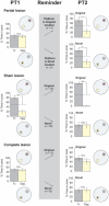
Percentage time in PT1 (left) and PT2 (right) spent within a zone, 20 cm in radius, centred on either the original training location (broken circle; grey) or an equivalent location in the opposite quadrant (broken circle; yellow), expressed as a percentage of the total time spent in both of the zones. The reminder treatment is again shown as the grey central lane and as the location where the hidden platform became available at the end of PT1 within these zones (original = red; novel = black). Consistent with Figure 4, rats with partial hippocampal lesions were amnesic in PT1 but could be reminded of the correct location, even by exposure to the platform in a novel location. *p < 0.05; **p < 0.01; n.s. = nonsignificant; comparisons with chance = 50%; one-sample t-tests.
Figure 6
Figure 6. Novel Context Learning
Percentage time spent in the target quadrant containing the escape platform during one-trial new learning in a different pool. *p < 0.05; n.s. = nonsignificant; comparison of percentage time spent in training zone during PTn1 and PTn2; paired-sample t-tests. New learning was observed only in sham-lesioned rats.

References

    1. Bannerman DM, Good MA, Butcher SP, Ramsay M, Morris RGM. Distinct components of spatial learning revealed by prior training and NMDA receptor blockade. Nature. 1995;378:182–186. - PubMed
    1. Cherkin A. Retrograde amnesia in the chick: Resistance to the reminder effect. Physiol Behav. 1972;8:949–955.
    1. Concannon JT, Carr C. Pre-test epinephrine injections reverse DDC-induced retrograde amnesia. Physiol Behav. 1982;9:443–448. - PubMed
    1. Davis HP, Squire LR. Protein synthesis and memory: A review. Psychol Bull. 1984;96:518–559. - PubMed
    1. de Hoz L, Knox J, Morris RGM. Longitudinal axis of the hippocampus: Both septal and temporal poles of the hippocampus support watermaze spatial learning depending on the training protocol. Hippocampus. 2003;13:587–603. - PubMed

Publication types