

AMP-activated protein kinase mediates ischemic glucose uptake and prevents postischemic cardiac dysfunction, apoptosis, and injury
- PMID: 15314686
- PMCID: PMC503766
- DOI: 10.1172/JCI19297
AMP-activated protein kinase mediates ischemic glucose uptake and prevents postischemic cardiac dysfunction, apoptosis, and injury
Abstract
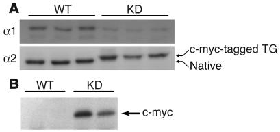
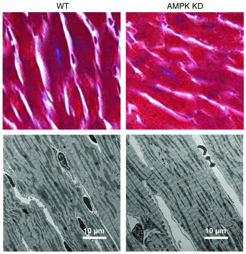
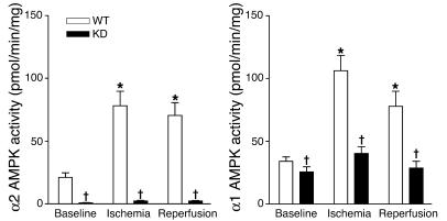
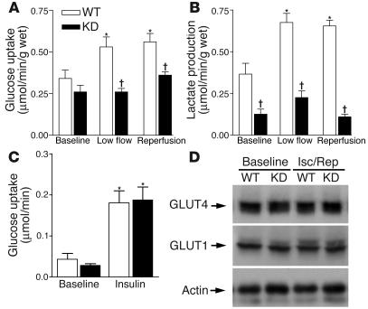
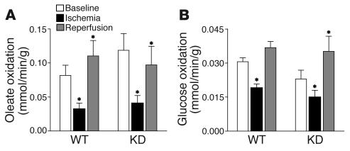
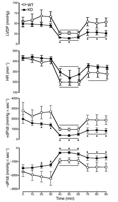
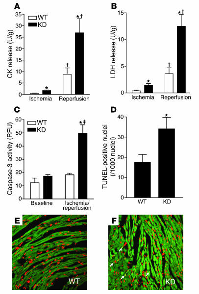
AMP-activated protein kinase (AMPK) is an important regulator of diverse cellular pathways in the setting of energetic stress. Whether AMPK plays a critical role in the metabolic and functional responses to myocardial ischemia and reperfusion remains uncertain. We examined the cardiac consequences of long-term inhibition of AMPK activity in transgenic mice expressing a kinase dead (KD) form of the enzyme. The KD mice had normal fractional shortening and no heart failure, cardiac hypertrophy, or fibrosis, although the in vivo left ventricular (LV) dP/dt was lower than that in WT hearts. During low-flow ischemia and postischemic reperfusion in vitro, KD hearts failed to augment glucose uptake and glycolysis, although glucose transporter content and insulin-stimulated glucose uptake were normal. KD hearts also failed to increase fatty acid oxidation during reperfusion. Furthermore, KD hearts demonstrated significantly impaired recovery of LV contractile function during postischemic reperfusion that was associated with a lower ATP content and increased injury compared with WT hearts. Caspase-3 activity and TUNEL-staining were increased in KD hearts after ischemia and reperfusion. Thus, AMPK is responsible for activation of glucose uptake and glycolysis during low-flow ischemia and plays an important protective role in limiting damage and apoptotic activity associated with ischemia and reperfusion in the heart.
Figures
Comment in
-
AMP-activated protein kinase: the guardian of cardiac energy status.J Clin Invest. 2004 Aug;114(4):465-8. doi: 10.1172/JCI22683. J Clin Invest. 2004. PMID: 15314681 Free PMC article. Review.
References
-
- Hardie D, Scott J, Pan D, Hudson E. Management of cellular energy by the AMP-activated protein kinase system. FEBS Lett. 2003;546:113–120. - PubMed
-
- Kemp BE, et al. AMP-activated protein kinase, super metabolic regulator. Biochem Soc. Trans. 2003;31:162–168. - PubMed
-
- Kudo N, et al. Characterization of 5’AMP-activated protein kinase activity in the heart and its role in inhibiting acetyl-CoA carboxylase during reperfusion following ischemia. . Biochim. Biophys. Acta. 1996; 1301:67–75. - PubMed
-
- Winder W, Hardie D. Inactivation of acetyl-CoA carboxylase and activation of AMP-activated protein kinase in muscle during exercise. Am. J. Physiol. 1996;270:E299–E304. - PubMed
-
- Russell R, Bergeron R, Shulman G, Young L. Translocation of myocardial GLUT4 and increased glucose uptake through activation of AMP-activated protein kinase by AICAR. Am. J. Physiol. 1999;277:H643–H649. - PubMed
Publication types
MeSH terms
Substances
Grants and funding
LinkOut - more resources
Full Text Sources
Other Literature Sources
Molecular Biology Databases
Research Materials
