. 2004 Aug;114(4):551-9.
doi: 10.1172/JCI21695.

Poor immunogenicity of a self/tumor antigen derives from peptide-MHC-I instability and is independent of tolerance

Affiliations

Poor immunogenicity of a self/tumor antigen derives from peptide-MHC-I instability and is independent of tolerance

Zhiya Yu et al. J Clin Invest. 2004 Aug.

Abstract

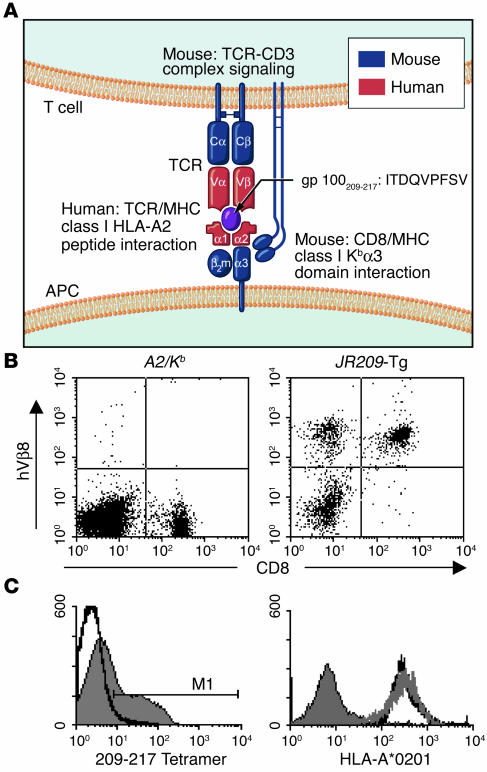
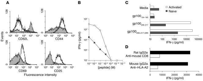
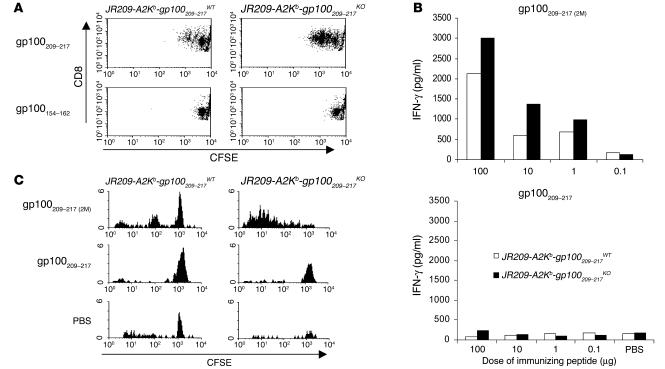
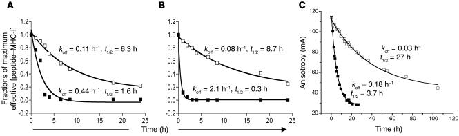
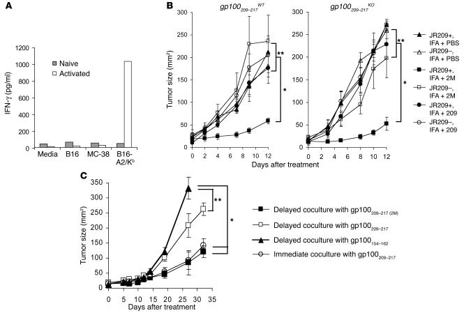
Understanding the mechanisms underlying the poor immunogenicity of human self/tumor antigens is challenging because of experimental limitations in humans. Here, we developed a human-mouse chimeric model that allows us to investigate the roles of the frequency and self-reactivity of antigen-specific T cells in determination of the immunogenicity of an epitope (amino acids 209-217) derived from a human melanoma antigen, gp100. In these transgenic mice, CD8+ T cells express the variable regions of a human T cell receptor (hTCR) specific for an HLA-A*0201-restricted gp100(209-217). Immunization of hTCR-transgenic mice with gp100(209-217) peptide elicited minimal T cell responses, even in mice in which the epitope was knocked out. Conversely, a modified epitope, gp100(209-217(2M)), was significantly more immunogenic. Both biological and physical assays revealed a fast rate of dissociation of the native peptide from the HLA-A*0201 molecule and a considerably slower rate of dissociation of the modified peptide. In vivo, the time allowed for dissociation of peptide-MHC complexes on APCs prior to their exposure to T cells significantly affected the induction of immune responses. These findings indicate that the poor immunogenicity of some self/tumor antigens is due to the instability of the peptide-MHC complex rather than to the continual deletion or tolerization of self-reactive T cells.


Figures

Figure 1
Figure 1
Characterization of the human-mouse TCR–HLA-A2/Kb transgenic mouse model. (A) Illustration of a hTCR–HLA-A*0201–gp100209–217 complex in the JR209-Tg mouse model. (B) hTCR Vβ8 and CD8 expression on lymphocyte-gated population from spleens of non–TCR-transgenic A2/Kb (A2/Kb) and JR209-Tg littermate mice. The percentage of hVβ8+CD8+ lymphocytes was approximately 0 in A2/Kb mice and 41 in JR209-Tg littermates. (C) Binding of HLA-A*0201–gp100209–217 tetramer to the lymphocyte-gated population from spleens of A2/Kb (area under thick black line) and JR209-Tg (gray area) mice. M1 (tetramer positive gate) represented 42% of the gated population. (D) HLA-A*0201 expression on lymphocyte-gated population from splenocytes of C57BL/6 (gray area), A2/Kb (area under gray line), and JR209-Tg (area under thick black line) mice.
Figure 2
Figure 2
T cells from JR209-Tg mice are functional. (A) Expression of CD62L, CD44, CD69, and CD25 on freshly isolated (gray area) and ex vivo peptide-stimulated JR209-Tg (area under thick black line) CD8+ T cells. The florescence intensities of cells labeled with isotype Ab’s were less than 102 (not shown). (B) IFN-γ release in 24-hour coculture of 1 × 10⁵ naive (filled squares) or gp100209–217 peptide–activated (open squares) CD8+ JR209-Tg T cells and 1 × 10⁵ A2/Kb splenocytes pulsed with titrated gp100209–217 peptides. Data represented the mean of duplicate testing samples. (C) IFN-γ release in 24-hour coculture of 1 × 10⁵ freshly isolated (gray bars) or ex vivo peptide-stimulated (white bars) CD8+ JR209-Tg T cells and 1 × 10⁵ A2/Kb splenocytes pulsed with 1 μM of gp100209–217(2M), gp100209–217, and gp100154–162 (irrelevant control) peptide. (D) IFN-γ release in 24-hour coculture of 1 μM gp100209–217 peptide–pulsed A2/Kb splenocytes and activated JR209-Tg T cells and their blockade by anti–HLA-A2 and anti-mCD8 mAb’s. Data represent the mean of duplicate testing samples.
Figure 3
Figure 3
Native gp100209–217 peptide fails to activate JR209-Tg T cells in both gp100209–217WT and gp100209–217KO mice. (A) Ex vivo antigen-specific proliferative responses of freshly isolated splenocytes from JR209-Tg mice with and without gp100209–217 epitope expression. CFSE-labeled splenocytes were cultured in media containing 1 μM of gp100209–217 or gp100154–162 peptide for 48 hours before FACS. The dot plots represent 10,000 total events in each sample. (B) gp100209–217 peptide–specific IFN-γ release in cells from draining lymph nodes (pooled from two mice in each group) after various doses of gp100209–217(2M) and gp100209–217 peptide immunization in JR209-Tg mice with (white bars) and without (black bars) the epitope expression. Draining lymph nodes were collected 7 days after immunization. One micromole of gp100209–217 peptide was added to 1 × 10⁵ cells in 200 μl of culture media and incubated for 24 hours. IFN-γ concentrations in the supernatant were determined by ELISA. (C) In vivo antigen-specific proliferative responses of adoptively transferred freshly isolated splenocytes from JR209-Tg mice with and without gp100209–217 epitope expression. CFSE-labeled splenocytes (1 × 10⁷) from JR209-Tg–gp100209–217WT or JR209-Tg–gp100209–217KO mice were intravenously injected into A2/Kb recipient mice, followed by immunization (into the footpad) with 100 μg of gp100209–217(2M), gp100209–217 peptide, or PBS (in IFA). Four days after immunization, the cells of draining lymph nodes were pooled from two mice in each group and gated on hVβ8+CD8+ T cells for FACS analysis.
Figure 4
Figure 4
Native gp100209–217–MHC-I complex is metastable. (A and B) Determination of peptide dissociation rates from target cell surface. Calculated amounts of gp100209–217 (filled squares) and gp100209–217(2M) (open squares) peptide bound to HLA-A2 molecules were plotted over time. Apparent koff values were determined by fitting to a single exponential decay of all points above an undetectable concentration of peptide (1 × 10⁻¹⁰ M using human T cell clone for A and 1 × 10⁻⁹ M using JR209-Tg T cells for B). t1/2 was determined from the relationship t1/2 = 0.693/koff. (C) Direct assay of peptide dissociation from purified peptide-MHC complexes using fluorescence anisotropy. Dissociation rates and t1/2of gp100209–217and gp100209–217(2M) peptides are indicated. Experiments were repeated in triplicate; experimental errors for the reported parameters were 3% for gp100209–217 and 7% for gp100209–217(2M). mA, millianisotropy.
Figure 5
Figure 5
Stable peptide–MHC-I complex is required for in vivo induction of antitumor responses in JR209-Tg mice. (A) Tumor-specific IFN-γ production by freshly isolated (gray bars) or ex vivo peptide-stimulated (white bars) CD8+ JR209-Tg T cells in 24-hour coculture with target cells from B16-A2/Kb, its parental B16 melanoma, and MC-38 murine adenocarcinoma. Data represent the mean of duplicate testing samples. (B) Treatment of B16-A2/Kb tumor in A2/Kb (open symbols) and JR209-Tg (filled symbols) mice by peptide immunization. One hundred micrograms of gp100209–217 (circles), gp100209–217(2M) (squares) peptides or PBS (triangles) in IFA was subcutaneously injected into mice 13 days after tumor inoculation. Five mice were in each group. *Significantly different (P < 0.05). **Not significantly different (P > 0.05). (C) Treatment of B16-A2/Kb tumor in A2/Kb transgenic mice receiving adoptive transfer of ex vivo immunized JR209-Tg splenocytes. Peptide-pulsed splenocytes were separated from JR209-Tg T cells for 24 hours before being cocultured overnight and then injected into tumor-bearing mice. In a control group, gp100209–217 peptide–pulsed splenocytes were directly cocultured with JR209-Tg T cells overnight and injected into tumor-bearing mice. Seven mice were in each group.

Comment in

References

    1. Van den Eynde, B., and van der Bruggen, P. 2003. Peptide database of T-cell defined tumor antigens. Cancer Immunity. [serial online]. http://www.cancerimmunity.org/peptidedatabase/Tcellepitopes.htm#text.
    1. Renkvist N, Castelli C, Robbins PF, Parmiani G. A listing of human tumor antigens recognized by T cells. Cancer Immunol. Immunother. 2001;50:3–15. - PMC - PubMed
    1. Kawakami Y, et al. Recognition of multiple epitopes in the human melanoma antigen gp100 by tumor-infiltrating T lymphocytes associated with in vivo tumor regression. J. Immunol. 1995;154:3961–3968. - PubMed
    1. Kawakami Y, et al. Identification of a human melanoma antigen recognized by tumor-infiltrating lymphocytes associated with in vivo tumor rejection. Proc. Natl. Acad. Sci. U. S. A. 1994;91:6458–6462. - PMC - PubMed
    1. Parkhurst MR, et al. Improved induction of melanoma-reactive CTL with peptides from the melanoma antigen gp100 modified at HLA-A*0201-binding residues. J. Immunol. 1996;157:2539–2548. - PubMed

Publication types

MeSH terms