. 2004 Sep;16(9):2380-93.
doi: 10.1105/tpc.104.022400. Epub 2004 Aug 17.

DNA replication licensing affects cell proliferation or endoreplication in a cell type-specific manner

Affiliations

DNA replication licensing affects cell proliferation or endoreplication in a cell type-specific manner

María del Mar Castellano et al. Plant Cell. 2004 Sep.

Abstract

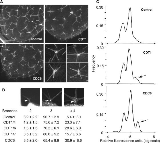
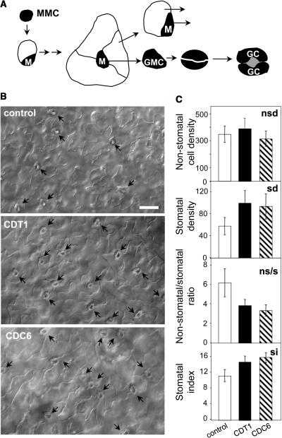
In eukaryotic cells, the function of DNA replication licensing components (Cdc6 and Cdt1, among others) is crucial for cell proliferation and genome stability. However, little is known about their role in whole organisms and whether licensing control interfaces with differentiation and developmental programs. Here, we study Arabidopsis thaliana CDT1, its regulation, and the consequences of overriding licensing control. The availability of AtCDT1 is strictly regulated at two levels: (1) at the transcription level, by E2F and growth-arresting signals, and (2) posttranscriptionally, by CDK phosphorylation, a step that is required for its proteasome-mediated degradation. We also show that CDC6 and CDT1 are key targets for the coordination of cell proliferation, differentiation, and development. Indeed, altered CDT1 or CDC6 levels have cell type-specific effects in developing Arabidopsis plants: in leaf cells competent to divide, cell proliferation is stimulated, whereas in cells programmed to undergo differentiation-associated endoreplication rounds, extra endocycles are triggered. Thus, we propose that DNA replication licensing control is critical for the proper maintenance of proliferative potential, developmental programs, and morphogenetic patterns.


Figures

Figure 1.
Figure 1.
Arabidopsis CDT1a and CDT1b Proteins. (A) Phylogenetic tree and domain organization of yeast, animal, and Arabidopsis CDT1 proteins. Two high homology regions define the central (hatched) and the C-terminal (gray) domains. Putative coiled-coil domains (lines and dots), PEST sequences (black), KEN sequences (open circles), and putative CDK phosphorylation sites (closed circles) are shown. (B) Alignment of the highly conserved central and C-terminal domains of CDT1 proteins of various sources. Identical and conserved residues appear in black and gray boxes, respectively. The asterisk indicates the R/K residue conserved in CDT1 proteins. (C) Interaction of in vitro transcribed and translated AtCDC6a protein (arrow) with purified protein GST-AtCDT1a. The bands below the full-length AtCDT1a protein correspond to partial translation products present in the bacterial lysate.
Figure 2.
Figure 2.
Expression Analysis of AtCDT1 Genes. (A) to (C) Arabidopsis pAtCDT1a:GUS seedlings at 20 (A), 40 (B), and 60 (C) h after transferring to light and 22°C. (D) Four-day-old seedlings grown in the light. (E) Detail of the cotyledon shown in (D). (F) Ten-day-old light-grown seedlings with developing lateral roots. (G) to (I) Cells of the stomatal lineage in 5- to 10-d-old cotyledons. Fully developed stomata in 5- (G) and 10-d-old (I) cotyledons. Primary (left) and secondary (right) meristemoids in 5-d-old cotyledons (H). (J) Leaves at different stages of development (15-d-old seedlings). (K) Detail of trichomes in leaf primordia. (L) Young flowers. (M) Mature flowers. (N) Siliques.
Figure 3.
Figure 3.
Regulation of AtCDT1a Gene Expression. (A) Real-time RT-PCR of Arabidopsis transgenic seedlings expressing a dominant negative version of wheat (Triticum aestivum) DP (DPΔBD). (B) and (C) Same as in (A) but with wild-type seedlings dehydrated for 6 h (B) or treated with 100 μM ABA (C). In (A), (B), and (C), values are referred to the fold change compared with untreated controls. (D) Detection of GUS activity in ABA-treated pAtCDT1a:GUS plants. Note that promoter activity in leaf primordia (top panels), but not in the root meristems (bottom panels), is inhibited.
Figure 4.
Figure 4.
AtCDT1a Is Subjected to Proteasome-Mediated Degradation. (A) Scheme of the construct used to generate transgenic plants ectopically expressing AtCDT1a-Myc-His (black) protein under the constitutive 35S promoter (hatched) of Cauliflower mosaic virus (top panel). Detection of AtCDT1a-Myc-His transgene expression by RT-PCR (middle panel) and protein gel blot (WB) with anti-Myc antibody (bottom panel) in whole trichloroacetic acid extracts of control (lane 1) and several transgenic lines (CDT1/4, 6, and 7; lanes 2 to 4) treated with the proteasome inhibitor MG132 (6-d-old seedlings treated with inhibitor for 8 h). The expression of DHR1 was used as loading control for RT-PCR. The asterisk indicates an unspecific protein used as loading control in protein gel blot analysis. (B) Levels of AtCDT1a-Myc-His detected by protein gel blot analysis with anti-Myc antibody in whole TCA extracts of 6-d-old Arabidopsis seedlings of control (lanes 1 and 2) or transgenic plants (CDT1; lanes 3 to 6) treated with MG132 or aphidicolin as indicated. (C) Levels of AtCDT1a-Myc-His in whole TCA extracts of 6-d-old Arabidopsis seedlings of transgenic plants (CDT1) treated with the CDK inhibitor roscovitine.
Figure 5.
Figure 5.
AtCDT1a Interacts with Arabidopsis CDKA, Cyclin D, and Cyclin A. (A) Detection of AtCDT1a-Myc-His in immunoprecipitates of AtCDKA with anti-PSTAIRE antibody of extracts of control and transgenic plants (CDT1) expressing AtCDT1a. (B) Detection of AtCDKA with anti-PSTAIRE antibodies in pull-down assays (top panel) of Arabidopsis cultured cell extracts incubated with GST-AtCDT1a or GST, as indicated, and kinase activity on GST-AtCDT1a of the bound material (bottom panel). GST-AtCDT1a-p indicates the phosphorylated protein. (C) Interaction of GST and GST-AtCDT1a with AtCDKA (top panel), His-AtCycD2;1 (middle panel), and His-AtCycA2;2 (bottom panel) by pull-down assays of baculovirus-infected insect cell extracts expressing the indicated Arabidopsis proteins and subsequent detection by protein gel blot analysis with anti-PSTAIRE or anti-His antibodies. (D) Kinase assays of baculovirus-infected insect cell extracts expressing the AtCDKA, AtCycD2;1, AtCycA2;2, or CDK/cyclin combinations, as indicated, using purified GST-AtCDT1a as substrate.
Figure 6.
Figure 6.
Ectopic Expression of AtCDT1a Increases Endoreplication Level. (A) Distribution of leaf nuclei with different DNA content in control, three independent CDT1 transgenic lines (CDT1/4, CDT1/6, and CDT1/7), and a CDC6 transgenic line (87-5; Castellano et al., 2001) at different times during leaf development. Flow cytometry profiles of leaf nuclei of 9-, 15-, and 28-d-old 1/2 leaves are shown. Note the increase in the 8C peak by 15 d and in the 16C peak by 28 d in CDT1 and CDC6 transgenic leaves. (B) Summary of each ploidy peak distribution at different leaf developmental stages. Note that CDT1 and CDC6 decrease the amount of 2C nuclei concomitantly with an increase in 8C and 16C nuclei from day 15 onwards.
Figure 7.
Figure 7.
Ectopic Expression of AtCDT1a Increases the Trichome Nuclear Ploidy and Branching Number. (A) Scanning electron micrographs of the adaxial surface of the 3rd rosette leaf (14-d-old plants) in control, AtCDC6a (line 87-5; Castellano et al., 2001), and AtCDT1a (line CDT1/4) transgenic plants. Detail of abnormal trichomes with more than four branches and emerging from neighbor cells found in the AtCDT1a and AtCDC6a transgenic plants is shown in the smaller panels. Bar = 100 μm. (B) Percentage of trichomes with two, three, and more than four branches of control and AtCDT1a (lines CDT1/4, CDT1/6, and CDT1/7) and AtCDC6a (line 87-5; Castellano et al., 2001) transgenic plants calculated on samples of 600 to 1000 trichomes in each case. DAPI-stained nuclei (arrows) of two-, three-, and four-branched trichomes are shown. (C) DNA content distribution of individual trichome nuclei in the 3rd to 4th leaves of control and transgenic 14-d-old plants ectopically expressing AtCDT1a or AtCDC6a. Arrows point to trichome nuclei having undergone an extra endocycle.
Figure 8.
Figure 8.
Ectopic Expression of AtCDT1a Increases the Amount of Stomata but Not the Nonstomatal Cell Number. (A) Overview of cell proliferation and differentiation of different cell types during stomata development. GC, guard cell; GMC, guard mother cell. (B) Adaxial epidermis (1st rosette leaf, 20-d-old plants) of control, AtCDT1a (line CDT1/4), and AtCDC6a (line 87-5; Castellano et al., 2001) ectopically expressing plants. Arrows point to stomata. Bar = 50 μm. (C) Nonstomatal cell density (nsd; number of nonstomatal cells per mm2), stomatal density (sd; number of stomata per mm2), nonstomatal epidermal cells/stomata ratio (ns/s), and stomatal index [si; (sd/sd + epidermal cell density)*100] for control (white), AtCDT1a (black), and AtCDC6a (hatched) ectopically expressing plants. In each case, three different first leaves were analyzed and at least 1600 cells were scored. In all cases, the differences between control and transgenic plants were statistically significant (P < 0.001), except for the nonstomatal density.
Figure 9.
Figure 9.
Model of DNA Replication Licensing Control Operating in Different Cell Types Based on Our Results (in Bold) in Leaves of Plants Ectopically Expressing AtCDT1 and AtCDC6. Cells whose differentiation program involves the occurrence of endoreplication cycles (e.g., trichomes) are induced to undergo extra DNA replication rounds, and as a consequence, their morphogenesis is altered (overbranching) in cells competent to divide (e.g., secondary meristemoids). The sites where some genes relevant for this discussion can be putatively located are also indicated.

Similar articles

Cited by

References

    1. Arentson, E., Faloon, P., Seo, J., Moon, E., Studts, J.M., Fremont, D.H., and Choi, K. (2002). Oncogenic potential of the DNA replication licensing protein CDT1. Oncogene 21, 1150–1158. - PubMed
    1. Baulcombe, D.C., Saunders, G.R., Bevan, M.W., Mayo, M.A., and Harrison, B.D. (1986). Expression of biologically active viral satellite RNA from the nuclear genome of transformed plants. Nature 321, 446–449.
    1. Bell, S.P., and Dutta, A. (2002). DNA replication in eukaryotic cells. Annu. Rev. Biochem. 71, 333–374. - PubMed
    1. Berger, D., and Altmann, T. (2000). A subtilisin-like serine protease involved in the regulation of stomatal density and distribution in Arabidopsis thaliana. Genes Dev. 14, 1119–1131. - PMC - PubMed
    1. Bermejo, R., Vilaboa, N., and Cales, C. (2002). Regulation of CDC6, geminin, and CDT1 in human cells that undergo polyploidization. Mol. Biol. Cell 13, 3989–4000. - PMC - PubMed

Publication types

MeSH terms

Associated data