. 2004 Sep 13;91(6):1166-73.
doi: 10.1038/sj.bjc.6602080.

Effects of gemcitabine on APE/ref-1 endonuclease activity in pancreatic cancer cells, and the therapeutic potential of antisense oligonucleotides

Affiliations

Effects of gemcitabine on APE/ref-1 endonuclease activity in pancreatic cancer cells, and the therapeutic potential of antisense oligonucleotides

J P Lau et al. Br J Cancer. .

Abstract

Apurinic/apyrimidinic endonuclease (APE) is a key enzyme involved in DNA base excision repair (BER) that is often expressed at elevated levels in human cancers. Pancreatic cancer cells treated with the nucleoside analogue gemcitabine (2', 2'-difluoro-2'deoxycytidine) showed increases in APE/redox effector factor (ref-1) protein levels (approximately two-fold for Panc-1 and six-fold for MiaPaCa-2), with corresponding increases in endonuclease activity. These results suggested that the activation of APE/ref-1 might be an adaptive response that contributes to gemcitabine resistance by facilitating BER. To test this hypothesis, we examined the effects of disrupting APE/ref-1 using antisense on gemcitabine toxicity. Antisense oligonucleotides decreased protein levels three-fold in MiaPaCa-2 and five-fold in Panc-1 in comparison to controls, associated with reduced endonuclease activity. Combination treatments with antisense oligonucleotides and gemcitabine partially suppressed the increase in APE/ref-1 activity seen in cells exposed to gemcitabine alone. While clonogenic assays showed only slight decreases in colony formation in cells treated with either antisense oligonucleotides or gemcitabine alone, the combination with APE/ref-1 antisense resulted in a 2-log enhancement of gemcitabine toxicity in Panc-1 cells. Overall these findings suggest that APE/ref-1 plays a significant role in gemcitabine resistance in some pancreatic cancer cells, and support the further investigation of novel treatments that target this protein.


Figures
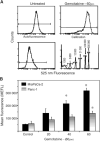
Figure 1
Figure 1
APE/ref-1 protein levels in control and gemcitabine-treated MiaPaCa and Panc-1 monolayers. (A) Representative flow cytometry data for MiaPaCa cells treated with 60 μM gemcitabine for 48 h compared to untreated control, autofluorescence background and calibration beads. Gated on light scatter to exclude dead cells. (B) APE/ref-1 protein levels in MiaPaCa and Panc-1 controls and cells treated with 20, 40 and 60  μm gemcitabine for 48 h, expressed as mean equivalent fluorescein (MEFL) values obtained from the calibration beads. Bars represent means from three separate experiments±s.e. Stars indicate results that are statistically significant (P<0.05) with respect to the nontreated samples.
Figure 2
Figure 2
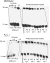
Endonuclease activity present in gemcitabine-treated cells. (A) Film images of 26-mer substrate vs 14-mer product in MiaPaCa and Panc-1 cells after various doses of gemcitabine for 48 h. Positive controls consisted of purified recombinant APE/ref-1 protein and negative controls included the BER inhibitor methoxyamine. (B) Range of gemcitabine concentrations vs the fraction of 14-mer product formed. Stars indicate results that are statistically significant (P<0.05) with respect to the nontreated samples. The concentration of gemcitabine is expressed as μM. All values are means of three independent experiments±s.e.
Figure 3
Figure 3
Relationship between APE/ref-1 protein level vs endonuclease activity postgemcitabine exposure. MiaPaCa (diamonds) and Panc-1 (squares) cells were treated with various doses of gemcitabine for 48 h. Samples from the same flask were collected and assessed using both flow cytometry and endonuclease assay. Points represent mean values from three separate experiments and error bars are standard error of the mean; r=0.951 (MiaPaCa) and 0.999 (Panc-1).
Figure 4
Figure 4
Subcellular localisation of fluorescence-labelled antisense oligonucleotides. That targeting the exon–intron junction of APE/ref-1 pre-mRNA is localised in the nucleus, whereas the translational start is cytoplasmic.
Figure 5
Figure 5
Endonuclease activity of MiaPaCa and Panc-1 cells treated with antisense oligonucleotides. The MiaPaCa and Panc-1 cell lines were exposed to antisense or sense oligonucleotides for various time points. Cell lysates were collected and analysed using the endonuclease assay. C – control; AS – antisense; S – sense.
Figure 6
Figure 6
APE/ref-1 protein levels (A) and endonuclease activity (B) post-treatment with antisense/sense oligonucleotide±gemcitabine. (A) Protein levels. MiaPaCa (dark bars) and Panc-1 (light bars) cells were treated with antisense or sense oligonucleotides±20 μM gemcitabine. APE/ref-1 protein levels were accessed at 12 or 24 h post-transfection of oligonucleotides or 24 h post-treatment with gemcitabine. As indicated, some samples were treated with gemcitabine prior to transfection with antisense oligonucleotides to determine whether the sequence of treatment affected outcome. Bars represent three individual experiments, with error bars representing standard error of the mean. AS – antisense; S – sense; Gem – gemcitabine.
Figure 7
Figure 7
Clonogenic survival of (A) Panc-1 and (B) MiaPaCa cells treated with gemcitabine alone, with antisense (AS) or sense oligonucleotide for 12 or 24 h or the combination. Bars represent at least three individual experiments, with standard error of the mean indicated by the error bars. aStatistically significant (P<0.05) compared to control samples. bStatistically significant (P<0.05) with respect to the gemcitabine-only treated samples.

Similar articles

Cited by

References

    1. Abbruzzese JL (2002) Past and present treatment of pancreatic adenocarcinoma: chemotherapy as a standard treatment modality. Semin Oncol 29: 2–8 - PubMed
    1. Arlt A, Gehrz A, Muerkoster S, Vorndamm J, Kruse ML, Folsch UR, Schafer H (2003) Role of NF-kappaB and Akt/PI3K in the resistance of pancreatic carcinoma cell lines against gemcitabine-induced cell death. Oncogene 22: 3243–3251 - PubMed
    1. Arrigo AP (1999) Gene expression and the thiol redox state. Free Radic Biol Med 27: 936–944 - PubMed
    1. Baldwin AS (2001) Control of oncogenesis and cancer therapy resistance by the transcription factor NF-kappaB. J Clin Invest 107: 241–246 - PMC - PubMed
    1. Ema M, Hirota K, Mimura J, Abe H, Yodoi J, Sogawa K, Poellinger L, Fujii-Kuriyama Y (1999) Molecular mechanisms of transcription activation by HLF and HIF1alpha in response to hypoxia: their stabilization and redox signal-induced interaction with CBP/p300. EMBO J 18: 1905–1914 - PMC - PubMed

Publication types

MeSH terms