

Subthreshold micropulse diode laser treatment in diabetic macular oedema
- PMID: 15317711
- PMCID: PMC1772323
- DOI: 10.1136/bjo.2003.040949
Subthreshold micropulse diode laser treatment in diabetic macular oedema
Abstract
Background/aim: Enlargement of laser scars after retinal argon laser photocoagulation can give rise to deterioration in visual acuity. Subthreshold micropulse diode laser may decrease this risk. The aim of this study was to compare the effectiveness of subthreshold micropulse diode laser (810 nm) and conventional argon laser (514 nm) photocoagulation for the treatment of clinically significant macular oedema in diabetic patients.
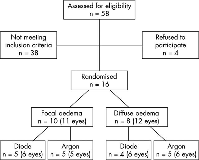
Methods: 23 eyes of 16 patients were randomised to either treatment. Follow up was conducted for a minimum of 5 months. Changes in visual acuity and macular oedema measured by optical coherence tomography were examined.
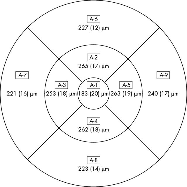
Results: Visual acuity remained stable in all treatment groups throughout the observation period. Changes in retinal thickness were small both foveally and perifoveally. In patients with focal macular oedema a significant reduction in retinal thickness (9% approximately -26 microm, p = 0.02) was seen foveally 3 months after diode laser photocoagulation.
Conclusion: Subthreshold micropulse diode laser and conventional argon laser treatment showed an equally good effect on visual acuity. Subthreshold micropulse diode laser showed a stabilising or even improving effect on macular oedema. The combination of primary diode laser and supplementary argon laser might be particularly favourable in reducing diabetic macular oedema.
Figures
Similar articles
-
Microperimetry and fundus autofluorescence in diabetic macular edema: subthreshold micropulse diode laser versus modified early treatment diabetic retinopathy study laser photocoagulation.Retina. 2010 Jun;30(6):908-16. doi: 10.1097/IAE.0b013e3181c96986. Retina. 2010. PMID: 20168272 Clinical Trial.
-
Subthreshold diode micropulse photocoagulation for the treatment of clinically significant diabetic macular oedema.Br J Ophthalmol. 2005 Jan;89(1):74-80. doi: 10.1136/bjo.2004.051540. Br J Ophthalmol. 2005. PMID: 15615751 Free PMC article.
-
Prospective randomised controlled trial comparing sub-threshold micropulse diode laser photocoagulation and conventional green laser for clinically significant diabetic macular oedema.Br J Ophthalmol. 2009 Oct;93(10):1341-4. doi: 10.1136/bjo.2008.146712. Epub 2008 Dec 3. Br J Ophthalmol. 2009. PMID: 19054831 Clinical Trial.
-
Monotherapy laser photocoagulation for diabetic macular oedema.Cochrane Database Syst Rev. 2018 Oct 15;10(10):CD010859. doi: 10.1002/14651858.CD010859.pub2. Cochrane Database Syst Rev. 2018. PMID: 30320466 Free PMC article.
-
[Angiopathy and the eye].Vnitr Lek. 2010 Apr;56(4):333-9. Vnitr Lek. 2010. PMID: 20465107 Review. Czech.
Cited by
-
Subthreshold Micropulse Laser in Diabetic Macular Edema: 1-Year Improvement in OCT/OCT-Angiography Biomarkers.Transl Vis Sci Technol. 2020 Sep 30;9(10):31. doi: 10.1167/tvst.9.10.31. eCollection 2020 Sep. Transl Vis Sci Technol. 2020. PMID: 33062394 Free PMC article.
-
Efficacy and Safety of a Dexamethasone Implant in Patients with Diabetic Macular Edema at Tertiary Centers in Korea.J Ophthalmol. 2016;2016:9810270. doi: 10.1155/2016/9810270. Epub 2016 May 17. J Ophthalmol. 2016. PMID: 27293879 Free PMC article.
-
Micropulse and continuous wave diode retinal photocoagulation: visible and subvisible lesion parameters.Br J Ophthalmol. 2006 Jun;90(6):709-12. doi: 10.1136/bjo.2005.086942. Epub 2006 Mar 10. Br J Ophthalmol. 2006. PMID: 16531424 Free PMC article.
-
Treatment of Diabetic Macular Edema with Aflibercept and Micropulse Laser (DAM Study).Clin Ophthalmol. 2022 Apr 8;16:1109-1115. doi: 10.2147/OPTH.S360869. eCollection 2022. Clin Ophthalmol. 2022. PMID: 35422607 Free PMC article. Clinical Trial.
-
The Evolving Treatment of Diabetic Retinopathy.Clin Ophthalmol. 2020 Mar 4;14:653-678. doi: 10.2147/OPTH.S236637. eCollection 2020. Clin Ophthalmol. 2020. PMID: 32184554 Free PMC article. Review.
References
-
- Patz A, Schatz H, Berkow JW, et al. Macular edema—an overlooked complication of diabetic retinopathy. Trans Am Acad Ophthalmol Otolaryngol 1973;77:OP34–42. - PubMed
-
- Blankenship GW. Diabetic macular edema and argon laser photocoagulation: a prospective randomized study. Ophthalmology 1979;86:69–78. - PubMed
-
- Olk RJ. Argon green (514 nm) versus krypton red (647 nm) modified grid laser photocoagulation for diffuse diabetic macular edema. Ophthalmology 1990;97:1101–12. - PubMed
-
- Early Treatment Diabetic Retinopathy Study Research Group. Photocoagulation for diabetic macular edema. Early Treatment Diabetic Retinopathy Study Report Number 1. Arch Ophthalmol 1985;103:1796–806. - PubMed
-
- Olk RJ. Modified grid argon (blue-green) laser photocoagulation for diffuse diabetic macular edema. Ophthalmology 1986;93:938–50. - PubMed
Publication types
MeSH terms
LinkOut - more resources
Full Text Sources
Other Literature Sources
Medical