

Colour vision testing as an aid to diagnosis and management of age related maculopathy
- PMID: 15317712
- PMCID: PMC1772298
- DOI: 10.1136/bjo.2003.033480
Colour vision testing as an aid to diagnosis and management of age related maculopathy
Abstract
Aim: To provide a simple test that detects the onset of age related maculopathy (ARM), and can be used to monitor its severity.
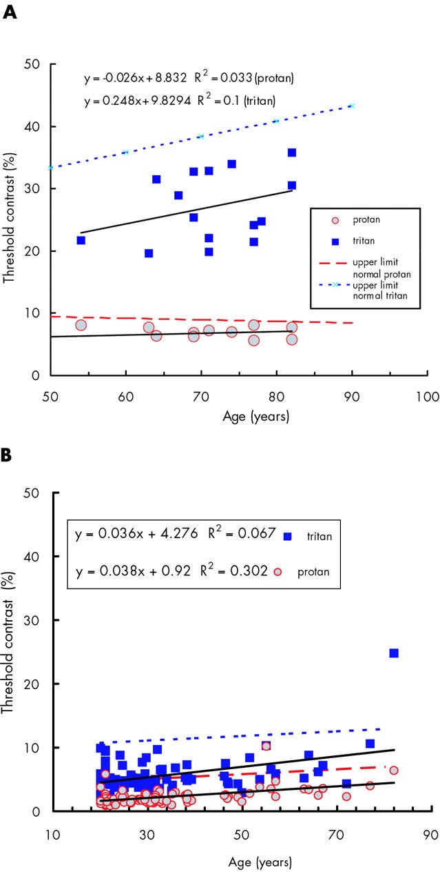
Methods: Colour contrast sensitivity was measured using computer graphics techniques. Colour thresholds were measured along tritan and protan colour confusion axes in the presence of dynamic luminance noise. Thresholds were determined separately for two sizes of optotypes (6.5 degrees and 1.5 degrees). Natural pupils were used. Normal values for the test have been established.
Results: In all patients with unilateral age related macular degeneration, the smaller optotype was invisible in that eye and in almost all, the larger optotype could not be seen. In the symptomless fellow eyes (with ARM) the larger optotype thresholds were raised. The degree of loss was larger for tritan. For the smaller optotype, protan thresholds were elevated in the majority of patients. Tritan losses were greater and disproportionate to the loss seen with the larger optotype. Every person including those with minimal fundal changes had tritan test results for 1.5 degree optotypes >2 SD above the normal mean. Tritan thresholds varied with the severity of the ARM.
Conclusions: The test is sensitive, simple and quick to administer, and easy for patients. Therefore, it should be useful in detecting and monitoring elderly people with age related changes in their fundi before irreversible loss of vision has occurred.
Figures


Similar articles
-
Colour contrast sensitivity in patients with age-related Bruch's membrane changes.Ger J Ophthalmol. 1995 Nov;4(6):336-41. Ger J Ophthalmol. 1995. PMID: 8751098
-
Colour contrast sensitivity in patients with soft drusen, an early stage of ARM.Doc Ophthalmol. 1995;90(4):377-86. doi: 10.1007/BF01268123. Doc Ophthalmol. 1995. PMID: 8620820
-
Colour contrast thresholds are normal in functional amblyopia.Eye (Lond). 1996;10 ( Pt 4):479-84. doi: 10.1038/eye.1996.106. Eye (Lond). 1996. PMID: 8944103
-
Luminance noise and the rapid determination of discrimination ellipses in colour deficiency.Vision Res. 1994 May;34(10):1279-99. doi: 10.1016/0042-6989(94)90203-8. Vision Res. 1994. PMID: 8023437
-
Assessment of age-related maculopathy using subjective vision tests.Clin Exp Optom. 2005 Sep;88(5):292-303. doi: 10.1111/j.1444-0938.2005.tb06713.x. Clin Exp Optom. 2005. PMID: 16255688 Review.
Cited by
-
Safety monitoring in bevacizumab (Avastin) treatment: retinal function assessed by psychophysical (visual fields, colour vision) and electrophysiological (ERG/EOG) tests in two subgroups of patients.Int Ophthalmol. 2008 Apr;28(2):101-9. doi: 10.1007/s10792-007-9122-1. Epub 2007 Jul 20. Int Ophthalmol. 2008. PMID: 17634860
-
New iPAD-based test for the detection of color vision deficiencies.Graefes Arch Clin Exp Ophthalmol. 2018 Dec;256(12):2349-2360. doi: 10.1007/s00417-018-4154-y. Epub 2018 Oct 6. Graefes Arch Clin Exp Ophthalmol. 2018. PMID: 30291435
-
The burden of neovascular age-related macular degeneration: a patient's perspective.Clin Ophthalmol. 2018 Dec 4;12:2483-2491. doi: 10.2147/OPTH.S185052. eCollection 2018. Clin Ophthalmol. 2018. PMID: 30584267 Free PMC article.
-
Color vision and neuroretinal function in diabetes.Doc Ophthalmol. 2015 Apr;130(2):131-9. doi: 10.1007/s10633-014-9476-4. Epub 2014 Dec 17. Doc Ophthalmol. 2015. PMID: 25516428 Free PMC article.
-
Enhanced S-Cone Syndrome: Spectrum of Clinical, Imaging, Electrophysiologic, and Genetic Findings in a Retrospective Case Series of 56 Patients.Ophthalmol Retina. 2021 Feb;5(2):195-214. doi: 10.1016/j.oret.2020.07.008. Epub 2020 Jul 15. Ophthalmol Retina. 2021. PMID: 32679203 Free PMC article.
References
-
- Lim JI, Enger C, Fine SL. Foveomacular dystrophy. Am J Ophthalmol 1994;117:1–6. - PubMed
-
- Treatment of Age Related Macular Degeneration with Photodynamic Therapy (TAP) Study Group. Photodynamic therapy of subfoveal choroidal neovascularization in age related macular degeneration with verteporfin: one year results of randomized clinical trials—TAP report. Arch Ophthalmol 1999;117:1329–45. - PubMed
-
- Bressler NM. Photodynamic therapy of subfoveal choroidal neovascularization in age-related macular degeneration with verteporfin: two-year results of two randomized clinical trials—TAP report 2. Arch Ophthalmol 2001;119:198–207. - PubMed
-
- Verteporfin in Photodynamic Therapy Study Group. Verteporfin therapy of subfoveal choroidal neovascularization in age-related macular degeneration: two-year results of a randomized clinical trial including lesions with occult with no classic choroidal neovascularization—Verteporfin in Photodynamic Therapy Report 2. Am J Ophthalmol 2001;131:541–60. - PubMed
-
- Blumenkranz MS, Bressler NM, Bressler SB, et al. Verteporfin therapy for subfoveal choroidal neovascularization in age-related macular degeneration: three-year results of an open-label extension of 2 randomized clinical trials—TAP report no. 5. Arch Ophthalmol 2002;120:1307–14. - PubMed
MeSH terms
LinkOut - more resources
Full Text Sources
Medical