. 2004 Sep;186(17):5692-8.
doi: 10.1128/JB.186.17.5692-5698.2004.

The Bvg virulence control system regulates biofilm formation in Bordetella bronchiseptica

Affiliations

The Bvg virulence control system regulates biofilm formation in Bordetella bronchiseptica

Yasuhiko Irie et al. J Bacteriol. 2004 Sep.

Abstract

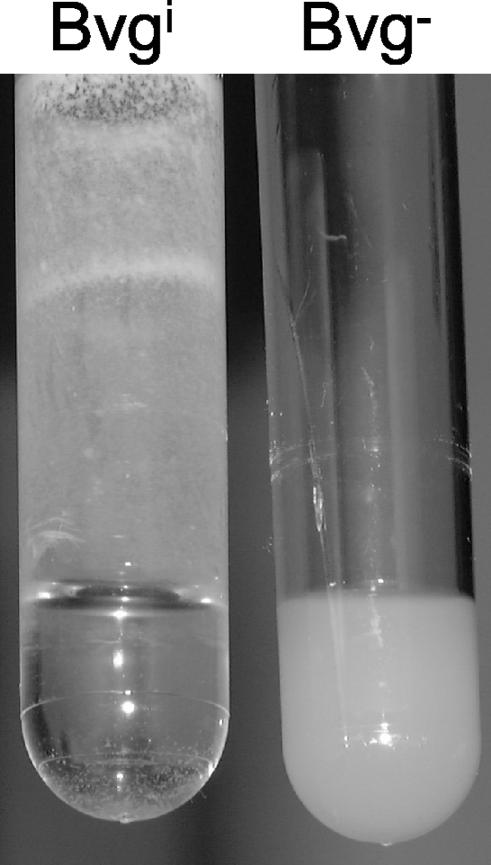
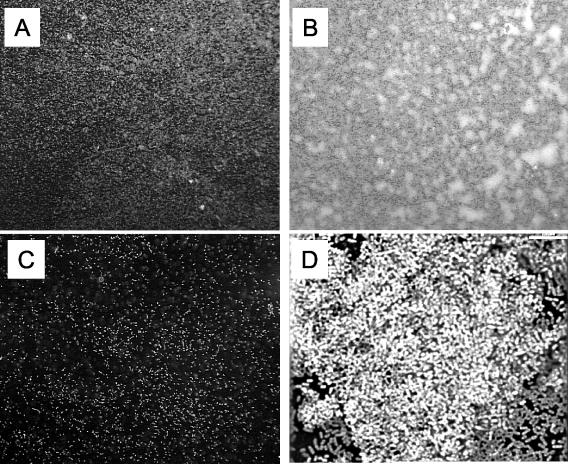
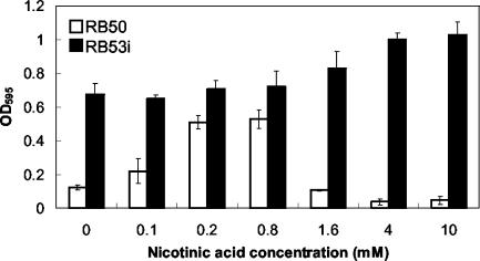
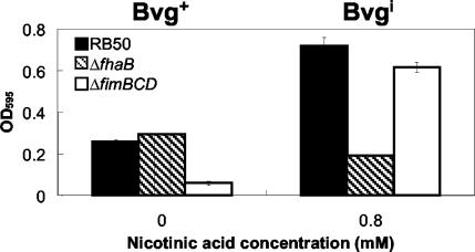
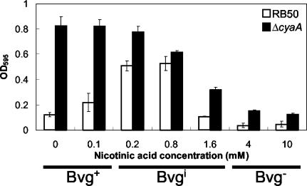
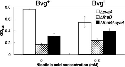
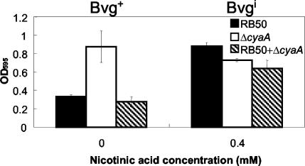
Bordetella species utilize the BvgAS (Bordetella virulence gene) two-component signal transduction system to sense the environment and regulate gene expression among at least three phases: a virulent Bvg+ phase, a nonvirulent Bvg- phase, and an intermediate Bvgi phase. Genes expressed in the Bvg+ phase encode known virulence factors, including adhesins such as filamentous hemagglutinin (FHA) and fimbriae, as well as toxins such as the bifunctional adenylate cyclase/hemolysin (ACY). Previous studies showed that in the Bvgi phase, FHA and fimbriae continue to be expressed, but ACY expression is significantly downregulated. In this report, we determine that Bordetella bronchiseptica can form biofilms in vitro and that the generation of biofilm is maximal in the Bvgi phase. We show that FHA is required for maximal biofilm formation and that fimbriae may also contribute to this phenotype. However, expression of ACY inhibits biofilm formation, most likely via interactions with FHA. Therefore, the coordinated regulation of adhesins and ACY expression leads to maximal biofilm formation in the Bvgi phase in B. bronchiseptica.


Figures

FIG. 1.
FIG. 1.
Biofilm formation by B. bronchiseptica grown in the Bvgi phase. Overnight liquid cultures of B. bronchiseptica were grown in the Bvgi phase (0.8 mM nicotinic acid, left) or Bvg phase (4 mM nicotinic acid, right) in polystyrene culture tubes in continuous rotation on roller drums. In the Bvgi phase, a majority of the bacteria were adherent to the test tube wall, in contrast to bacteria that was grown in Bvg phase (or Bvg+ phase; data not shown) in which most bacterial cells remained in the liquid media.
FIG. 2.
FIG. 2.
Formation of microcolonies by B. bronchiseptica on glass coverslips. Wild-type B. bronchiseptica organisms were grown on glass coverslips and then were stained with Syto Red 17 and observed under a fluorescent microscope (20× objective and 10× eyepiece). (A) Culture medium with no nicotinic acid (Bvg+ phase). (B) Culture medium supplemented with 0.8 mM nicotinic acid (Bvgi phase). (C) Culture medium supplemented with 4 mM nicotinic acid (Bvg phase). Bacteria grown in Bvg+ phase (A) appear to form small aggregates, whereas microcolonies formed by bacteria grown in Bvgi phase are large and distinct (B). Bacteria in Bvg phase (C) displayed little adherence to the coverslip with no aggregative properties. (D) Deconvolution micrograph of a microcolony depicted in panel B displaying the cellular architecture of the microcolony. Bar, 7 μm.
FIG. 3.
FIG. 3.
Quantitative assay of biofilm formation by wild-type (RB50) and Bvgi-phased-locked (RB53i) B. bronchiseptica at different nicotinic acid concentrations. Bacteria were grown in 96-well polystyrene plates, and biofilm formation was quantified by absorbance of solubilized crystal violet stains, as described in Materials and Methods. Biofilm formation in the wild-type bacteria peaked in the Bvgi phase (0.2 to 0.8 mM nicotinic acid). The Bvgi-phase-locked mutant formed high levels of biofilm at all nicotinic acid concentrations. Bvg+-phase growth condition is 0 to 0.1 mM nicotinic acid, 0.2 to 1.6 mM is Bvgi phase, and 4.0 mM (and above) is Bvg phase. OD595, optical density at 595 nm.
FIG. 4.
FIG. 4.
Quantitative assay of biofilm formation in wild-type B. bronchiseptica (RB50), ΔfhaB mutant, and ΔfimBCD mutant in the Bvg+ phase (0 mM nicotinic acid) and Bvgi phase (0.8 mM nicotinic acid). Bacteria were grown in 96-well polystyrene plates, and biofilm formation was quantified by absorbance of solubilized crystal violet stains, as described in Materials and Methods. The amount of biofilm formed by the ΔfhaB mutant in the Bvg+ phase was similar to that of the wild-type but was significantly decreased in the Bvgi phase. The ΔfimBCD mutant appears to form almost no biofilm in the Bvg+ phase, but the amount of biofilm formed by this mutant in the Bvgi phase was comparable to that of the wild-type bacteria. OD595, optical density at 595 nm.
FIG. 5.
FIG. 5.
Quantitative assay of biofilm formation by wild-type B. bronchiseptica (RB50) and ΔcyaA mutant at different nicotinic acid concentrations. Bacteria were grown in 96-well polystyrene plates, and biofilm formation was quantified by absorbance of solubilized crystal violet stains, as described in Materials and Methods. The ΔcyaA mutant formed high levels of biofilm in both Bvg+ and Bvgi phases compared to that of wild-type bacteria, which formed high levels of biofilm only in the Bvgi phase. OD595, optical density at 595 nm.
FIG. 6.
FIG. 6.
Comparative quantitative assay of biofilm formation by the ΔcyaA mutant, ΔfhaB mutant, and ΔfhaBΔcyaA double mutant in the Bvg+ phase (0 mM nicotinic acid) and Bvgi phase (0.8 mM nicotinic acid). Bacteria were grown in 96-well polystyrene plates, and biofilm formation was quantified by absorbance of solubilized crystal violet stains, as described in Materials and Methods. The double mutant formed higher levels of biofilm than the ΔfhaB mutant but formed lower levels than the ΔcyaA mutant. OD595, optical density at 595 nm.
FIG. 7.
FIG. 7.
Quantitative assay of biofilm formation in cocultures containing both wild-type B. bronchiseptica (RB50) and the ΔcyaA mutant in the Bvg+ phase (0 mM nicotinic acid) and Bvgi phase (0.4 mM nicotinic acid). Bacteria were grown in 96-well polystyrene plates, and biofilm formation was quantified by absorbance of solubilized crystal violet stains, as described in Materials and Methods. Coculture of RB50 with the ΔcyaA mutant results in a low level of biofilm (comparable to that of RB50 alone) in the Bvg+ phase, but no significant reduction of biofilm formation in the coculture was observed in the Bvgi phase. OD595, optical density at 595 nm.

References

    1. Akerley, B. J., P. A. Cotter, and J. F. Miller. 1995. Ectopic expression of the flagellar regulon alters development of the Bordetella-host interaction. Cell 80:611-620. - PubMed
    1. Akerley, B. J., D. M. Monack, S. Falkow, and J. F. Miller. 1992. The bvgAS locus negatively controls motility and synthesis of flagella in Bordetella bronchiseptica. J. Bacteriol. 174:980-990. - PMC - PubMed
    1. Allison, D. G., B. Ruiz, C. SanJose, A. Jaspe, and P. Gilbert. 1998. Extracellular products as mediators of the formation and detachment of Pseudomonas fluorescens biofilms. FEMS Microbiol. Lett. 167:179-184. - PubMed
    1. Austin, J. W., G. Sanders, W. W. Kay, and S. K. Collinson. 1998. Thin aggregative fimbriae enhance Salmonella enteritidis biofilm formation. FEMS Microbiol. Lett. 162:295-301. - PubMed
    1. Costerton, J. W., P. S. Stewart, and E. P. Greenberg. 1999. Bacterial biofilms: a common cause of persistent infections. Science 284:1318-1322. - PubMed

Publication types

MeSH terms

LinkOut - more resources