. 2004 Oct;15(10):4682-94.
doi: 10.1091/mbc.e04-04-0353. Epub 2004 Aug 18.

A critical role of tropomyosins in TGF-beta regulation of the actin cytoskeleton and cell motility in epithelial cells

Affiliations

A critical role of tropomyosins in TGF-beta regulation of the actin cytoskeleton and cell motility in epithelial cells

Andrei V Bakin et al. Mol Biol Cell. 2004 Oct.

Abstract

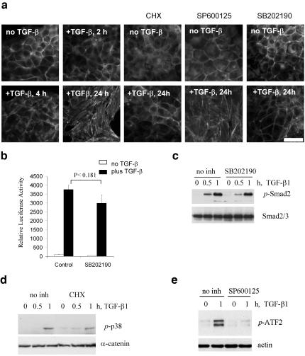
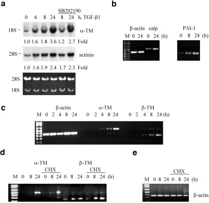
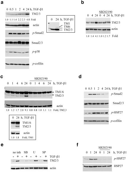
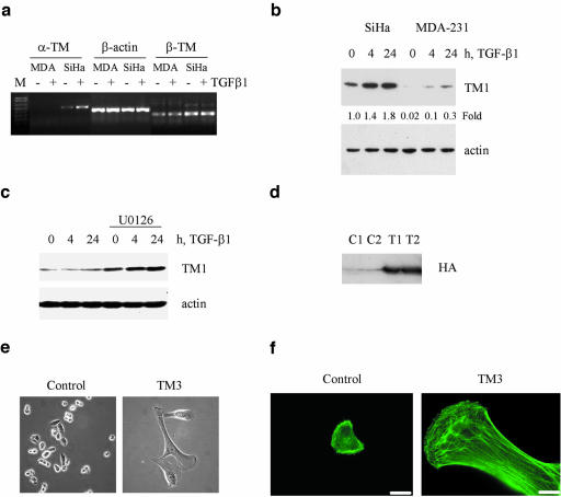
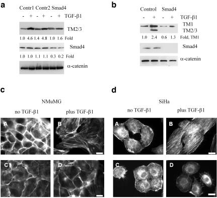
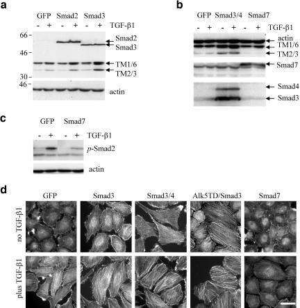
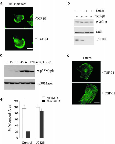
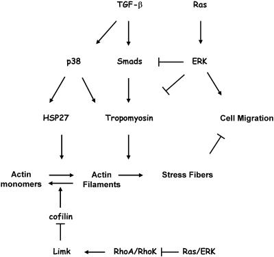
We have investigated transforming growth factor beta (TGF-beta)-mediated induction of actin stress fibers in normal and metastatic epithelial cells. We found that stress fiber formation requires de novo protein synthesis, p38Mapk and Smad signaling. We show that TGF-beta via Smad and p38Mapk up-regulates expression of actin-binding proteins including high-molecular-weight tropomyosins, alpha-actinin and calponin h2. We demonstrate that, among these proteins, tropomyosins are both necessary and sufficient for TGF-beta induction of stress fibers. Silencing of tropomyosins with short interfering RNAs (siRNAs) blocks stress fiber assembly, whereas ectopic expression of tropomyosins results in stress fibers. Ectopic-expression and siRNA experiments show that Smads mediate induction of tropomyosins and stress fibers. Interestingly, TGF-beta induction of stress fibers was not accompanied by changes in the levels of cofilin phosphorylation. TGF-beta induction of tropomyosins and stress fibers are significantly inhibited by Ras-ERK signaling in metastatic breast cancer cells. Inhibition of the Ras-ERK pathway restores TGF-beta induction of tropomyosins and stress fibers and thereby reduces cell motility. These results suggest that induction of tropomyosins and stress fibers play an essential role in TGF-beta control of cell motility, and the loss of this TGF-beta response is a critical step in the acquisition of metastatic phenotype by tumor cells.


Figures

Figure 1.
Figure 1.
TGF-β1–induced actin stress fibers require p38Mapk and a novel protein synthesis. (a) Actin filaments staining with phalloidin-Alexa Green in cells treated with 2 ng/ml TGF-β1 in the absence or presence of inhibitors. Where it is indicated, cells were treated with 10 μg/ml cycloheximide (CHX) starting at 6 h after addition of TGF-β1. Kinase inhibitors (15 μM SP600125, a JNK inhibitor, and 10 μM SB202190, a p38Mapk inhibitor) were added 1 h before addition of TGFβ1. Scale bar, 15 μm. (b) Luciferase activity in NMuMG transfected with Smad-dependent reporter pSBE-Lux and pCMV-Rl vectors and treated with 1 ng/ml TGF-β1 for 16 h in the absence or presence of 15 μM SB202190. Each bar represents the mean ± SD of three wells. P value was determined by t test. The difference in luciferase activity in control and SB202190-treated cells is not statistically significant. (c) Immunoblot analysis of Smad2 phosphorylation in protein extracts (35 μg/well) from NMuMG cells treated with 2 ng/ml TGF-β1 in the absence or presence of 10 μM SB202190. (d) p38Mapk phosphorylation in response to TGF-β1 in protein extracts (35 μg/well) from SiHa cells cotreated with 10 μg/ml cycloheximide (CHX). (e) Inhibition of ATF2 phosphorylation by JNK inhibitors in protein extracts (35 μg/well) from NMuMG cells treated with 2 ng/ml TGF-β1.
Figure 2.
Figure 2.
TGF-β regulates expression of genes encoding actin-binding proteins in NMuMG cells. (a) Northern blot analysis of TM2/3 and α-actinin1 mRNA levels in total RNA samples (15 μg/lane) from NMuMG cells treated with 2 ng/ml TGF-β1 in the absence or presence of 10 μM SB202190. Blots were quantified using PhosphorImager. Bottom panel shows ethidium bromide staining of total RNA. (b–e) Analysis of β-actin, PAI-1, calponin2, and α- and β-tropomyosin transcripts by PCR with reverse transcription in total RNA samples from NMuMG cells treated with 2 ng/ml TGF-β1. Where it is indicated, cells were treated with 10 μg/ml cycloheximide (CHX) 1 h before addition of TGF-β1.
Figure 3.
Figure 3.
TGF-β regulates tropomyosins and HSP27 phosphorylation in epithelial cells. (a and b) Immunoblot analysis of actin and tropomyosins as well as phosphorylation of Smad2, HSP27, and cofilin using phosphospecific antibodies in NMuMG cells treated with 2 ng/ml TGF-β1. Where it is indicated 10 μM SB202190 was added 1 h before TGF-β treatment. Inset shows induction of TM1 and TM6 on a longer exposed film of the same immunoblot. (c–f) Immunoblot analysis of tropomyosin expression and phosphorylation of Smad2, p38Mapk, and cofilin in SiHa cells treated with 2 ng/ml TGF-β1. Where it is indicated, the cells were cotreated 1 h before addition of TGF-β with 10 μM SB202190 (SB), 5 μM U0126 (U), or 15 μM SP600125 (SP). Insert shows immunoblot with TM311 antibody using the same protein extracts as in c. Fold difference of tropomyosin levels relative to actin was estimated using NIH ImageJ software (http://rsb.info.nih.gov/nih-image/).
Figure 8.
Figure 8.
TGF-β and ERK signaling differentially regulates expression of tropomyosins. (a) Analysis of α-TM and β-TM transcripts by RT-PCR in total RNA samples from MDA-MB-231 (MDA) and SiHa cells treated with 2 ng/ml TGF-β1 for 24h. (b) Immunoblot analysis of tropomyosin expression in protein extracts (35 μg/lane) from SiHa and MDA-MB-231 cells treated with 2 ng/ml TGF-β1. (c) Tropomyosin protein expression in MDA-MB-231 cells cotreated with TGF-β1 and 5 μM U0126 for 24h. (d) Detection of HA-tagged rat TM3 with anti-HA antiserum in protein extracts from two independent transfections of MDA-MB-231 cells with expression vector encoding HA-tagged rat TM3 (T1 and T2) or a control empty vector (C1 and C2). (e) Phase-contrast images show flattening and size increase in TM3-transfected MDA-MB-231 cells compared with control cells. (f) Immunofluorescence images show a marked increase in actin stress fibers in TM3-transfected MDA-MB-231 cells. Scale bar, 20 μM. Fold differences in tropomyosin levels relative to actin were estimated using NIH ImageJ software.
Figure 4.
Figure 4.
Smad signaling is required for TGF-β–induced expression of tropomyosins and stress fiber formation in epithelial cells. (a and b) Immunoblot analysis of tropomyosins and Smad4 in NMuMG cells (a) and SiHa cells (b) transfected with siRNAs against Smad4. (c and d) Actin filament staining with phalloidin-Alexa Green in NMuMG and SiHa cells transfected with control scramble siRNA (A and B) or siR-NAs to Smad4 (C and D). The cells were treated with 2 ng/ml TGF-β1 for 24 h. Scale bar, 10 μm. Fold differences in tropomyosin and Smad4 levels relative to α-catenin were estimated using NIH ImageJ software.
Figure 5.
Figure 5.
Smads mediate TGF-β–induced tropomyosin expression and stress fiber formation. (a and b) Tropomyosin expression in cells infected with adenoviruses encoding EGFP (GFP) and Flag-tagged Smad2, Smad3, Smad4, and Smad7. Cells were treated with 2 ng/ml TGF-β1 for 24 h. (c) Inhibition of TGF-β1–induced phosphorylation of Smad2 by adenoviral expression of Smad7 in SiHa cells. (d) Actin filaments staining in SiHa cells infected with adenoviruses: EGFP, Smad3, Smad4, Smad7, constitutively active Alk5T204D, or their combinations. The cells were treated with 2 ng/ml TGF-β1 for 24 h. Scale bars, 15 μm.
Figure 6.
Figure 6.
Tropomyosins are required for TGF-β–induced stress fiber formation. (a) Localization of tropomyosins (TM) to stable actin filaments (actin) resistant to 0.05% Triton X-100 treatment in SiHa cells untreated or treated with TGF-β1 for 24 h. Scale bar, 10 μM. (b) Suppression of tropomyosin expression in SiHa cells transfected with siRNA against TMs (si-TM) compared with a scrambled control. (c) Actin filaments (A, B, E, and F) and tropomyosin (C, D, G, and H) in SiHa cells, transfected with siRNA against tropomyosins (E–H) and a scrambled control siRNA (A–D). The cells were treated with 2 ng/ml TGF-β1 for 24 h. Scale bar, 10 μM. (d) Actin filaments in NMuMG and SiHa cells expressing HA-tagged TM3. Cells were stained with phalloidin-Texas Red and fluorescein-labeled anti-HA antibody (A–F). Overlay images are shown in panels E and F. Panels G and H show actin filaments and tropomyosins (TM311 antibody) in cells transfected with empty vector control. Scale bar, 15 μm.
Figure 7.
Figure 7.
TGF-β and ERK signaling differentially regulate stress fiber formation. (a) Actin filaments staining in MDA-MB-231 cells treated with TGF-β1 for 24 h. (B) Phosphorylation of cofilin and ERK1/2 in MDA-MB-231 cells cotreated with TGF-β1 for 24 h and 5 μM U0126. (c) Immunoblot analysis of p38Mapk phosphorylation in MDA-MB-231 cells treated with 2 ng/ml TGF-β1. (d) Actin filaments staining in MDA-MB-231 cells cotreated with TGF-β1 and 5 μM U0126 for 24 h. (e) Wound closure in MDA-MB-231 cells treated with TGF-β1 in the absence or presence of 5 μM U0126. The experiment was done in triplicates and repeated at least two times. Scale bar, 20 μM.
Figure 9.
Figure 9.
Opposing roles of the TGF-β and Ras-ERK signaling pathways in the regulation of actin filament dynamics and cell motility.

Similar articles

Cited by

References

    1. Ayscough, K.R. (1998). In vivo functions of actin-binding proteins. Curr. Opin. Cell Biol. 10, 102-111. - PubMed
    1. Bakin, A.V., and Curran, T. (1999). Role of DNA 5-methylcytosine transferase in cell transformation by fos. Science 283, 387-390. - PubMed
    1. Bakin, A.V., Rinehart, C., Tomlinson, A.K., and Arteaga, C.L. (2002). p38 mitogen-activated protein kinase is required for TGF{beta}-mediated fibroblastic transdifferentiation and cell migration. J. Cell Sci. 115, 3193-3206. - PubMed
    1. Bakin, A.V., Tomlinson, A.K., Bhowmick, N.A., Moses, H.L., and Arteaga, C.L. (2000). Phosphatidylinositol 3-kinase function is required for TGFbeta-mediated epithelial to mesenchymal transition and cell migration. J. Biol. Chem. 275, 36803-36810. - PubMed
    1. Bamburg, J.R. (1999). Proteins of the ADF/cofilin family: essential regulators of actin dynamics. Annu. Rev. Cell Dev. Biol. 15, 185-230. - PubMed

Publication types

MeSH terms

LinkOut - more resources