. 2004 Nov;15(11):5130-44.
doi: 10.1091/mbc.e04-05-0405. Epub 2004 Aug 18.

Toc12, a novel subunit of the intermembrane space preprotein translocon of chloroplasts

Affiliations

Toc12, a novel subunit of the intermembrane space preprotein translocon of chloroplasts

Thomas Becker et al. Mol Biol Cell. 2004 Nov.

Abstract

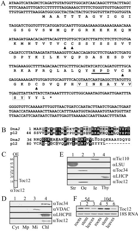
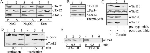
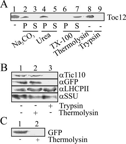
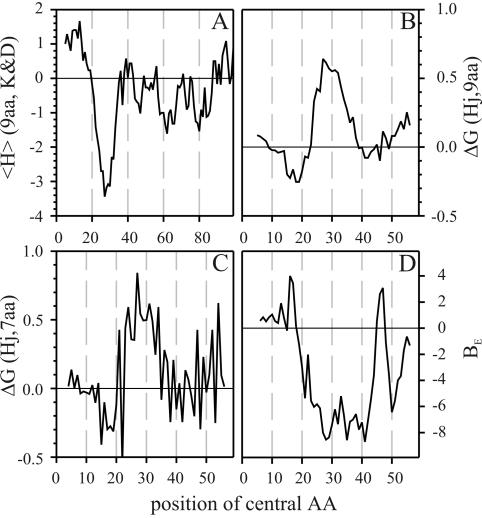
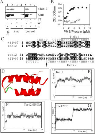
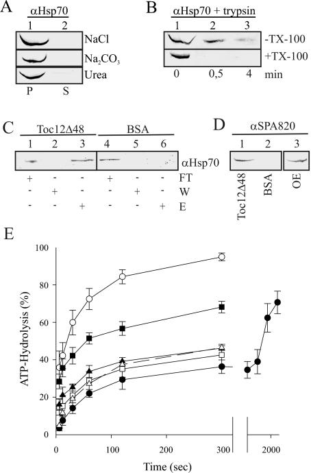
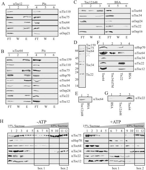
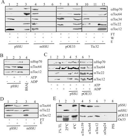
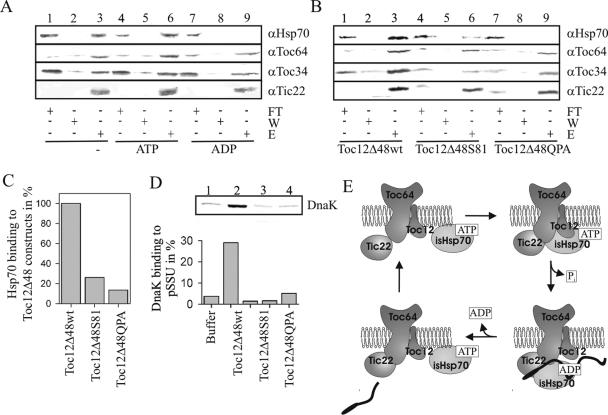
Translocation of proteins across membranes is essential for the biogenesis of each cell and is achieved by proteinaceous complexes. We analyzed the translocation complex of the intermembrane space from chloroplasts and identified a 12-kDa protein associated with the Toc machinery. Toc12 is an outer envelope protein exposing a soluble domain into the intermembrane space. Toc12 contains a J-domain and stimulates the ATPase activity of DnaK. The conformational stability and the ability to stimulate Hsp70 are dependent on a disulfide bridge within the loop region of the J-domain, suggesting a redox-regulated activation of the chaperone. Toc12 is associated with Toc64 and Tic22. Its J-domain recruits the Hsp70 of outer envelope membrane to the intermembrane space translocon and facilitates its interaction to the preprotein.


Figures

Figure 1.
Figure 1.
A novel DnaJ homologue in the outer envelope of chloroplasts of P. sativum. (A) The identified cDNA (acc. number AY357119) and amino acid sequence is shown. The peptide identified by amino acid sequencing is framed. Arrowheads point to the four cysteines. The HPD motive is underlined. (B) An alignment of the amino acid sequence of the C-terminal portion of the identified p12 to the J domain of the E. coli DnaJ (acc. number P08622) using clustal X is shown. Essential amino acids for the function of the J-domain are marked on top. (C) OEVs were separated by SDS-PAGE followed by immunodecoration using p12 (Toc12) antisera. (D) Leaves from 10-d-old plants (P. sativum) were harvested, lysed, and fractionated. Cytosol (lane 1), microsomal (lane 2), mitochondrial and chloroplast proteins (30 μg each) were separated by SDS-PAGE, transferred to nitrocellulose, and incubated with antiserum against Toc34, VDAC, LHCP, and p12 (Toc12). (E) Chloroplasts from P. sativum were fractionated into stroma (lane 1), outer envelope (lane 2), inner envelope (lane 3), and thylakoids (lane 4). Fractions were separated by SDS-PAGE, transferred to nitrocellulose, and incubated with indicated antisera. (F) The transcript level of p12 (Toc12, top panel) was tested in roots (lanes 1 and 4), stems (lanes 2 and 5), and leaves (lanes 3 and 6) of 5- (lanes 1-3) and 10-d-old (lanes 4-6) plants (P. sativum). For control, the amount of 18S RNA was analyzed (bottom panel).
Figure 2.
Figure 2.
p12 is an integral outer membrane protein of chloroplasts containing a soluble region facing the intermembrane space. (A) Isolated OEVs were incubated with 1 M NaCl (lanes 1 and 2), 100 mM Na2CO3, pH 11.4 (lanes 3 and 4) or 4 M urea (lanes 5 and 6). The pellet (lanes 1, 3, and 5) and supernatant (lanes 2, 4, and 6) were separated and subjected to SDS-PAGE followed by immunodecoration using antisera against Toc75 (top panel), Tic22 (middle panel), and p12 (Toc12, bottom panel). (B and C) Isolated chloroplasts (40 μg chlorophyll, lane 1) were incubated with thermolysin (250 μg, lane 2, B) or trypsin (25 μg, C) after (lane 2) or before addition of trypsin inhibitor (lane 2, C). After inhibition of the protease (lane 2, B; lane 3, C) chloroplasts were reisolated and proteins separated by SDS-PAGE followed by immunodecoration by indicated antisera. (D) Isolated OEVs, 30 μg (lanes 1 and 4), were incubated with 12.5 μg trypsin for 0.5 (lanes 2 and 5) or 4 min (lanes 3 and 6) without (lanes 1-3) or with sonication (lanes 4-6). After inhibition of the protease, the envelope was subjected to SDS-PAGE, transferred to nitrocellulose, and decorated with antibodies against Toc75 (top panel) or p12 (Toc12, bottom panel). (E) Outer envelope vesicles treated as described in B were subjected to SDS-PAGE analysis, transferred to nitrocellulose, and immunodecorated with antibodies against DnaJ.
Figure 3.
Figure 3.
p12 is targeted in vitro and in vivo to chloroplasts. (A) Isolated chloroplasts (40 μg) from P. sativum were incubated with in vitro-translated and 35S-labeled p12 for 15 min (lane 1) followed by reisolation of chloroplasts and subsequent incubation with 100 mM Na2CO3, pH 11.4 (lanes 2 and 3), 8 M urea (lanes 4 and 5), or 0.5% Triton X-100, followed by separation of the pellet (lanes 2, 4, and 6) and supernatant (lanes 3, 5, and 7). Further, chloroplasts were treated with thermolysin (250 μg, lane 8) or trypsin (25 μg, lane 9). Samples were separated and subjected to SDS-PAGE followed by visualization by autoradiography. (B) Chloroplasts isolated from tobacco protoplasts expressing the p12-GFP fusion (lane 1) were incubated with 100 μg thermolysin (lane 2) or 10 μg trypsin (lane 3) for 5 min at 25°C. After inhibition of the proteases chloroplasts were subjected to SDS-PAGE, transferred to nitrocellulose, and incubated with antibodies against the inner envelope protein Tic110 (top lane), GFP (panel 2), LHCP II (panel 3), or SSU (bottom panel). (C) In vitro-translated GFP (lane 1) was incubated with 2 μg thermolysin for 5 min at 25°C (lane 2), subjected to SDS-PAGE, and visualized by autoradiography.
Figure 4.
Figure 4.
Transmembrane prediction of Toc12. (A) The hydrophobicity distribution for the whole sequence using a window of 9 amino acids was calculated using the Kyte & Doolittle scale. (B) The average energy for the transfer of a 9-amino acid stretch into an octanol layer was calculated according to Wimley and White (2000). (C) The average alternating energy for the transfer of a β-barrel segment of 9 amino acids into the octanol layer was calculated. (D) The exact β-barrel score (Schleiff et al., 2003a) was calculated using a window of 10 amino acids.
Figure 5.
Figure 5.
The cysteines of p12 do not promote zinc binding but might stabilize the secondary structure. (A) A chelate column (lanes 5-7) was coated with zinc (lanes 2-4) and incubated with purified p12 (Toc12, top panel) or ALDH (bottom panel). Lane 1 shows the amount of protein loaded. The flow-through (lanes 2 and 5), wash (lanes 3 and 6), and elution (lanes 4 and 7) were subjected to SDS-PAGE followed by immunodecoration using p12 antibodies (top panel) or silver staining (bottom panel). (B) The release of zinc from p12 (○) or ALDH (•) after incubation with indicated amounts of PMB was determined. A representative result is shown. (C) The alignment of the C-terminus of the 12-kDa protein to the J-domain of Hsp40 is shown. The structural features of Hsp40 are indicated; H represents a helical structure and T a turn structure. The proline in Hsp40 is marked by an arrow. (D) The average structure of the last two nanoseconds of the molecular dynamic simulation of the p12 J-domain structure is shown. The disulfide bridge between cysteine 81 and cysteine 86 is shown in gray and the localization of the HPD motif in dark gray. (E) The RMSD during molecular dynamic simulation in comparison to the initial structure derived by amino acid replacement using the amino acids of Toc12 and the structural model of Hsp40 (hdj) is given. (F) As in E with the substitution of the his and asp amino acid in the HPD domain by gln and ala. (G) As in E with substitution of the both cys by ser.
Figure 6.
Figure 6.
p12 interacts with the membrane inserted intermembrane space facing Hsp70 and stimulates ATPase activity of DnaK. (A) OEVs were incubated with 1 M NaCl (top), 100 mM Na2CO3, pH 11.4 (middle), or 4 M urea (bottom). The pellet (lane 1) and the supernatant (lane 2) were separated and subjected to SDS-PAGE followed by immunodecoration using Hsp70-antiserum. (B) Isolated OEVs, 30 μg (lane 1), were incubated with 12.5 μg trypsin for 0.5 (lane 2) or 4 min (lane 3) in the absence (top panel) or presence (bottom panel) of 1% Triton X-100. After inhibition of the protease, the envelope was subjected to SDS-PAGE, transferred to nitrocellulose, and decorated with Hsp70-antibodies. (C) A Ni2+-NTA matrix (lanes 4-6) was coated with p12Δ48 fused to a C-terminal hexa-histidine extension (Toc12Δ48, lanes 1-3), and incubated with solubilized OEVs (75 μg). The flow-through (5%, lanes 1 and 4), wash (5%, lanes 2 and 5), and eluted fractions (100%, lanes 3 and 6) were subjected to SDS-PAGE followed by blotting and immunodecoration using Hsp70 antibodies. For controls see Figure 7C. (D) As in C, but elution fractions of the p12Δ48 (Toc12Δ48, lane 1) or BSA-coated column (lane 2) were immunodecorated by SPA820 antibodies. For comparison OEVs (7.5 μg) are shown in lane 3. (E) DnaK, 6.5 μM (•, solid line), was preloaded with ATP followed by incubation with 1 μM of DnaJ (○, solid line) or 5 μM of p12 (▪, solid lines), p12 HD/QA (□, solid lines), p12 C/S (▴, solid line), or denatured Oep16 (▵, dashed line). ATP hydrolysis was determined and quantified as described. Data reflect the average of at least three independent measurements.
Figure 7.
Figure 7.
p12 is a Toc component and involved in formation of the intermembrane space translocon. (A) Solubilized OEVs (400 μg) were incubated with Toyopearl material coated with p12 antibodies (Toc12, lanes 1-3) or preimmune serum (Pis, lanes 4-6). The flow-through (5%, lanes 1 and 4), wash (5%, lanes 2 and 5) and eluted fractions (100%, lanes 3 and 6) were subjected to SDS-PAGE followed by blotting and immunodecoration using indicated antibodies. (B) Solubilized OEVs (400 μg) were incubated with Toyopearl coated with Toc64-antibodies (αToc64, lanes 1-3) or Pis (lanes 4-6). The flow-through (5%, lanes 1 and 4), wash (5%, lanes 2 and 5) and eluate (100%, lanes 3 and 6) were subjected to SDS-PAGE followed by blotting and immunodecoration using indicated antibodies. (C) Ni2+-NTA coated with either Toc12Δ48 (lanes 1-3) or BSA (lanes 4-6) and incubated with solubilized OEVs (75 μg). The flow-through (5%, lanes 1 and 4), wash (5%, lanes 2 and 5), and eluted fractions (100%, lanes 3 and 6) were subjected to SDS-PAGE followed by blotting and immunodecoration using indicated antibodies. (D) Ni2+-NTA coated with Toc12Δ48 (lanes 1-3) was incubated with solubilized OEVs (75 μg). Fifty percent of the elution fraction was subjected to SDS-PAGE followed by silver-staining. The major protein band was marked according to immunodecoration results from C. Unspecifically bound large subunit of the RubisCo is marked by asterisk. (E) Expressed Toc64 was incubated with Toyopearl matrix (lane 2) coated with 50 μg Toc12Δ48 (lane 1). The bound protein was eluted and subjected to SDS-PAGE followed by immunodecoration using Toc64 antibodies. (F) Expressed and purified Toc64, 100 μg (lane 1), Toc34ΔTM (lane 2), or Tic22 (lane 3) were coupled to a Ni-NTA affinity matrix (lane 4) followed by incubation with solubilized OEVs. The bound proteins were eluted and subjected to SDS-PAGE followed by immunodecoration using indicated antibodies. (G) Tic22 was extracted from inner envelope membranes by treatment with 0.1 M sodium carbonate and dialyzed. Aggregated proteins were removed by centrifugation (2 h, 300,000 × g). Subsequently, purified Tic22 was incubated with an affinity matrix (lane 1) coated with 100 μg Toc12Δ48 (lane 2) or Toc64 (lane 3). The bound protein was eluted and subjected to SDS-PAGE followed by immunodecoration using Tic22 antibodies. (H) OEVs (150 μg protein) were solubilized with 1.5% decylmaltoside and subjected to sucrose density centrifugation in the absence (left panel) or in the presence (right panel) of 1 mM ATP. The outer membrane proteins were separated by SDS-PAGE, transferred to nitrocellulose-membrane, and immunodecorated with the indicated antisera. The intermembrane space complex containing fraction and the Toc core complex fraction are marked by box 1 or box 2, respectively.
Figure 8.
Figure 8.
The intermembrane space translocon interacts with pSSU. (A) OEVs (75 μg protein) were solubilized and incubated with a Ni-NTA coated with 100 μg pSSU (lanes 1-3), mSSU (lanes 4-6), pOE33 (lanes 7-9), or Tic32 (lanes 10-12). The flow-through (5%, lanes 1, 4, 7, and 10), wash (5%, lanes 2, 5, 8, and 11), and eluate (100%, lanes 3, 6, 9, and 12) was collected and subjected to SDS-PAGE followed by blotting and immunodecoration using indicated antibodies. (B) As in A, in the absence (lane 1) or presence of 1 mM ATP (lane 2) or ADP (lane 3). (C) OEVs (150 μg protein) were incubated with 0.5 mM MgCl2, (lanes 1-5) and 1 mM ATP (lanes 2, 4, and 6), 1 mM ADP (lanes 3, 5, 7), 10 μg pSSU (lanes 4 and 5), 10 μg pOE33 (lanes 6 and 7) solubilized and subjected on top of a sucrose gradient as in Figure 7G (25-70%). A fraction containing the intermembrane space complex (compare Figure 7G) were collected and subjected to SDS-PAGE analysis followed by blotting and incubation with indicated antibodies. (D) Expressed and purified Toc64, Tic22, or Toc12, 5 μg, was incubated with a Toyopearl matrix (lanes 3 and 4) coated with 6 μg pSSU (lanes 1 and 2) or mSSU (lanes 3 and 4). The flow-through (5%, lanes 1 and 3) and eluate (100%, lanes 2 and 4) were subjected to SDS-PAGE followed by blotting and immunodecoration by indicated antibodies. (E) Isolated chloroplasts were incubated with pSSU (top panel) or pOE33 (bottom panel) for 10 min at 4°C (lanes 2) in the presence of chemical cross-linker DSP (lanes 4-8) followed by lysis of the chloroplasts and incubation with indicated antisera. After immunoprecipitation, the cross-linker was cleaved by DTT, the proteins were separated on SDS-PAGE, and pSSU or mSSU was visualized by phosphorimaging. Lane 3 shows an import at 25°C. In lane 1, 1% of translation product (TP) is loaded.
Figure 9.
Figure 9.
Toc12 recruits Hsp70 to the intermembrane space translocon. (A) Ni2+-NTA coated with 200 μg Toc12Δ48 and incubated with solubilized OEVs (75 μg) in the absence (lanes 1-3) or presence of 0.5 mM ATP (lanes 4-6) or 0.5 mM ADP (lanes 7-9). The flow-through (5%, lanes 1, 4, and 7), wash (5%, lanes 2, 5, and 8) and eluted fractions (100%, lanes 3, 6, and 9) were subjected to SDS-PAGE followed by blotting and immunodecoration using indicated antibodies. (B) Ni2+-NTA coated with 200 μg of either Toc12Δ48 (lanes 1-3), Toc12Δ48S81 (lanes 4-6), or Toc12Δ48QPA (lanes 7-9) were incubated with solubilized OEVs (75 μg) in the presence of 0.5 mM ATP. The flow-through (5%, lanes 1, 4, and 7), wash (5%, lanes 2, 5, and 8), and eluted fractions (100%, lanes 3, 6, and 9) were subjected to SDS-PAGE followed by blotting and immunodecoration using indicated antibodies. One representative results of three experiments is shown. (C) Quantification of the results shown in B. The relative amount of bound Hsp70 to Toc12Δ48 constructs is depicted in percentage of the binding to Toc12Δ48 wild type. (D) DnaK, 1 μM, was incubated in the absence (lane 1) or presence of either 2 μM Toc12Δ48 (lane 2), Toc12Δ48S81 (lane 3), or Toc12Δ48QPA (lane 4) with on Toyopearl column material immobilized pSSU as described (Brychzy et al., 2003). After sufficient washing, bound proteins were eluted by 8 M urea, subjected to SDS-PAGE, and subsequently silver-stained. The amount of bound DnaK was quantified using AIDA software. (E) Dynamic model of the intermembrane space complex action. For detailed description see Discussion.

Similar articles

Cited by

References

    1. Akita, M., Nielsen, E., and Keegstra, K. (1997). Identification of protein transport complexes in the chloroplastic envelope membranes via chemical cross-linking. J. Cell Biol. 136, 983-994. - PMC - PubMed
    1. Alexandrov, N.N., Nussinov, R., and Zimmer, R.M. (1995). Fast protein fold recognition via sequence to structure alignment and contact capacity potentials, Singapore: World Scientific Publishing, 53-72. - PubMed
    1. Berendsen, H.J.C., Postma, J.P.M., van Gunsteren, W.F., and Hermans, J. (1981). Interaction models for water in relation to protein hydratation. In: Intermolecular Forces, ed. B. Pullman, Dordrecht: D. Reidel Publishing, 331-342.
    1. Berendsen, H.J.C., Postma, J.P.M., DiNola, A., and Haak, J.R. (1984). Molecular dynamics with coupling to an external bath. J. Chem. Phys. 81, 3684-3690.
    1. Berendsen, H.J.C., van der Spoel, D., and van Drunen, R. (1995). GROMACS: a message-passing parallel molecular dynamics implementation. Comput. Phys. Comm. 95, 43-56.

Publication types

MeSH terms

LinkOut - more resources