

BetaIV spectrins are essential for membrane stability and the molecular organization of nodes of Ranvier
- PMID: 15317849
- PMCID: PMC6729762
- DOI: 10.1523/JNEUROSCI.2125-04.2004
BetaIV spectrins are essential for membrane stability and the molecular organization of nodes of Ranvier
Abstract
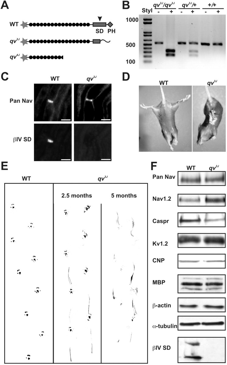
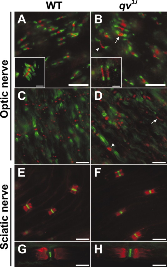
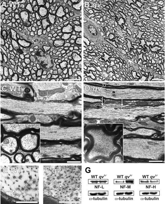
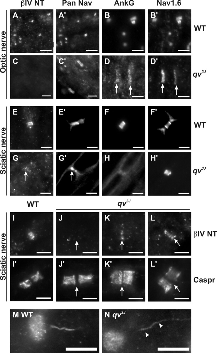
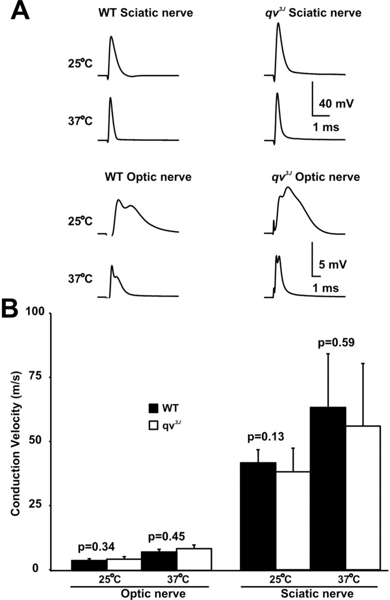
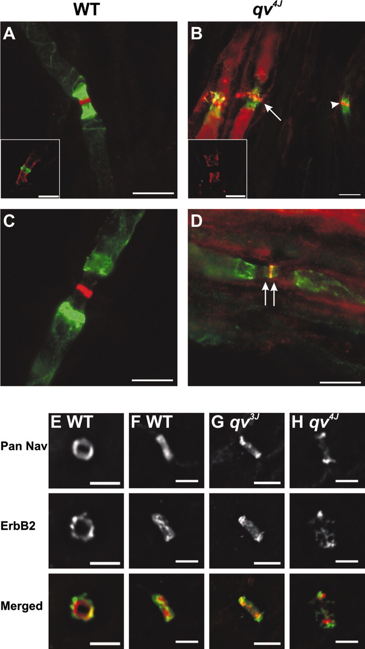
High densities of sodium channels at nodes of Ranvier permit action potential conduction and depend on betaIV spectrins, a family of scaffolding proteins linked to the cortical actin cytoskeleton. To investigate the molecular organization of nodes, we analyzed qv(3J)"quivering" mice, whose betaIV spectrins have a truncated proline-rich "specific" domain (SD) and lack the pleckstrin homology (PH) domain. Central nodes of qv(3J) mice, which lack betaIV spectrins, are significantly broader and have prominent vesicle-filled nodal membrane protrusions, whereas axon shape and neurofilament density are dramatically altered. PNS qv(3J) nodes, some with detectable betaIV spectrins, are less affected. In contrast, a larger truncation of betaIV spectrins in qv(4J) mice, deleting the SD, PH, and ankyrinG binding domains, causes betaIV spectrins to be undetectable and causes dramatic changes, even in peripheral nodes. These results show that quivering mutations disrupt betaIV spectrin retention and stability at nodes and that distinct protein domains regulate nodal structural integrity and molecular organization.
Figures

Similar articles
-
betaIV spectrin, a new spectrin localized at axon initial segments and nodes of ranvier in the central and peripheral nervous system.J Cell Biol. 2000 Nov 27;151(5):985-1002. doi: 10.1083/jcb.151.5.985. J Cell Biol. 2000. PMID: 11086001 Free PMC article.
-
betaIV spectrin is recruited to axon initial segments and nodes of Ranvier by ankyrinG.J Cell Biol. 2007 Feb 12;176(4):509-19. doi: 10.1083/jcb.200610128. Epub 2007 Feb 5. J Cell Biol. 2007. PMID: 17283186 Free PMC article.
-
The C-terminal domain of ßIV-spectrin is crucial for KCNQ2 aggregation and excitability at nodes of Ranvier.J Physiol. 2010 Dec 1;588(Pt 23):4719-30. doi: 10.1113/jphysiol.2010.196022. Epub 2010 Oct 20. J Physiol. 2010. PMID: 20962009 Free PMC article.
-
Spectrin and ankyrin-based cytoskeletons at polarized domains in myelinated axons.Exp Biol Med (Maywood). 2008 Apr;233(4):394-400. doi: 10.3181/0709-MR-243. Exp Biol Med (Maywood). 2008. PMID: 18367627 Review.
-
Accumulation of synaptic vesicle proteins and cytoskeletal specializations at the peripheral node of Ranvier.Microsc Res Tech. 1996 Aug 1;34(5):462-73. doi: 10.1002/(SICI)1097-0029(19960801)34:5<462::AID-JEMT6>3.0.CO;2-O. Microsc Res Tech. 1996. PMID: 8837022 Review.
Cited by
-
Axon initial segment-associated microglia.J Neurosci. 2015 Feb 4;35(5):2283-92. doi: 10.1523/JNEUROSCI.3751-14.2015. J Neurosci. 2015. PMID: 25653382 Free PMC article.
-
Reassembly of Excitable Domains after CNS Axon Regeneration.J Neurosci. 2016 Aug 31;36(35):9148-60. doi: 10.1523/JNEUROSCI.1747-16.2016. J Neurosci. 2016. PMID: 27581456 Free PMC article.
-
GABA neuron alterations, cortical circuit dysfunction and cognitive deficits in schizophrenia.Neural Plast. 2011;2011:723184. doi: 10.1155/2011/723184. Epub 2011 Sep 5. Neural Plast. 2011. PMID: 21904685 Free PMC article. Review.
-
Schwannomin-interacting Protein 1 Isoform IQCJ-SCHIP1 Is a Multipartner Ankyrin- and Spectrin-binding Protein Involved in the Organization of Nodes of Ranvier.J Biol Chem. 2017 Feb 10;292(6):2441-2456. doi: 10.1074/jbc.M116.758029. Epub 2016 Dec 15. J Biol Chem. 2017. PMID: 27979964 Free PMC article.
-
Disruption of the axon initial segment cytoskeleton is a new mechanism for neuronal injury.J Neurosci. 2009 Oct 21;29(42):13242-54. doi: 10.1523/JNEUROSCI.3376-09.2009. J Neurosci. 2009. PMID: 19846712 Free PMC article.
References
-
- Bekele-Arcuri Z, Matos MF, Manganas L, Strassle BW, Monaghan MM, Rhodes KJ, Trimmer JS (1996) Generation and characterization of subtype-specific monoclonal antibodies to K+ channel alpha- and beta-subunit polypeptides. Neuropharmacology 35: 851-865. - PubMed
-
- Bennett V, Baines AJ (2001) Spectrin and ankyrin-based pathways: metazoan inventions for integrating cells into tissues. Physiol Rev 81: 1353-1392. - PubMed
-
- Berghs S, Aggujaro D, Dirkx R, Maksimova E, Stabach P, Hermel JM, Zhang JP, Philbrick W, Slepnev V, Ort T, Solimena M (2000) BetaIV spectrin, a new spectrin localized at axon initial segments and nodes of Ranvier in the central and peripheral nervous system. J Cell Biol 151: 985-1002. - PMC - PubMed
-
- Berthold C-H (1978) Morphology of normal peripheral axons. In: Physiology and pathobiology of axons (Waxman SG, ed), pp 3-63. New York: Raven.
Publication types
MeSH terms
Substances
LinkOut - more resources
Full Text Sources
Molecular Biology Databases