. 2004 Aug 18;24(33):7324-34.
doi: 10.1523/JNEUROSCI.2111-04.2004.

Regulation of Cu/Zn-superoxide dismutase expression via the phosphatidylinositol 3 kinase/Akt pathway and nuclear factor-kappaB

Affiliations

Regulation of Cu/Zn-superoxide dismutase expression via the phosphatidylinositol 3 kinase/Akt pathway and nuclear factor-kappaB

Ana I Rojo et al. J Neurosci. .

Abstract

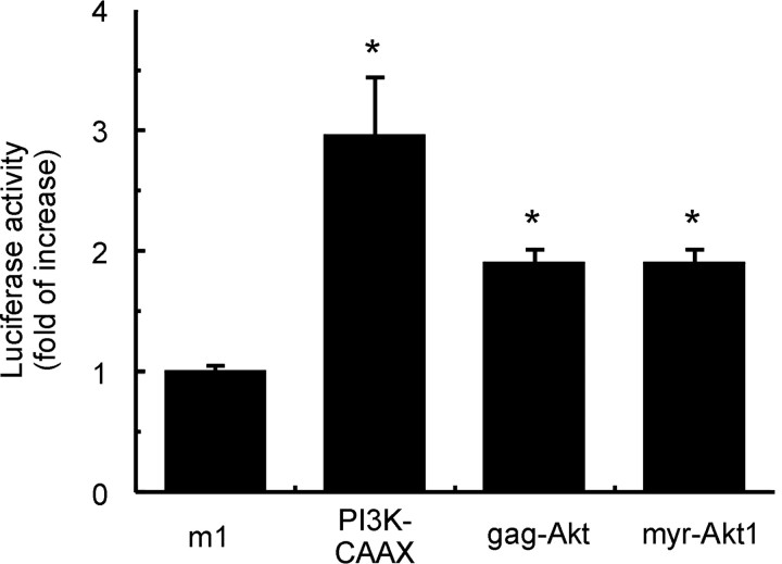
Aerobic cells adjust the expression of antioxidant enzymes to maintain reactive oxygen species within tolerable levels. In addition, phosphatidylinositol 3 kinase (PI3K) and its downstream protein kinase effector Akt adapt cells to survive in the presence of oxidative stress. Here we provide evidence for an association between these two defense systems via transcriptional regulation of Cu/Zn-superoxide dismutase (Cu/Zn-SOD). PC12 pheochromocytoma cells expressing active Akt1 exhibit lower ROS levels in response to hydrogen peroxide, as determined with the superoxide-sensitive probe hydroethidine. Transfection of constitutive or 4-hydroxytamoxifen-inducible versions of Akt1 results in higher messenger RNA and protein levels of Cu/Zn-SOD. Luciferase reporter constructs, carrying different length fragments of the human sod1 gene promoter, have identified a region between -552 and -355 that is targeted by PI3K and Akt and that contains a putative site of regulation by nuclear factor-kappaB (NF-kappaB). Nerve growth factor (NGF) and Akt augment the transactivating activity and produce higher nuclear levels of p65-NF-kappaB. Electrophoretic mobility shift assays indicate that the putative NF-kappaB regulatory sequence binds p65-NF-kappaB more efficiently in nuclear extracts from these cells. A dominant-negative mutant of IkappaBalpha further demonstrates that the PI3K/Akt axis targets the sod1 promoter at the level of the newly characterized NF-kappaB site. These results illustrate a new mechanism by which the PI3K/Akt pathway protects cells against oxidative stress, involving the upregulation of Cu/Zn-SOD gene expression, and the results identify NF-kappaB as a key mediator in the regulation of this gene.


Figures

Figure 1.
Figure 1.
H2O2 increases intracellular ROS as determined by HEt staining. PC12 cells were maintained in low-serum conditions for 16 hr and then untreated or incubated with 0.5 mm H2O2 for 3 hr. During the last hour of the experiment, 2 μm HEt was added. A, Confocal microscopy pictures of representative fields from untreated and H2O2-treated cells stained with HEt (HE) and Hoechst 33258. B, Flow cytometry analysis of H2O2-induced intracellular ROS production in HEt-stained cells. A representative sample of 10,000 cells is shown for untreated or 0.5 mm H2O2-treated cells. C, Analysis by flow cytometry of the HEt incorporation rate in cells submitted to 0.5 mm H2O2 in the absence and in the presence of 10 mm GSH.
Figure 2.
Figure 2.
PC12 cells expressing active Akt1 exhibit oxidant and apoptotic protection. PC12 cells were stably transfected with expression vectors for EGFP or myr-EGFP-Akt1. A, Immunodetection of endogenous Akt and ectopically expressed myr-EGFP-Akt1 with anti-Akt antibodies. B, Immunodetection of active, phosphorylated Akt with anti-phospho-Akt antibodies. C, Flow cytometry determination of ROS in PC12 cells submitted for 3 hr to the indicated H2O2 concentrations as described in Figure 1; *p < 0.001. D, Determination by flow cytometry of H2O2-induced apoptosis and necrosis in cells stained with Annexin-V-PE and 7-AAD. EGFP and myr-EGFP-Akt1 cells were treated with 0.5 or 1.5 mm H2O2 for 6 hr and then stained for 15 min with Annexin-V-PE (1:20v/v) and 2 μm 7-AAD. A representative sample of 10,000 cells is shown for each experimental condition. E, Comparison of the percentages of dead cells, determined from Annexin-V-PE staining at the bottom right and top right quadrants in D, which represent the total number of cells in apoptosis and necrosis. *p < 0.001, comparing H2O2-treated EGFP and myr-EGFP-Akt1 groups.
Figure 3.
Figure 3.
PC12 cells expressing active Akt1 exhibit higher levels of Cu/Zn-SOD messenger RNA and protein. A, Semi-quantitative RT-PCR of EGFP and myr-EGFP-Akt1 cells showing induction of Cu/Zn-SOD mRNA. Top, Cu/Zn-SOD mRNA; bottom, β-actin mRNA showing a similar amount of RNA per lane. B, Immunoblot showing the levels of Cu/Zn-SOD protein in the same cells. Top, Blot with anti-Cu/Zn-SOD antibodies; bottom, blot with anti-PDI antibodies showing a similar amount of protein per lane. C, Flow cytometry analysis of Cu/Zn-SOD protein levels in EGFP and myr-EGFP-Akt1 cells after fixation, permeabilization, and staining with anti-Cu/Zn-SOD antibodies.
Figure 4.
Figure 4.
Analysis of Cu/Zn-SOD levels in PC12 cells expressing a conditionally active version of Akt1. PC12 cells, stably transfected with an expression vector for myr-Akt1-ER*, were submitted to 1 μm 4-HT for 16 hr under low-serum conditions. A, Immunodetection of endogenous Akt and ectopically expressed myr-Akt1-ER* with anti-Akt antibodies. B, Immunodetection of active, phosphorylated Akt with anti-phospho-Akt antibodies. C, Semi-quantitative RT-PCR of vector- and myr-Akt1-ER*-transfected cells showing the levels of Cu/Zn-SOD mRNA in the presence and absence of 4-HT. Top, Cu/Zn-SOD mRNA; bottom, β-actin mRNA showing a similar amount of RNA per lane. D, Immunoblot showing the levels of Cu/Zn-SOD protein in the same cells in the presence and absence of 4-HT. Top, Blot with anti-Cu/Zn-SOD antibodies; bottom, blot with anti-PDI antibodies showing a similar amount of protein per lane.
Figure 5.
Figure 5.
Time course effect of H2O2 on Akt and Cu/Zn-SOD protein levels in control EGFP- and myr-EGFP-Akt1-transfected cells. A, Cells incubated in low serum for 16 hr were submitted to 0.5 mm H2O2 for the indicated times. Top, Immunoblots with anti-phospho-Akt antibodies. Top middle, Immunoblots with anti-Akt antibodies. Bottom middle, Immunoblots with anti-Cu/Zn-SOD antibodies. Bottom, Immunoblots with anti-PDI antibodies of the same lysates showing similar amounts of protein per lane. B-D, Densitometric quantitation of phospho-Akt, total Akt, and Cu/Zn-SOD levels relative to PDI levels after treatment with 0.5 mm H2O2, as shown in A.
Figure 6.
Figure 6.
Activation of the human sod1 promoter by the PI3K/Akt axis. PC12 cells were transiently cotransfected with a luciferase reporter construct containing 1499 bp of the 5′ regulatory sequence of human sod1 (pGL3basic-sod1-1499) and expression vectors for muscarinic receptor m1 as a negative control or for active PI3K-CAAX, gag-Akt, or myr-Akt1. After 24 hr from transfection, the cells were maintained in low serum for 16 hr and then lysed and analyzed for luciferase activity. *p < 0.001, comparing m1 versus the other treatments.
Figure 7.
Figure 7.
Identification of the sod1 promoter region that is regulated by PI3K. A, Schematic showing candidate regulatory elements for PI3K in the human sod1 promoter and a detail of a putative NF-κB site at a position between -552 and -355 from the transcription start site. B, PC12 cells were cotransfected with an expression vector for active PI3K-CAAX and luciferase reporter constructs containing the indicated fragments of the human sod1 promoter. After 24 hr from transfection, the cells were maintained in low serum for 16 hr and then lysed and analyzed for luciferase activity. The figure shows folds of increase in luciferase activity over the promoterless vector pGL3basic. *p < 0.001, comparing vector-transfected versus PI3K(CAAX)-transfected groups.
Figure 8.
Figure 8.
Regulation of NF-κB activity by NGF and Akt signaling. A, Effect on the transactivating capacity of p65-NF-κB. Left, PC12 cells were cotransfected with an expression vector for a fusion protein consisting of the transactivation domain of p65-NF-κB and the DNA-binding domain of Gal4 (pGal4-p65) and a luciferase reporter under the control of Gal4 (pGal4-LUC). After 24 hr from transfection, the cells were submitted to low-serum conditions for 16 hr and then pretreated for 15 min with 10 μm LY294002 (LY) or treated with 25 ng/ml NGF for 6 hr, as indicated. Right, EGFP and myr-EGFP-Akt1 cells were cotransfected with the same plasmids (pGal4-p65 and pGal4-LUC), incubated in low serum for 16 hr, and analyzed for luciferase activity. *Statistically significant differences, with p < 0.001, comparing NGF-treated groups and EGFP and myr-EGFP-Akt1 groups. B, Comparison of the nuclear and cytosolic p65-NF-κB and IκBα protein levels. EGFP and myr-EGFP-Akt1 cells were submitted to low-serum conditions for 16 hr and then stimulated with 25 ng/ml NGF for the indicated times. Cell fractions were resolved in SDS-PAGE and immunoblotted with anti-p65-NF-κB and anti-IκBα antibodies. The same blots were immunoblotted with anti-Sp1 and anti-PDI antibodies to demonstrate similar protein load per lane.
Figure 9.
Figure 9.
The NF-κB site is sufficient to confer PI3K inducibility to the human sod1 promoter. A, EMSA, using a double-stranded oligonucleotide with the putative NF-κB sequence (wild type) identified in the human sod1 promoter or a mutated control sequence (mutated) and nuclear extracts from PC12 cells expressing EGFP or myr-EGFP-Akt1. The arrow points to the position of a band that was not formed when the complexes were incubated in the presence of anti-p65-NF-κB antibody. B, Luciferase activity of PC12 cells cotransfected with PI3K-CAAX or empty vector and reporter constructs comprising three wild-type tandem or three mutated tandem sequences for the NF-κB site of human sod1. In addition, the cells also were cotransfected with a luciferase reporter construct for NF-κB from human immunodeficiency virus (NF-κB-HIV), with p29-sod1, or with pGL3basic. *p < 0.001, comparing vector-transfected versus PI3K(CAAX)-transfected groups. C, Blockage of PI3K induction of the NF-κB-responsive element by cotransfection with dominant-negative IκBα(S32A/S36A) mutant. Cells were cotransfected as in B plus the expression vector for IκBα(S32A/S36A). After 24 hr from transfection, the cells were submitted to low-serum conditions for 16 hr and then analyzed for luciferase activity. *p < 0.001, comparing vector-transfected versus IκBα(S32A/S36A)-transfected groups.

Similar articles

Cited by

References

    1. Asanuma M, Hirata H, Cadet JL (1998) Attenuation of 6-hydroxydopamine-induced dopaminergic nigrostriatal lesions in superoxide dismutase transgenic mice. Neuroscience 85: 907-917. - PubMed
    1. Bindokas VP, Jordán J, Lee CC, Miller RJ (1996) Superoxide production in rat hippocampal neurons: selective imaging with hydroethidine. J Neurosci 16: 1324-1336. - PMC - PubMed
    1. Brunet A, Bonni A, Zigmond MJ, Lin MZ, Juo P, Hu LS, Anderson MJ, Arden KC, Blenis J, Greenberg ME (1999) Akt promotes cell survival by phosphorylating and inhibiting a Forkhead transcription factor. Cell 96: 857-868. - PubMed
    1. Brunet A, Datta SR, Greenberg ME (2001) Transcription-dependent and -independent control of neuronal survival by the PI3K-Akt signaling pathway. Curr Opin Neurobiol 11: 297-305. - PubMed
    1. Bui NT, Livolsi A, Peyron JF, Prehn JH (2001) Activation of nuclear factor-κB and bcl-x survival gene expression by nerve growth factor requires tyrosine phosphorylation of IκBα. J Cell Biol 152: 753-764. - PMC - PubMed

Publication types

MeSH terms