. 2004 Aug 18;32(15):4439-47.
doi: 10.1093/nar/gkh776. Print 2004.

Selectivity and affinity of triplex-forming oligonucleotides containing 2'-aminoethoxy-5-(3-aminoprop-1-ynyl)uridine for recognizing AT base pairs in duplex DNA

Affiliations

Selectivity and affinity of triplex-forming oligonucleotides containing 2'-aminoethoxy-5-(3-aminoprop-1-ynyl)uridine for recognizing AT base pairs in duplex DNA

Sadie D Osborne et al. Nucleic Acids Res. .

Abstract

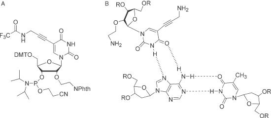
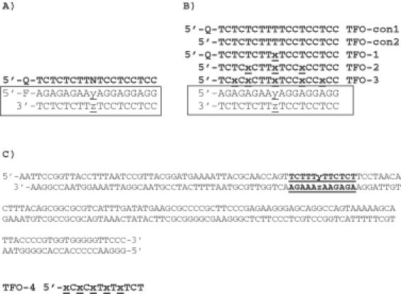
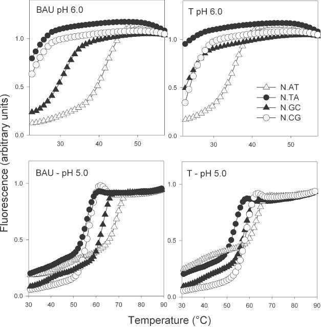
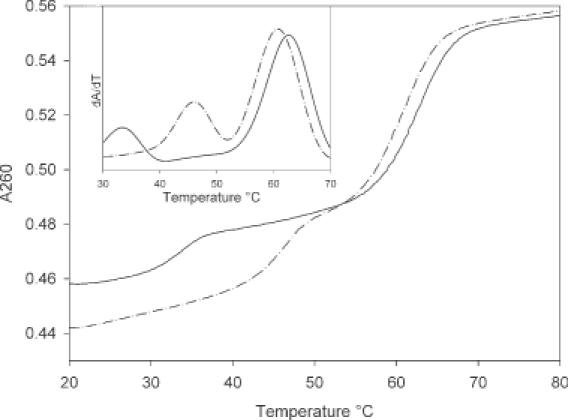
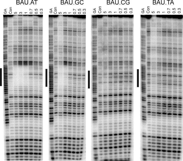
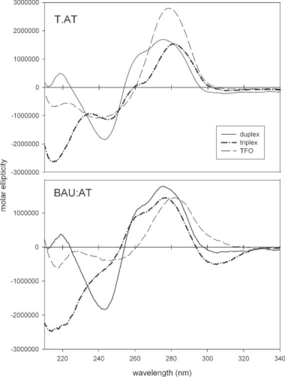
We have used DNase I footprinting, fluorescence and ultraviolet (UV) melting experiments and circular dichroism to demonstrate that, in the parallel triplex binding motif, 2'-aminoethoxy-5-(3-aminoprop-1-ynyl)uridine (bis-amino-U, BAU) has very high affinity for AT relative to all other Watson-Crick base pairs in DNA. Complexes containing two or more substitutions with this nucleotide analogue are stable at pH 7.0, even though they contain several C.GC base triplets. These modified triplex-forming oligonucleotides retain exquisite sequence specificity, with enhanced discrimination against YR base pairs (especially CG). These properties make BAU a useful base analogue for the sequence-specific creation of stable triple helices at pH 7.0.


Figures

Figure 1
Figure 1
Chemical structures of (A) bis-amino uridine phosphoramidite (BAU), (B) BAU.AT triplet.
Figure 2
Figure 2
Sequence of oligonucleotides used in this work. In each case the third strand is shown in bold and the duplex target is boxed. (A) Oligonucleotides used for fluorescence melting experiments, Q, methyl red serinol; F, 6-amidohexylfluorescein; N, BAU T, A, G or C; y.z = each base pair in turn TA, AT, CG or GC. (B) Oligonucleotides used for UV melting studies x, BAU; y.z, each base pair in turn TA, AT, CG, or GC. TFO-1 (containing one BAU residue) was identical to one of the oligonucleotides used for the fluorescence melting studies. (C) Sequence of the footprinting substrates derived from tyrT(43–59); y.z = each base pair in turn in different fragments. The DNA was labelled at the 3′ end of the EcoRI site (lower strand). The triplex target site is underlined and in bold and was targeted with TFO-4 (x = BAU).
Figure 3
Figure 3
Representative fluorescence melting curves for the triplexes shown in Figure 2A. The data for pH 6.0 (upper panels) were obtained by heating at a rate of rate of 0.2°C/min, while the data at pH 5.0 (lower panels) were obtained by heating at a rate of 0.067°C/min. In these curves, the third strands contained BAU as the central base (N) targeted against duplexes containing each base pair (y.z) in turn (AT, open triangles; TA, closed circles; GC, closed triangles; CG, open circles). These experiments were performed in 50 mM sodium acetate (pH 5.0 or 6.0) containing 200 mM NaCl.
Figure 4
Figure 4
UV-melting curves for the triplex formed between TFO-3 and the duplex containing a central AT base pair (dashed line) and a central GC base pair (solid line) determined in 10 mM sodium phosphate, pH 6.5 containing 1 mM EDTA and 200 mM NaCl. The inset shows the first derivative of the melting profiles.
Figure 5
Figure 5
DNase I footprints showing the interaction of TFO-4 [d(xCxCxTxTxTCT-3′), x = BAU] with DNA fragments that contain each base pair in turn in the centre of the oligopurine tract, opposite the base shown in boldface. The TFO concentration (μM) is shown at the top of each lane. The experiments were performed in 10 mM Tris–HCl, pH 7.0, containing 50 mM sodium chloride and the complexes were left to equilibrate for 30 min before digestion with DNase I. The lanes labelled ‘GA’ and ‘con’ represent Maxim–Gilbert markers specific for purines and DNase I cleavage of duplex DNA in the absence of TFO, respectively.
Figure 6
Figure 6
CD spectra of TFO-con2 (upper panel) and TFO-3 (lower panel) together with the target duplex and the triplex as indicated. The spectra were recorded in 10 mM sodium phosphate pH 6.0 containing 1 mM EDTA and 200 mM NaCl.
None
Scheme 1. Synthesis of 2′-aminoethoxy-5-(3-aminoprop-1-ynyl)uridine phosphoramidite 1 (BAU monomer). Reagents and conditions: (i) acetyl chloride, MeOH, rt, 3 h, quant.; (ii) TIPDSCl (1.2 eq), py, rt, 16 h, 72%; (iii) methyl bromoacetate (5 eq), NaH (4.4 eq), DMF, −5°C to rt, 6 h, 61%; (iv) LiBH4 (2 eq), THF, rt, 30 min, 86%; (v) Phthalimide (1.4 eq), PPh3 (1.4 eq), DEAD (1.4 eq), THF, rt, 2 h, 91%; (vi) AcOH:Ac2O (1:1), H2SO4, rt, 2 h, 79%; (vii) (a) HMDS, 5-iodouracil, TMSCl (1.0 eq), 120°C, 16 h; (b) 7 (0.3 eq), TMSOTf (3.2 eq), DCE, 0°C to rt, 2 h, 52%; (viii) NaOMe (3 eq), MeOH, rt, 2 h, quant.; (ix) DMTrCl (1.3 eq), py, rt, 3 h, 76%; (x) 3-trifluoroacetylamidoprop-1-yne (1.1 eq), CuI (0.3 eq), TEA (3 eq), Pd(PH3)4 (0.1 eq), DMF, rt, dark, 5 h, 90%; (xi) 2-cyanoethoxy-(N,N-diisopropylamino)chlorophosphine (1.2 eq), DIPEA (2.6 eq), THF, rt, 5 h, 70%.

Similar articles

Cited by

References

    1. Chan P.P. and Glazer,P.M. (1997) Triplex DNA: fundamentals, advances and potential applications for gene therapy. J. Mol. Med., 75, 267–282. - PubMed
    1. Gorman L. and Glazer,P.M. (2001) Directed gene modification by triple helix formation. Curr. Mol. Med., 1, 391–399. - PubMed
    1. Praseuth D., Guieysse,A.L. and Hélène,C. (1999) Triple helix formation and the antigene strategy for sequence-specific control of gene expression. Biochim. Biophys. Acta, 1489, 181–206. - PubMed
    1. Neidle S. (1997) Recent developments in triple-helix regulation of gene expression. Anticancer Drug Des., 12, 433–442. - PubMed
    1. Vasquez K.M. and Wilson,J.H. (1998) Triplex-directed modification of genes and gene activity. Trends Biochem. Sci., 23, 4–9. - PubMed

Publication types