

Fusion-independent expression of functional ACh receptors in mouse mesoangioblast stem cells contacting muscle cells
- PMID: 15319417
- PMCID: PMC1665253
- DOI: 10.1113/jphysiol.2004.070607
Fusion-independent expression of functional ACh receptors in mouse mesoangioblast stem cells contacting muscle cells
Abstract
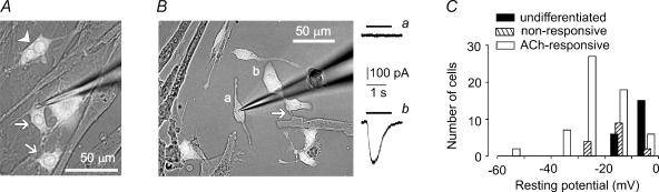
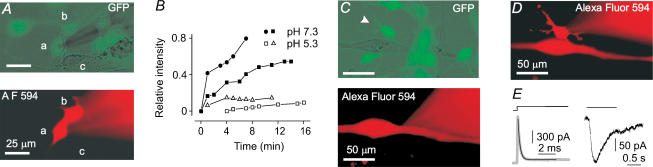
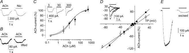
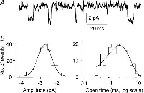
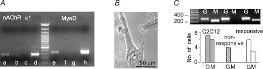
Mesoangioblasts are vessel-associated fetal stem cells that can be induced to differentiate into skeletal muscle, both in vitro and in vivo. Whether this is due to fusion or to transdifferentiation into bona fide satellite cells is still an open question, for mesoangioblasts as well as for other types of stem cells. The early steps of satellite cell myogenic differentiation involve MyoD activation, membrane hyperpolarization and the appearance of ACh sensitivity and gap junctional communication. If mesoangioblasts differentiate into satellite cells, these characteristics should be observed in stem cells prior to fusion into multinucleated myotubes. We have investigated the functional properties acquired by mononucleated green fluorescent protein (GFP)-positive mesoangioblasts co-cultured with differentiating C2C12 myogenic cells, using the patch-clamp technique. Mesoangioblasts whose membrane contacted myogenic cells developed a hyperpolarized membrane resting potential and ACh-evoked current responses. Dye and electrical coupling was observed among mesoangioblasts but not between mesoangioblasts and myotubes. Mouse MyoD was detected by RT-PCR both in single, mononucleated mesoangioblasts co-cultured with C2C12 myotubes and in the total mRNA from mouse mesoangioblasts co-cultured with human myotubes, but not in human myotubes or stem cells cultured in isolation. In conclusion, when co-cultured with muscle cells, mesoangioblasts acquire many of the functional characteristics of differentiating satellite cells in the absence of cell fusion, strongly indicating that these stem cells undergo transdifferentiation into satellite cells, when exposed to a myogenic environment.
Figures
Similar articles
-
Green fluorescent protein incorporation by mouse myoblasts may yield false evidence of myogenic differentiation of human haematopoietic stem cells.Acta Physiol (Oxf). 2008 Jul;193(3):249-56. doi: 10.1111/j.1748-1716.2008.01833.x. Epub 2008 Feb 18. Acta Physiol (Oxf). 2008. PMID: 18284377
-
Effective myotube formation in human adipose tissue-derived stem cells expressing dystrophin and myosin heavy chain by cellular fusion with mouse C2C12 myoblasts.Biochem Biophys Res Commun. 2011 Apr 29;408(1):167-73. doi: 10.1016/j.bbrc.2011.04.002. Epub 2011 Apr 5. Biochem Biophys Res Commun. 2011. PMID: 21473854
-
Contribution of human bone marrow stem cells to individual skeletal myotubes followed by myogenic gene activation.Exp Cell Res. 2005 Jul 1;307(1):174-82. doi: 10.1016/j.yexcr.2005.03.008. Epub 2005 Apr 8. Exp Cell Res. 2005. PMID: 15922737
-
[Electrophysiological properties of stem cells].Herz. 2006 Apr;31(2):123-6. doi: 10.1007/s00059-006-2793-y. Herz. 2006. PMID: 16738835 Review. German.
-
Mesoangioblasts of inclusion-body myositis: a twofold tool to study pathogenic mechanisms and enhance defective muscle regeneration.Acta Myol. 2011 Jun;30(1):24-8. Acta Myol. 2011. PMID: 21842589 Free PMC article. Review.
Cited by
-
Biological behaviors of muscarinic receptors in mesenchymal stem cells derived from human placenta and bone marrow.Iran J Basic Med Sci. 2020 Jan;23(1):124-132. doi: 10.22038/IJBMS.2019.38582.9151. Iran J Basic Med Sci. 2020. PMID: 32405354 Free PMC article.
-
Differentiation of control and ALS mutant human iPSCs into functional skeletal muscle cells, a tool for the study of neuromuscolar diseases.Stem Cell Res. 2016 Jul;17(1):140-7. doi: 10.1016/j.scr.2016.06.003. Epub 2016 Jun 8. Stem Cell Res. 2016. PMID: 27318155 Free PMC article.
-
Physiological characterization of human muscle acetylcholine receptors from ALS patients.Proc Natl Acad Sci U S A. 2011 Dec 13;108(50):20184-8. doi: 10.1073/pnas.1117975108. Epub 2011 Nov 29. Proc Natl Acad Sci U S A. 2011. PMID: 22128328 Free PMC article.
-
Acetylcholine induces mesenchymal stem cell migration via Ca2+ /PKC/ERK1/2 signal pathway.J Cell Biochem. 2012 Aug;113(8):2704-13. doi: 10.1002/jcb.24148. J Cell Biochem. 2012. PMID: 22441978 Free PMC article.
-
Myogenic potential of canine craniofacial satellite cells.Front Aging Neurosci. 2014 May 13;6:90. doi: 10.3389/fnagi.2014.00090. eCollection 2014. Front Aging Neurosci. 2014. PMID: 24860499 Free PMC article.
References
-
- Balogh S, Naus SS, Merrifield PA. Expression of gap junctions in cultured rat L6 cells during myogenesis. Dev Biol. 1993;155:351–360. - PubMed
-
- Brazelton TR, Rossi FM, Keshet GI, Blau HM. From marrow to brain: expression of neuronal phenotypes in adult mice. Science. 2000;290:1775–1779. - PubMed
-
- Broccolini A, Ricci E, Pescatori M, Papacci M, Gliubizzi C, D'Amico A, Servidei S, Tonali P, Mirabella M. Insulin-like growth factor I in inclusion-body myositis and human muscle cultures. J Neuropathol Exp Neurol. 2004;63:650–659. - PubMed
Publication types
MeSH terms
Substances
LinkOut - more resources
Full Text Sources
Medical
Miscellaneous