. 2004 Dec;18(12):3064-75.
doi: 10.1210/me.2004-0187. Epub 2004 Aug 19.

Multiple kinase cascades mediate prolactin signals to activating protein-1 in breast cancer cells

Affiliations

Multiple kinase cascades mediate prolactin signals to activating protein-1 in breast cancer cells

Jennifer H Gutzman et al. Mol Endocrinol. 2004 Dec.

Abstract

The importance of prolactin (PRL) in physiological proliferation and differentiation of the mammary gland, together with high levels of PRL receptors in breast tumors, the association of circulating PRL with incidence of breast cancer, and the recognition of locally produced PRL, point to the need for greater understanding of PRL actions in mammary disease. Although PRL has been shown to activate multiple kinase cascades in various target cells, relatively little is known of its signaling pathways in the mammary gland apart from the Janus kinase 2/ signal transducer and activator of transcription 5 pathway, particularly in tumor cells. Another potential effector is activating protein-1 (AP-1), a transcription complex that regulates processes essential for neoplastic progression, including proliferation, survival and invasion. We demonstrate that PRL activates AP-1 in MCF-7 cells, detectable at 4 h and sustained for at least 24 h. Although Janus kinase 2 and ERK1/2 are the primary mediators of PRL-induced signals, c-Src, phosphatidylinositol 3'-kinase, protein kinase C, and other MAPKs contribute to maximal activity. PRL activation of these pathways leads to increased c-Jun protein and phosphorylation, JunB protein, and phosphorylation of c-Fos, elevating the levels of AP-1 complexes able to bind DNA. These active AP-1 dimers may direct expression of multiple target genes, mediating some of PRL's actions in mammary disease.


Figures

Fig. 1
Fig. 1
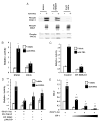
PRL Induces AP-1 Activity in PRL-Deficient MCF-7 Cells A, Representative time course of AP-1 activation. Cells were cotransfected with 4XAP-1-luc, lPRLR, and β-galactosidase. After transfection, cells were washed and treated ± 4 nM PRL, and cell lysates were harvested at the times indicated. All lysates were assayed for luciferase and β-galactosidase activity as described in Materials and Methods with each data point in triplicate ± SD. RLU, Relative luciferase units. B (left), Effect of PRLR isoform on AP-1 activity. Cells were cotransfected with 4XAP-1-luc or the enhancer-less control vector, pXP2-luc; lPRLR or iPRLR; and β-galactosidase. After transfection, cells were treated ± 4 nM PRL for 6 h and assayed as in panel A. B (right), To confirm expression of transfected PRLR, lysates from nontransfected (non-tr) cells and cells transfected with the iPRLR or lPRLR were subjected to Western analysis using the PRLR antibody recognizing the extracellular domain. C, Cell cycle controls for AP-1 activation. Cells were pretreated for 24 h with 1 mM hydroxyurea or 100 ng/ml nocodazole in serum-free media, and then cotransfected with 4XAP-1-luc, lPRLR, and ± -galactosidase. After transfection, cells were treated ± 4 nM PRL, or ± 4 nM PRL in the presence of 1 mM hydroxyurea or 100 ng/ml nocodazole in serum-free media for 6 h and assayed as in panel A. In B and C, relative activity represents the mean of the corrected luciferase activity from at least three independent experiments, represented as mean fold change relative to the vehicle-treated control transfection ± SEM. For statistical analyses, PRL- and vehicle-treated samples similarly transfected were compared. Asterisks denote significant differences between vehicle and PRL-treated cells (P > 0.05) using Student’s t test. In panel C, cell cycle inhibitors did not significantly affect the PRL response.
Fig. 2
Fig. 2
PRL Activates AP-1 via Multiple Proximal Signaling Pathways Cells were cotransfected with 4XAP-1-luc, lPRLR, β-ga-lactosidase, and DN Jak2 or DN Src as indicated. After trans-fection, cells were washed and treated in serum-free media ± 4 nM PRL for 6 h. Cells to be treated with 10 μM LY294002 (which inhibited phosphorylation of Akt without altering cell viability) or 10 μM Bis II were washed after transfection and pretreated for 1 h with inhibitor, then treated ± 4 nM PRL for 6 h in the presence of inhibitor. Cells were harvested after treatment and assayed for luciferase and β-galactosidase activity. Relative activity represents the mean of the corrected luciferase activity from at least three independent experiments, represented as mean fold change relative to the vehicle-treated cells in the absence of inhibitor or dominant negative construct ± SEM. For statistical analyses, PRL- and vehicle-treated cells transfected and/or treated with the same plasmid or inhibitor were compared. Asterisks denote significant differences between vehicle- and PRL-treated cells (P < 0.04) using Student’s t test.
Fig. 3
Fig. 3
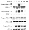
PRL Activates Multiple MAPKs Cells were plated as described in Materials and Methods, serum starved for 24 h, and then treated with 4 nM PRL for the times indicated. Cell lysates were harvested and analyzed by Western blot using the antibodies shown.
Fig. 4
Fig. 4
PRL Signals to AP-1 via ERK1 and ERK2 Cells were grown and plated as described in Materials and Methods. A, Cells were pretreated for 1 h with vehicle, 20 μM PD98059, or 10 μM U0126, and then treated ± 4 nM PRL for 15 min. Cell lysates were harvested, and equal amounts of protein were analyzed by Western blot for phospho-ERK1/2, phospho-ERK5, and phospho-JNK1/2. B, Cells were cotransfected with 4XAP-1-luc, lPRLR, and β-galactosidase. After transfection, cells were pretreated for 1 h with vehicle or 10 μM U0126, and then treated ± 4 nM PRL for 6 h in the presence of inhibitor. C, D, and E, Cells were cotransfected with 4XAP-1-luc, lPRLR, β-galactosidase, and DN ERK1 and DN ERK2 or vector (C) or DN JNK1 and DN JNK2, DN ERK5, p38α KD, or vector (D) or increasing concentrations of JIP-1 (34) or vector (E). After transfection, cells were allowed to recover overnight in serum-free media and then treated ± 4 nM PRL for 6 h. All lysates were assayed for luciferase and β-galactosidase activity as described in Materials and Methods. In B, C, and D, relative activity represents the mean of the corrected luciferase activity from at least three independent experiments, represented as mean fold change relative to the vehicle-treated cells in the absence of inhibitor or dominant negative construct ± SEM. For statistical analyses, PRL- and vehicle-treated samples similarly transfected, or treated with inhibitor, were compared. Asterisks denote significant differences between vehicle- and PRL-treated cells (P < 0.03) using Student’s t test.+, Significant difference from vehicle-treated control transfection (P < 0.05) using Student’s t test. E is a representative experiment with each data point in triplicate ± SD and graphed as relative luciferase units (RLUs).
Fig. 5
Fig. 5
PRL Induces AP-1 DNA Binding of Jun/Fos Family Members A, Cells were serum starved for 24 h and then treated ± 4 nM PRL for the times indicated. Nuclear proteins were harvested after PRL treatment and analyzed by EMSA as described in Materials and Methods. For the lane labeled “cold,” an excess of unlabeled oligonucleotide was included. B, Nuclear proteins were harvested after treatment ± 4 nM PRL for 3 h and were preincubated in the presence or absence of specific antibody to c-Jun (sc-45X), JunB (sc-46X), c-Fos (sc-52X), JunD (sc-74X), or FosB (sc-48X), as indicated before EMSA analysis. C, Cells were cotransfected with 4XAP-1-luc, lPRLR, β-galactosidase, and increasing concentrations of the dominant negative c-Jun, TAM-67 (37). After transfection, cells were allowed to recover overnight and then treated in serum-free media ± 4 nM PRL for 6 h. All lysates were assayed for luciferase and β-galactosidase activity as described in Materials and Methods, with each data point in triplicate ± SD. RLU, Relative luciferase units.
Fig. 6
Fig. 6
PRL Induces Modifications of c-Jun, JunB, and c-Fos in PRL-Deficient MCF-7 Cells A, Cells were serum starved for 24 h and then treated ± 4 nM PRL for the times indicated. Cellular lysates were harvested, and equal amounts of protein were analyzed by Western blot for c-Jun, phospho-c-Jun, JunB, JunD, c-Fos, or FosB. Solid arrowhead indicates a modification of c-Fos retarding mobility; the open arrowhead indicates a modification increasing mobility that was sometimes also observed. B, Lysates were harvested after 1 h ± 4 nM PRL treatment and incubated in the presence or absence of λ-PPase as described in Materials and Methods. After λ-PPase incubation, equal amounts of protein were analyzed by Western blot for c-Fos and phospho-ERK1/2 (positive control). C, Cells were serum starved for 24 h and then pretreated for 1 h with vehicle, 20 μM PD98059, or 10 μM U0126. After pretreatment with inhibitor, cells were treated ± 4 nM PRL for an additional hour along with inhibitor, and lysates were analyzed by Western blot using the antibodies shown.
Fig. 7
Fig. 7
Model for PRL Activation of AP-1 PRL binds to the PRLR and activates multiple proximal kinases including Jak2, c-Src, PI3K, and PKC. This results in phosphorylation and subsequent activation of multiple MAPKs, which increases phosphorylation and/or protein levels and leads to activation of AP-1 family members, including c-Jun, JunB, and c-Fos. These modifications in AP-1 components facilitate an increase in AP-1 transcriptional activity. In this PRL-deficient MCF-7 cell model, we have shown that Jak2 and ERK1/2 play primary roles in activation of AP-1 by PRL. However, other proximal kinases and MAPKs are also involved.

Similar articles

Cited by

References

    1. Clevenger CV, Furth PA, Hankinson SE, Schuler LA. Role of prolactin in mammary carcinoma. Endocr Rev. 2003;24:1–27. - PMC - PubMed
    1. Vonderhaar BK. Prolactin in human breast cancer development. In: Ethier SP, editor. Endocrine oncology. Totowa, NJ: Humana Press; 2000. pp. 101–120.
    1. Goffin V, Touraine P, Pichard C, Bernichtein S, Kelly PA. Should prolactin be reconsidered as a therapeutic target in human breast cancer. Mol Cell Endocrinol. 1999;151:79–87. - PubMed
    1. Hankinson SE, Willett WC, Michaud S, Manson JE, Colditz GA, Longcope C, Rosner B, Speizer FE. Plasma prolactin levels and subsequent risk of breast cancer in postmenopausal women. J Natl Cancer Inst. 1999;91:629–634. - PubMed
    1. Boyd NF, Stone J, Martin LJ, Jong R, Fishell E, Yaffe M, Hammond G, Minkin S. The association of breast mitogens with mammographic densities. Br J Cancer. 2002;87:876–882. - PMC - PubMed

Publication types

MeSH terms