. 2005 Jan 1;385(Pt 1):173-80.
doi: 10.1042/BJ20040650.

A cryptic matrix targeting signal of the yeast ADP/ATP carrier normally inserted by the TIM22 complex is recognized by the TIM23 machinery

Affiliations

A cryptic matrix targeting signal of the yeast ADP/ATP carrier normally inserted by the TIM22 complex is recognized by the TIM23 machinery

Maïlys A S Vergnolle et al. Biochem J. .

Abstract

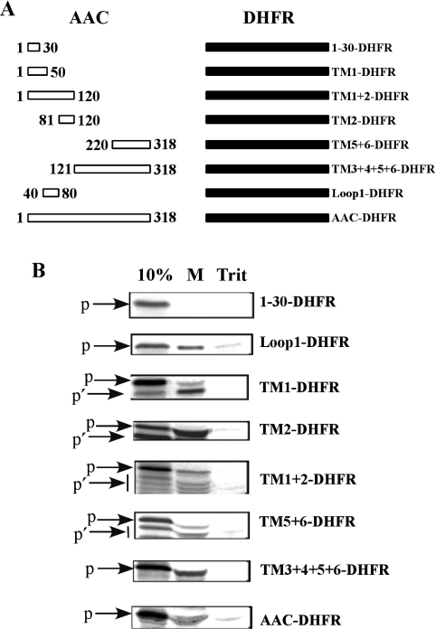
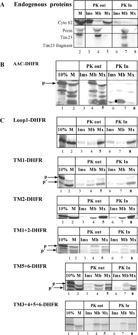
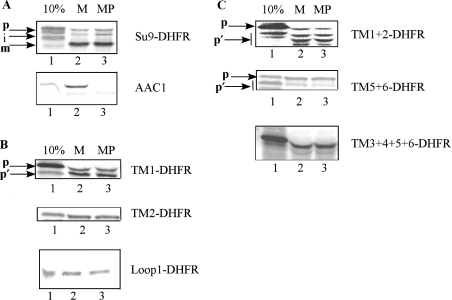
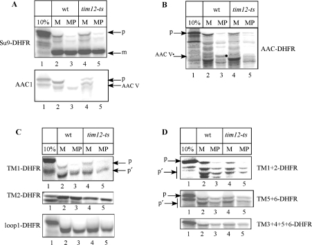
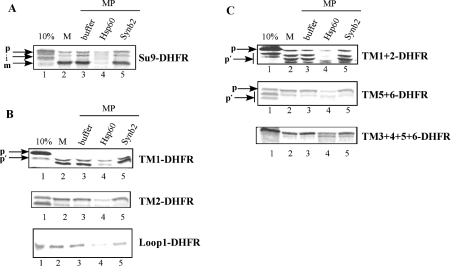
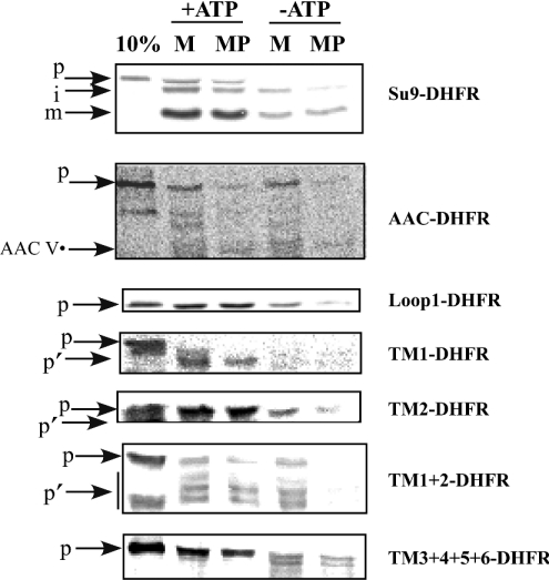
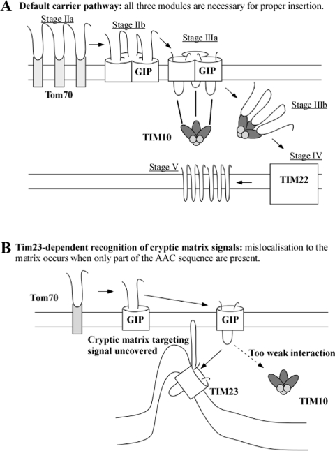
The yeast ADP/ATP carrier (AAC) is a mitochondrial protein that is targeted to the inner membrane via the TIM10 and TIM22 translocase complexes. AAC is devoid of a typical mitochondrial targeting signal and its targeting and insertion are thought to be guided by internal amino acid sequences. Here we show that AAC contains a cryptic matrix targeting signal that can target up to two thirds of the N-terminal part of the protein to the matrix. This event is coordinated by the TIM23 translocase and displays all the features of the matrix-targeting pathway. However, in the context of the whole protein, this signal is 'masked' and rendered non-functional as the polypeptide is targeted to the inner membrane via the TIM10 and TIM22 translocases. Our data suggest that after crossing the outer membrane the whole polypeptide chain of AAC is necessary to commit the precursor to the TIM22-mediated inner membrane insertion pathway.


Figures

Figure 1
Figure 1. All fusion proteins except 1-30-DHFR are efficiently imported into mitochondria
(A) The AAC fragments are represented by white boxes and the DHFR moiety by black boxes. The amino-acid length of the AAC fragments is indicated. (B) Import of the different fusion proteins into isolated mitochondria. Proteins were synthesized in a rabbit reticulocyte lysate in the presence of [35S]methionine. Radiolabelled proteins (10 μl) were imported into 100 μg of purified, energized mitochondria for 10 min at 30 °C. After import, mitochondria were re-isolated and treated with 0.1 mg/ml of proteinase K for 15 min on ice in the presence (Trit) or absence (M) of Triton-X100. Proteinase K was inhibited with 1 mM of PMSF for 10 min on ice. Triton samples were then precipitated with TCA and the pellets were solubilized in 10 μl of sample buffer. Mitochondria were re-isolated and resuspended in 10 μl of sample buffer. Samples were separated by SDS/PAGE (12% gel) and analysed using a phosphorimager. 10%, 10% of the input 35S-labelled protein used in the assay; p, full-length precursor; p′, secondary translation products.
Figure 2
Figure 2. Fusion proteins are localized in the matrix
(A) Western blotting of a mitochondrial subfractionation using antibodies against cytochrome b2 (‘cyto b2’), porin and Tim23. (B) Import and subfractionation using 35S-labelled AAC-DHFR. The black dot indicates the AAC stage V fully inserted form. (C) as in (B), but the precursors used were the different deletions of AAC fused to DHFR as indicated. Proteins were synthesized in a rabbit reticulocyte lysate in presence of [35S]methionine. 10 μl of radiolabelled protein were imported into 100 μg of purified energized mitochondria for 10 min at 30 °C. Lanes 1, 10% of the input in vitro translated precursor. Lanes 2, intact mitochondria after import were re-isolated and treated with 0.1 mg/ml of proteinase K for 15 min on ice. Proteinase K was inhibited with 1 mM of PMSF for 10 min on ice. Mitochondria were re-isolated and resuspended in 10 μl of 2×sample buffer. Lanes 3, 4, 5, after import mitochondria were proteinase K and PMSF treated. After re-isolation mitochondria were osmotically shocked and centrifuged. The supernatant corresponding to the IMS fraction was precipitated with TCA (lanes 3) and the pellet was sodium carbonate extracted. The sodium carbonate sample was centrifuged: pellet corresponding to the membrane fraction was resuspended in sample buffer (lanes 4) and supernatant corresponding to the matrix fraction was precipitated with TCA (lanes 5). Lanes 6, 7, 8: The same subfractionation as in lanes 3, 4, 5 with the exception that proteinase K treatment was made during the osmotic shock, followed by addition of PMSF. Samples were analysed by SDS/PAGE (12% gel) and using a phosphorimager. p, full-length precursor; p′, secondary translation products.
Figure 3
Figure 3. Import of all fusion proteins into mitoplasts is similar to import into intact mitochondria
(A) Control imports for Su9-DHFR (matrix-targeted) and AAC1 (IM-inserted). (B) Imports for the constructs TM1-DHFR, TM2-DHFR and Loop1-DHFR containing segments of only the first AAC2 repeat. (C) Imports of the longer constructs TM1+2-DHFR, TM5+6-DHFR and TM3+4+5+6-DHFR. Import was performed as in Figure 1(B) in intact mitochondria (M) or into mitoplasts (MP). After import, proteinase K treatment was done followed by PMSF. Mitochondria and mitoplasts were analysed by SDS/PAGE (12%) followed by autoradiography. p, full-length precursor; p′, secondary translation products; m, mature form; i, intermediate form.
Figure 4
Figure 4. Fusion proteins retain their capacity to import into mitochondria from a tim12-ts strain
(A) Import of control proteins Su9-DHFR and AAC1. (B) Import of full-length AAC2 fused to DHFR (AAC-DHFR). (C) Import of segments of the first repeat of AAC fused to DHFR (TM1-DHFR, TM2-DHFR, loop1-DHFR). (D) Import of the longer constructs TM1+2-DHFR, TM5+6-DHFR and TM3+4+5+6-DHFR. Lanes 1, 10% of the input precursor. Import was into intact mitochondria using either wt mitochondria (lanes 2,3) or tim12-ts mitochondria (lanes 4,5). After import, proteinase K treatment was performed on mitochondria (M, lanes 2,4) or mitoplasts (MP, lanes 3,5). p, full-length precursor; p′, secondary translation products; m, mature form; AAC V (indicated by a black dot), stage V of wt AAC imported form.
Figure 5
Figure 5. Import of fusion proteins is competed by the Hsp60 presequence peptide
Import of 35S-labelled precursor was performed for 10 min at 30 °C into mitochondria (M, lanes 2) or mitoplasts (MP, lanes 3–5). Import was done either in the presence of 3 μg of synthetic peptide (Hsp60, lanes 4 or Synb2, lanes 5) or 50 mM Tris pH 8 buffer alone (buffer, lanes 3), Lanes 1, 10% of the input precursor. p, full-length precursor; p′, secondary translation products; m, mature form; i, intermediate form. (A) Import of control protein Su9-DHFR. (B) Import of the first repeat of AAC fused to DHFR (TM1-DHFR, TM2-DHFR, loop1-DHFR). (C) Import of the longer constructs TM1+2-DHFR, TM5+6-DHFR and TM3+4+5+6-DHFR.
Figure 6
Figure 6. Import of fusion proteins is affected by matrix ATP depletion in the same manner as the matrix targeted Su9-DHFR precursor
The different fusion proteins were imported into isolated mitochondria in presence (+ATP) or absence (−ATP) of intramitochondrial ATP (see Materials and methods). After import samples were halved. The first half (M) was proteinase K treated followed by PMSF. The other half was first osmotically shocked (MP) then proteinase K and PMSF treated. Finally mitochondria (M) and mitoplasts (MP) were solubilized in sample buffer and analysed by SDS/PAGE followed by autoradiography. p, full-length precursor; p′, secondary translation products, i, intermediate form of the SU9-DHFR and m, mature form of the Su9-DHFR.
Scheme 1
Scheme 1. Targeting of the AAC to the IM after crossing the OM
(A) Physiological, ‘en-bloc’ recognition by the TIM10 and TIM22 complexes that mediate targeting and insertion at the IM. (B) Mistargeting to the TIM23 machinery and matrix mislocalization occur when only part of the AAC sequence is present and the cryptic-matrix targeting signal(s) become functional.

Similar articles

Cited by

References

    1. Truscott K. N., Brandner K., Pfanner N. Mechanisms of protein import into mitochondria. Curr. Biol. 2003;13:R326–R337. - PubMed
    1. El Moualij B., Duyckaerts C., Lamotte-Brasseur J., Sluse F. E. Phylogenetic classification of the mitochondrial carrier family of Saccharomyces cerevisiae. Yeast. 1997;13:573–581. - PubMed
    1. Pfanner N., Neupert W. Distinct steps in the import of ADP/ATP carrier into mitochondria. J. Biol. Chem. 1987;262:7528–7536. - PubMed
    1. Zimmermann R., Neupert W. Transport of proteins into mitochondria. Posttranslational transfer of ADP/ATP carrier into mitochondria in vitro. Eur. J. Biochem. 1980;109:217–229. - PubMed
    1. Ryan M. T., Muller H., Pfanner N. Functional staging of ADP/ATP carrier translocation across the outer mitochondrial membrane. J. Biol. Chem. 1999;274:20619–20627. - PubMed

Publication types

MeSH terms

Substances