. 2004 Sep;137(3):469-77.
doi: 10.1111/j.1365-2249.2004.02547.x.

CD40-CD154 interactions between macrophages and natural killer cells during sepsis are critical for macrophage activation and are not interferon gamma dependent

Affiliations

CD40-CD154 interactions between macrophages and natural killer cells during sepsis are critical for macrophage activation and are not interferon gamma dependent

M J Scott et al. Clin Exp Immunol. 2004 Sep.

Abstract

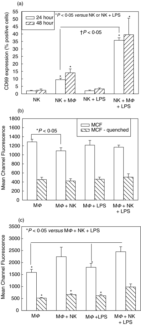
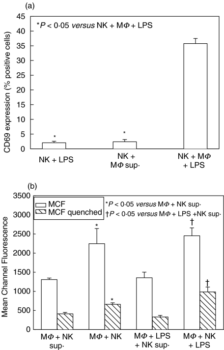
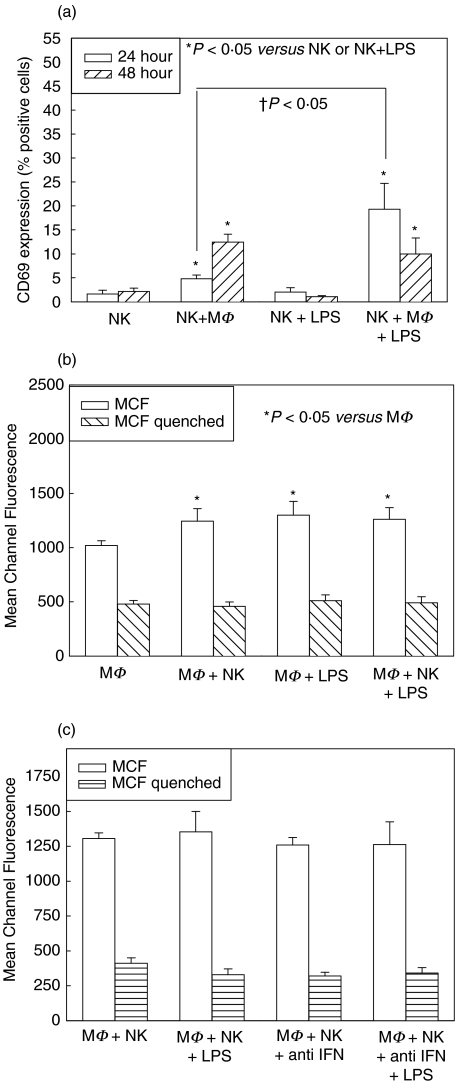
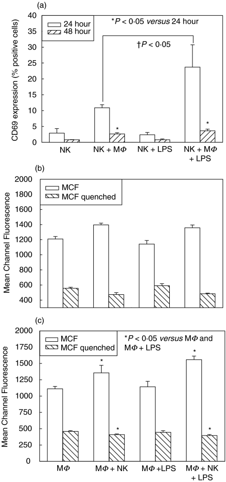
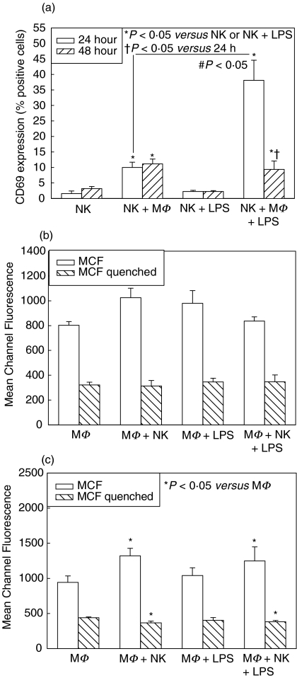
Natural killer (NK) cell interactions with macrophages have been shown to be important during bacterial sepsis in activating macrophages to improve bacterial clearance. The mechanism for this increased activation, however, is unclear. This study determines the relative roles of interferon (IFN)-gamma and CD40/CD154 direct cell interactions on macrophage and NK cell activation in an experimental model of sepsis. Splenic NK cells and peritoneal macrophages were isolated and cultured alone or in coculture, with and without LPS. CD69 expression on NK cells, phagocytosis ability of macrophages, and cell cytokine production was assessed at 24 and 48 h. Coculture of NK cells and macrophages significantly increased activation levels of both cell types, and through experiments culturing NK cells with supernatants from stimulated macrophages and macrophages with supernatants from stimulated NK cells, this activation was determined to be cell-contact-dependent. Similar experiments were conducted using NK cells from IFN-gamma deficient (-/-) mice, as well as anti-IFN-gamma neutralizing antibody. These experiments determined that IFN-gamma is not required for NK or macrophage activation, although it did augment activation levels. Experiments were again repeated using peritoneal macrophages from CD40-/- mice or splenic NK cells from CD154-/- mice. CD40/CD154 interactions were important in the ingestion of bacteria by macrophages, but did not affect NK cell activation at 24 h. There was, however, a protective effect of CD40/CD154 interactions on NK cell activation-induced cell death that occurred at 48 h. CD40/CD154 interactions between macrophages and NK cells are therefore important in macrophage phagocytosis, and are not dependent on IFN-gamma.


Figures

Fig. 1
Fig. 1
Splenic NK cell activation at 24 and 48 h (a) and peritoneal macrophage (MΦ) phagocytosis at 24 h (b) and 48 h (c) in single cell-type and mixed-cell cocultures from wild type C57BL/6 mice. Isolated splenic NK cells were cultured alone and with MΦ with and without LPS. CD69 expression was determined after 24 and 48 h culture, and levels were significantly increased in cocultures with MΦ (NK + MΦ, NK + MΦ+ LPS) (a). MΦ were similarly cultured alone or with NK cells, with and without LPS, and levels of phagocytosis of fluorescent-labeled E. coli determined at 24 h and 48 h. No differences were seen in phagocytosis after 24 h (b). Phagocytosis mean channel fluorescence (MCF) was significantly increased in cocultures of MΦ+NK cells +LPS at 48 h compared with other groups (c). n = 6 per experimental group and data are representative of separate repeated experiments. Data presented as mean ± s.e.m. * or †P < 0.05, Kruskal–Wallis (CD69 percentages), anova (MCF).
Fig. 2
Fig. 2
NK cell activation after 24 h culture with supernatant from stimulated macrophage (MΦ) culture (a), and MΦ phagocytosis after 48 h culture with supernatant from stimulated NK cell culture (b) in single cell-type and mixed-cell cocultures from wild type C57BL/6 mice. CD69 cell surface expression was determined on NK cells after 24 h culture with supernatant collected after 48 h culture of MΦ + 1 µg/ml LPS. Soluble mediators in the supernatant were not sufficient to increase NK cell activation to the level of mixed-cell cocultures (a). Levels of macrophage phagocytosis of fluorescent-labelled E. coliwere determined at 48 h after culture with supernatant collected after 24 h culture of NK cells with 10 ng/ml PMA and 500 ng/ml ionomycin. Soluble mediators in the supernatant were not sufficient to increase phagocytosis to the level of mixed-cell cocultures (b). n = 6 per experimental group and data are representative of separate repeated experiments. Data presented as mean ± s.e.m. * or †P < 0·05, Kruskal–Wallis (CD69 percentages), anova (MCF).
Fig. 3
Fig. 3
Role of IFN-γ on NK cell and macrophage (MΦ) activation in single cell-type and mixed-cell coculture. CD69 expression was determined on splenic NK cells isolated from IFN-γ–/– mice after 24 h culture alone or with MΦ from wild type (WT) C57BL/6, with and without LPS (a). Cocultures of NK cells with MΦ (NK + MΦ, NK + MΦ + LPS) showed increases in CD69 expression compared to culture alone (NK, NK + LPS) both with and without LPS, although these levels were significantly decreased from CD69 levels in NK cells from WT mice (compare with Fig. 1a). Phagocytosis level was determined at 48 h in WT MΦ with and without NK cells from IFN-γ–/– mice and with and without LPS (b). There were significant increases in phagocytosis mean channel fluorescence (MCF) of MΦ cocultured with NK cells (MΦ + NK, MΦ + NK + LPS), although these levels were again lower than in WT mice (compare with Figs 1b,c). There were no differences in phagocytosis MCF between MΦ cocultured with NK cells with and without 1 µg/ml anti-IFN-γ neutralizing antibody (c). n = 6 per experimental group and data are representative of separate repeated experiments. Data presented as mean ± s.e.m. * or †P < 0·05, Kruskal–Wallis (CD69 percentages), anova (MCF).
Fig. 4
Fig. 4
CD69 expression and macrophage phagocytosis in single cell-type and mixed-cell cocultures with macrophages (MΦ) from CD40-/– mice and splenic NK cells isolated from WT mice. CD69 expression was determined on NK cells at 24 and 48 h after culture with and without MΦ and LPS (a). There were significant increases in CD69 expression at 24 h in cocultured cells (NK + MΦ, NK + MΦ + LPS), although this increase was not maintained to 48 h (a). There were no differences in MΦ activation, as determined by phagocytosis ability, after 24 h culture with and without NK cells and LPS (b). There were significant increases mean channel fluorescence (MCF) (attached and ingested bacteria) between MΦ cocultures (MΦ + NK, MΦ + NK + LPS) and MΦ cultured alone (MΦ, MΦ + LPS) or with LPS for 48 h (c). However, there were significant decreases in quenched MCF (ingested bacteria alone) in the cocultured MΦ (MΦ + NK, MΦ + NK + LPS) compared with MΦ cultured alone (MΦ, MΦ + LPS) or with LPS at 48 h (c). n = 6 per experimental group and data are representative of repeated experiments. Data presented as mean ± s.e.m. * or †P < 0·05, Kruskal–Wallis (CD69 percentages), anova (MCF).
Fig. 5
Fig. 5
CD69 expression and macrophage phagocytosis in single cell-type and mixed-cell cocultures with macrophages (MΦ) from wild type (WT) mice and splenic NK cells isolated from CD154–/– mice. Cell surface CD69 expression was determined on NK cells at 24 and 48 h after culture with and without MΦ and LPS (a). There were significant increases in CD69 expression at 24 h in cocultured cells (NK + MΦ, NK + MΦ + LPS), although this increase was not maintained to 48 h (a). There were no differences in MΦ activation, as determined by phagocytosis ability, after 24 h culture with and without NK cells and LPS (b). There were significant increases mean channel fluorescence (MCF) (attached and ingested bacteria) between cocultured MΦ (MΦ + NK, MΦ + NK + LPS) and MΦ cultured alone (MΦ, MΦ + LPS) or with LPS for 48 h (c). However, there were significant decreases in quenched MCF (ingested bacteria alone) in the cocultured MΦ (MΦ + NK, MΦ + NK + LPS) compared with MΦ cultured alone (MΦ, MΦ + LPS) or with LPS at 48 h (c). n = 6 per experimental group and data are representative of separate repeated experiments. Data presented as mean ± s.e.m. * or † or P < 0·05, Kruskal–Wallis (CD69 percentages), anova (MCF).

References

    1. Kiessling R, Klein E, Wigzell H. ‘Natural’ killer cells in the mouse. I. Cytotoxic cells with specificity for mouse Moloney leukemia cells. Specificity and distribution according to genotype. Eur J Immunol. 1975;5:112–7. - PubMed
    1. Brutkiewicz RR, Sriram V. Natural killer T (NKT) cells and their role in antitumor immunity. Crit Rev Oncol Hematol. 2002;41:287–98. - PubMed
    1. Biron CA, Nguyen KB, Pien GC, Cousens LP, Salazar-Mather TP. Natural killer cells in antiviral defense: function and regulation by innate cytokines. Annu Rev Immunol. 1999;17:189–220. - PubMed
    1. Sharif S, Arreaza GA, Zucker P, et al. Activation of natural killer T cells by alpha-galactosylceramide treatment prevents the onset and recurrence of autoimmune Type 1 diabetes. Nat Med. 2001;7:1057–62. - PubMed
    1. Seaman WE. Natural killer cells and natural killer T cells. Arthritis Rheum. 2000;43:1204–17. - PubMed

Publication types

MeSH terms