. 2004 Sep;72(9):4985-95.
doi: 10.1128/IAI.72.9.4985-4995.2004.

Cryptococcal yeast cells invade the central nervous system via transcellular penetration of the blood-brain barrier

Affiliations

Cryptococcal yeast cells invade the central nervous system via transcellular penetration of the blood-brain barrier

Yun C Chang et al. Infect Immun. 2004 Sep.

Erratum in

  • Infect Immun. 2004 Nov;72(11):6753. Paul-Satyasee, Maneesh [corrected to Paul-Satyaseela, Maneesh]

Abstract

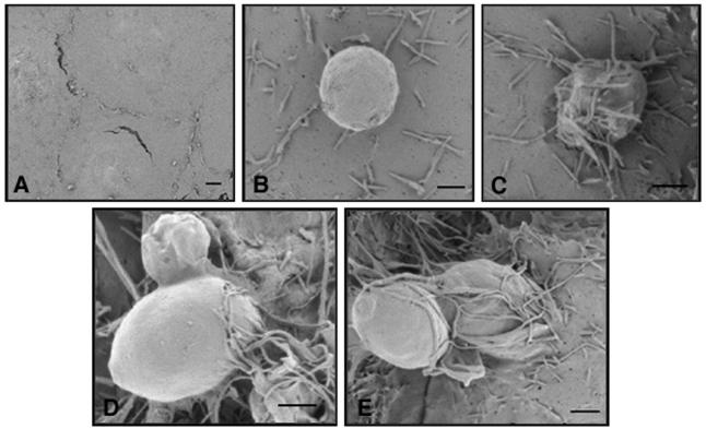
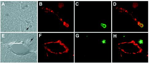
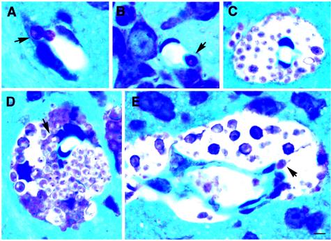
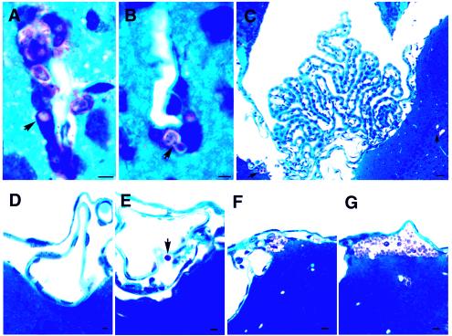
Cryptococcal meningoencephalitis develops as a result of hematogenous dissemination of inhaled Cryptococcus neoformans from the lung to the brain. The mechanism(s) by which C. neoformans crosses the blood-brain barrier (BBB) is a key unresolved issue in cryptococcosis. We used both an in vivo mouse model and an in vitro model of the human BBB to investigate the cryptococcal association with and traversal of the BBB. Exposure of human brain microvascular endothelial cells (HBMEC) to C. neoformans triggered the formation of microvillus-like membrane protrusions within 15 to 30 min. Yeast cells of C. neoformans adhered to and were internalized by the HBMEC, and they crossed the HBMEC monolayers via a transcellular pathway without affecting the monolayer integrity. The histopathology of mouse brains obtained after intravenous injection of C. neoformans showed that the yeast cells either were associated with endothelial cells or escaped from the brain capillary vessels into the neuropil by 3 h. C. neoformans was found in the brain parenchyma away from the vessels by 22 h. Association of C. neoformans with the choroid plexus, however, was not detected during up to 10 days of observation. Our findings indicate that C. neoformans cells invade the central nervous system by transcellular crossing of the endothelium of the BBB.


Figures

FIG. 1.
FIG. 1.
Association with and traversal of HBMEC by C. neoformans. (A) Association of strains B-3501 (encapsulated), B-4476FO5 (encapsulated), and TYCC-33 (Δcap59, acapsular) assessed at different times. The error bars indicate standard deviations. (B) Transcytosis of C. neoformans across HBMEC monolayers. HBMEC monolayers were exposed to B-3501, B-4476FO5, or TYCC33 (1 × 10⁵ cells/well), and the numbers of CFU transcytosed across the HBMEC at different times were determined. The values are the averages for two different samples of each strain at each time. The experiment was repeated, and similar results were obtained. (C) Effect of C. neoformans cells on the barrier function of HBMEC as measured by an ECIS apparatus after addition of B-3501 (4 × 10⁶ or 4 × 10⁵ cells/well) to an HBMEC monolayer.
FIG. 2.
FIG. 2.
SEM results. (A) Surface of the HBMEC at time zero (control). (B) Extensive microvillus-like projections observed 15 min after addition of B-3501 cells to an HBMEC monolayer. (C) Microvillus-like projections covering yeast cells at 30 min. (D and E) Penetration of yeast cells into HBMEC. Scale bars = 4 μm.
FIG. 3.
FIG. 3.
TEM results. (A) Formation of microvillus-like projection by HBMEC upon exposure to B-3501 cells. (B) TYCC33 yeast cell partially engulfed by HBMEC at 2 h. (C) Partially internalized budded cells of B-3501 at 6 h. (D and E) Cell of B-3501 completely internalized in the cytoplasm proximal to nuclei of HBMEC. The arrow in panel D indicates a vacuole. (F) Cell of B-3501 attached to the basolateral side of HBMEC after the HBMEC layer was crossed. Scale bars = 4 μm.
FIG. 4.
FIG. 4.
CFU recovered from the brain, lungs, and blood of mice (n = 3 for each time) injected with encapsulated cells (B-4476FO5) and isogenic acapsular cells (TYCC33). The CFU values are expressed as percentages of the counts at time zero. At time zero, the CFU from the brain and the lungs represent the CFU in the bloodstream circulating in the two organs. The error bars indicate standard deviations.
FIG. 5.
FIG. 5.
Immunostaining of cryosections prepared from mouse brain injected with B-3501. The sections were stained with a monoclonal anticapsular antibody (green) and anti-human factor VIII (red). (A) Cryosection prepared at 3 h postinjection (differential interference contrast microscopy). The arrow indicates a yeast cell. (B) Image matching the image in panel A, showing factor VIII-stained endothelium. (C) Field shown in panel B, showing a yeast cell stained with anticapsular antibody. (D) Superimposition of panels B and C, showing the yeast cell directly associated with endothelium. (E) Cryosection prepared at 22 h postinjection (differential interference contrast microscopy). The arrow indicates a yeast cell. (F) Fluorescent micrograph of panel E, showing a cross section of a capillary in the brain parenchyma. (G) Field shown in panel E, showing a yeast cell with a diffuse capsule. (H) Superimposition of panels G and H, showing a yeast cell in brain parenchyma outside the vessel.
FIG. 6.
FIG. 6.
Histological sections of mouse brain stained with toluidine blue. (A) Yeast cell (arrow) localized at the interface of a vessel and the neuropil at 3 h postinjection. The image suggests that the yeast cell is moving out of the vessel into the brain. (B) C. neoformans cell (arrow) closely associated with an endothelial cell (3 h postinjection) of a vessel in the brain parenchyma. (C) Perivascular cystic lesion formed by replication of C. neoformans (10 days). (D) Perivascular growth of C. neoformans (10 days). Some yeast cells are in the process of transmigrating from endothelial cells to the brain (the arrow indicates a yeast cell which is closely associated with an endothelial cell). (E) Cross section of a microvessel, showing the perivascular concentration of yeast cells and a yeast cell apparently leaving a vessel lumen (arrow). Scale bar = 4 μm.
FIG. 7.
FIG. 7.
Histological sections of mouse brain stained with toluidine blue. (A and B) C. neoformans cells lodged in endothelial cells of brain microvessels (10 days). (C) Choroid plexus remained normal and free of C. neoformans cells at 10 days postinjection, while cryptococcal lesions were seen in the nearby brain parenchyma (arrows). (D) Meninges free of C. neoformans 22 h postinjection. (E to G) C. neoformans was present in the subarachnoid space (arrow) or just under the pia matter of meninges by 10 days postinjection. Scale bars = 4 μm.

References

    1. Badger, J. L., M. F. Stins, and K. S. Kim. 1999. Citrobacter freundii invades and replicates in human brain microvascular endothelial cells. Infect. Immun. 67:4208-4215. - PMC - PubMed
    1. Bogaerts, J., D. Rouvroy, H. Taelman, A. Kagame, M. A. Aziz, D. Swinne, and J. Verhaegen. 1999. AIDS-associated cryptococcal meningitis in Rwanda (1983-1992): epidemiologic and diagnostic features. J. Infect. 39:32-37. - PubMed
    1. Buchanan, K. L., and J. W. Murphy. 1998. What makes Cryptococcus neoformans a pathogen. Emerg. Infect. Dis. 4:71-83. - PMC - PubMed
    1. Chang, Y. C., R. Cherniak, T. R. Kozel, D. L. Granger, L. C. Morris, L. C. Weinhold, and K. J. Kwon-Chung. 1997. Structure and biological activities of acapsular Cryptococcus neoformans 602 complemented with the CAP64 gene. Infect. Immun. 65:1584-1592. - PMC - PubMed
    1. Chang, Y. C., and K. J. Kwon-Chung. 1994. Complementation of a capsule-deficient mutation of Cryptococcus neoformans restores its virulence. Mol. Cell. Biol. 14:4912-4919. - PMC - PubMed

Publication types

MeSH terms