. 2004 Sep;72(9):5159-67.
doi: 10.1128/IAI.72.9.5159-5167.2004.

Proteus mirabilis ZapA metalloprotease degrades a broad spectrum of substrates, including antimicrobial peptides

Affiliations

Proteus mirabilis ZapA metalloprotease degrades a broad spectrum of substrates, including antimicrobial peptides

Robert Belas et al. Infect Immun. 2004 Sep.

Abstract

The 54-kDa extracellular metalloprotease ZapA is an important virulence factor of uropathogenic Proteus mirabilis. While ZapA has the ability to degrade host immunoglobulins (Igs), the dramatic attenuation of virulence in ZapA mutants suggests that this enzyme may have a broader spectrum of activity. This hypothesis was tested by in vitro assays with purified ZapA and an array of purified protein or peptide substrates. The data reveal that many proteins found in the urinary tract are substrates of ZapA proteolysis, including complement (C1q and C3), cell matrix (collagen, fibronectin, and laminin), and cytoskeletal proteins (actin and tubulin). Proteolysis of IgA and IgG was significantly enhanced by conditions that denatured the Igs. It was discovered that the antimicrobial peptides human beta-defensin 1 (hBD1) and LL-37 are readily cleaved by the enzyme. To the best of our knowledge, this is the first report of a bacterial protease capable of cleaving hBD1, a component of the human renal tubule innate immune response. Proteolysis of hBD1 resulted in ca. six peptides, while proteolysis of LL-37 resulted in at least nine products. Matrix-assisted laser desorption ionization-time of flight mass spectrometry analysis of the molecular masses of the reaction products indicated that ZapA preferred no distinct peptide bond. The antimicrobial activity of hBD1 and LL-37 was significantly reduced following ZapA treatment, suggesting that proteolysis results in inactivation of these peptides. The data suggest that a function of ZapA during urinary tract infections is the proteolysis of antimicrobial peptides associated with the innate immune response.


Figures
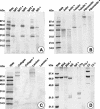
FIG. 1.
FIG. 1.
Qualitative measurement of the degradation of proteins associated with urinary tract epithelial cells by ZapA. A qualitative assessment of protein degradation by ZapA was made by an SDS-PAGE assay in which protein substrates by themselves and in the presence of ZapA (plus signs) were compared. The loss of a given protein band when reacted with the protease indicates that the protein is a substrate of ZapA. (A) IgA1, IgA2, and IgG. (B) Actin, tubulin, and fibronectin. (C) Collagen and laminin. (D) BSA (a negative control), IgA, C1q, and C3. All reaction mixtures were incubated at 37°C for 8 h in a reaction volume of 100 μl containing 20 nM ZapA and 100 to 125 μM respective protein substrate. Stds, standards.
FIG. 2.
FIG. 2.
Quantitative measurement of ZapA proteolysis of IgA and IgG by HPLC analysis. In this method, the decrease in the substrate protein peak (rightmost peak in each chromatogram; retention time of ca. 12 min) area and increases in the number and peak areas of product peaks (generally in the range of 2- to 10-min retention time) were used to assess proteolysis as a function of time of incubation and preincubation denaturation of the substrate. (A) Digestion of native IgA over time. The cascade of chromatograms from front to back is IgA without ZapA and in the presence of ZapA at 0, 0.5, 1, 2, 4, 6, and 16 h. (B) Digestion of IgA preincubated in 8 M urea. From front to back, the chromatograms are IgA without ZapA and in the presence of ZapA at 0, 0.5, 1, 2, 4, 6, and 16 h. (C) Digestion of heat-denatured IgA (preincubated at 85°C for 15 min). From front to back, the chromatograms are IgA without ZapA and in the presence of ZapA at 0, 0.5, 1, 2, 4, 6, and 16 h. (D) Digestion of IgG. From front to back, the chromatograms are IgG without ZapA (0 h), IgG plus ZapA (0 h) at 16 h and 37°C, heat-denatured IgG without ZapA, heat-denatured IgG with ZapA, urea-denatured IgG without ZapA, and urea-denatured IgG with ZapA. Reaction mixtures were incubated at 37°C for the specified times in a reaction volume of 100 μl containing 20 nM ZapA and 100 to 125 μM respective protein substrate.
FIG. 3.
FIG. 3.
HPLC chromatograms of the digestion of hBD1 by ZapA. Shown is cleavage of hBD1, part of the kidney innate defense mechanism, by the ZapA protease over time. Chromatograms are, in order from front to back, hBD1 at 0 h; hBD1 at 16 h of incubation; and hBD1 plus ZapA at 0, 1, 2, 4, 6, and 16 h of incubation. The reaction mixtures were incubated at 37°C for the specified times in a reaction volume of 100 μl containing 20 nM ZapA and 125 μM (50 μg) hBD1.
FIG. 4.
FIG. 4.
ZapA proteolysis of antimicrobial peptides. Shown are HPLC chromatograms of the digestion of hBD1 (A), hBD2 (B), human LL-37 (C), and porcine protegrin (D). In each panel the foremost chromatogram represents a reaction of the antimicrobial peptide without ZapA, while the rearmost chromatogram is the same substrate in the presence of the protease. In each, 20 nM ZapA and 125 μM respective antimicrobial peptide were incubated for 8 h at 37°C.
FIG. 5.
FIG. 5.
MALDI-TOF analysis of the cleavage of hBD1 by ZapA. (A) hBD1 alone. (B) hBD1 peptide fragments after incubation with ZapA under standard conditions (Materials and Methods). Arrows under the sequence of hBD1, potential sites of cleavage and peptide fragments deduced from the MALDI-TOF mass data.
FIG. 6.
FIG. 6.
MALDI-TOF analysis of the cleavage of LL-37 by ZapA. (A) LL-37 alone. (B) LL-37 peptide fragments after incubation with ZapA. Arrows under the sequence of LL-37, potential sites of cleavage and peptide fragments deduced from the MALDI-TOF mass data.
FIG. 7.
FIG. 7.
ZapA proteolysis of hBD1 and LL-37 reduces the antimicrobial activity of hBD1 and LL-37. The antimicrobial activity of each peptide was determined with E. coli D31, as described in Materials and Methods. (A) Antimicrobial activity, as LD50, of hBD1 prior to digestion (squares) and after digestion with ZapA (circles). (B) Antimicrobial activity, as LD50, of LL-37 prior to digestion (squares) and after digestion with ZapA (circles). The reaction mixtures were incubated at 37°C for 8 h.

Similar articles

Cited by

References

    1. Aneas, M. A., F. C. Portaro, I. Lebrun, L. Juliano, M. S. Palma, and B. L. Fernandes. 2001. ZapA, a possible virulence factor from Proteus mirabilis exhibits broad protease substrate specificity. Braz. J. Med. Biol. Res. 34:1397-1403. - PubMed
    1. Bals, R., X. Wang, Z. Wu, T. Freeman, V. Bafna, M. Zasloff, and J. M. Wilson. 1998. Human β-defensin 2 is a salt-sensitive peptide antibiotic expressed in human lung. J. Clin. Investig. 102:874-880. - PMC - PubMed
    1. Belas, R., D. Erskine, and D. Flaherty. 1991. Proteus mirabilis mutants defective in swarmer cell differentiation and multicellular behavior. J. Bacteriol. 173:6279-6288. - PMC - PubMed
    1. Belas, R., D. Erskine, and D. Flaherty. 1991. Transposon mutagenesis in Proteus mirabilis. J. Bacteriol. 173:6289-6293. - PMC - PubMed
    1. Bensch, K. W., M. Raida, H. J. Magert, P. Schulz-Knappe, and W. G. Forssmann. 1995. hBD-1: a novel beta-defensin from human plasma. FEBS Lett. 368:331-335. - PubMed

Publication types

MeSH terms

LinkOut - more resources