. 2004 Sep;72(9):5181-92.
doi: 10.1128/IAI.72.9.5181-5192.2004.

Helicobacter pylori infection targets adherens junction regulatory proteins and results in increased rates of migration in human gastric epithelial cells

Affiliations

Helicobacter pylori infection targets adherens junction regulatory proteins and results in increased rates of migration in human gastric epithelial cells

Victoria S Conlin et al. Infect Immun. 2004 Sep.

Abstract

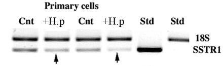
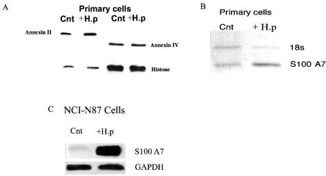
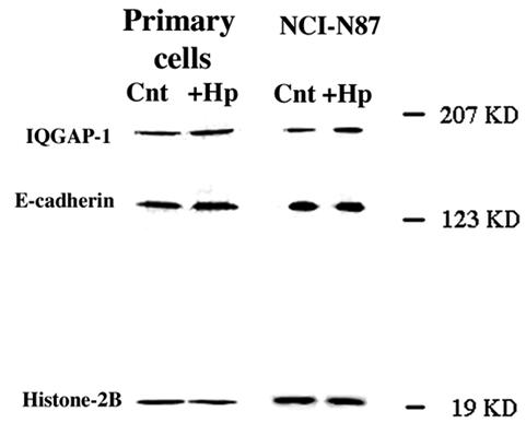
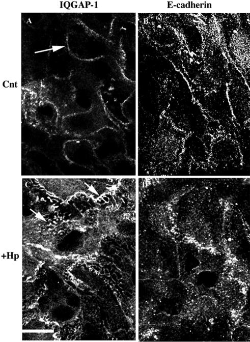
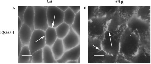
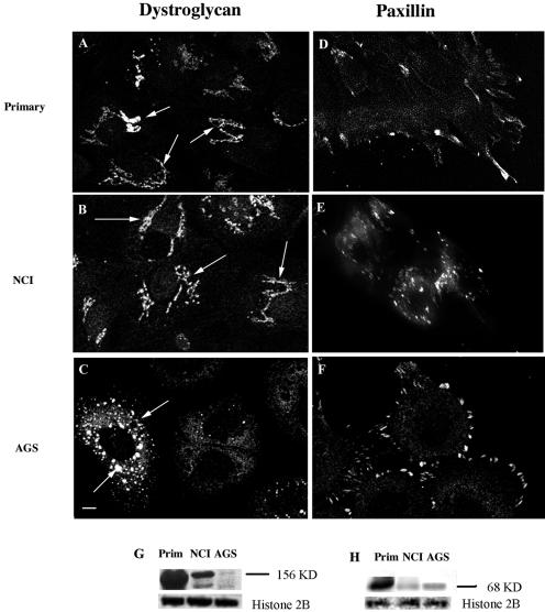
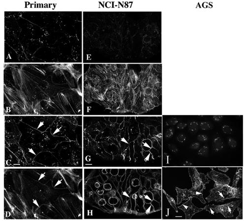
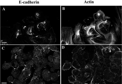
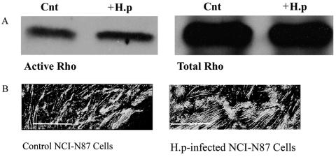
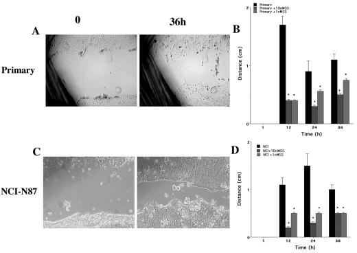
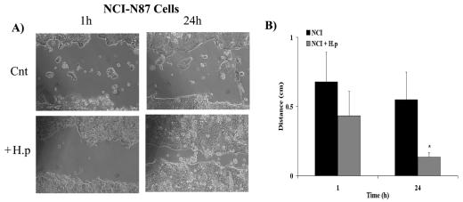
The human gastric pathogen Helicobacter pylori attaches to antral epithelial cells in vivo. Cultured human antral epithelial cells, AGS and NCI-N87 cell lines, were grown in the absence or presence of H. pylori and compared with respect to gene transcript levels, protein expression, organization of the actin cytoskeleton, and the regulation of cell migration. The Clontech Neurobiology array detected differentially expressed transcripts, while Western blots were used to investigate related changes in protein levels. Infection with H. pylori consistently upregulated annexin II, S100 A7, Rho-GTP, and IQGAP-1, whereas SSTR-1 was downregulated upon H. pylori infection. In the adherens junction, E-cadherin and IQGAP-1 were translocated from the plasma membrane to intracellular vesicles. The primary and NCI-N87 cells were similar with respect to cell-cell and cell-matrix adhesion and cell migratory behavior; in contrast the AGS cells were significantly different from the primary gastric epithelial cell preparations, and thus caution must be used when using this cell line for studies of gastric disease. These studies demonstrate a correlation between H. pylori infection and alterations to epithelial cell adhesion molecules, including increased levels of Rho-GTP and cell migration. These data indicate that destabilizing epithelial cell adherence is one of the factors increasing the risk of H. pylori-infected individuals developing gastric cancer.


Figures

FIG. 1.
FIG. 1.
Semiquantitative RT-PCR, using ribosomal 18S as the internal standard, confirmed that SSTR-1 transcripts were decreased in H. pylori-infected (+H.p) cells (arrow). The PCRs were carried out with 2 μl of cDNA in a 25-μl total volume of PCR buffer. SSTR-1 primers had the following sequences: forward, 5′GGAGGAGCCGGTTGACTATT3′; reverse, 5′AAGGTAGCCTGAAAGCCTTCC3′. The PCR products were electrophoresed in a 1.2% agarose gel. The DNA was visualized and photographed with the Eagle Eye II video system (Stratagene). A ratio of the band intensities between SSTR-1 and the internal standard for the uninfected sample was compared with that for the infected samples. Cnt, control uninfected cells. Results are representative of the results of nine different reactions.
FIG. 2.
FIG. 2.
H. pylori infection of primary-cell preparations and NCI-N87 cells induces increased levels of annexin II and S100 A7 protein expression. Ten microliters of H. pylori with an OD600 of 0.5 was used to inoculate 1 ml of growth medium. The infected epithelial cells were maintained in 10% CO2 at 37°C for 48 h prior to collection of protein. (A) Whole-cell lysates were prepared from control and infected-primary-cell preparations, and protein was analyzed by SDS-PAGE using 7.5% polyacrylamide gels. Annexin II protein expression was analyzed by blotting with a monoclonal anti-annexin II antibody. Annexin IV protein was analyzed by blotting with a monoclonal anti-annexin IV antibody. The density of the bands was related to that of the housekeeping protein histone 2B. Cnt, control uninfected cells; +H.p, H. pylori-infected cells. (B) Semiquantitative RT-PCR, using ribosomal 18S as the internal standard, confirmed that S100 A7 transcripts were upregulated in H. pylori-infected primary cells. The PCRs were carried out with 2 μl of cDNA in a 25-μl total volume of PCR buffer. S100 A7 primers had the following sequences: forward, 5′GAAAGCAAAGATGAGCAACACTC3′; reverse, 5′TTGGTGGGGCTGGGTCACTG3′. The PCR products were electrophoresed in a 1.2% agarose gel. The DNA was visualized and photographed with the Eagle Eye II video system (Stratagene). The ratio of the band intensity for SSTR-1 to that for the internal standard for the uninfected sample was compared with the corresponding ratio for the infected samples. (C) Total RNA (25 ng) was extracted from NCI-N87 cells. The RNA was then electrophoresed in a denatured 1% formaldehyde agarose gel and transferred to a nylon membrane. The membrane was then hybridized with a 32P-labeled probe against S100 A7 and GAPDH. After 2 h the filter was washed and autoradiographed with Kodak XAR5 film for 16 h at −70°C. GAPDH was used as the internal standard and confirmed that S100 A7 levels were upregulated in H. pylori-infected NCI-N87 cells. Data are representative of six independent experiments.
FIG. 3.
FIG. 3.
H. pylori infection of primary-cell preparations and NCI-N87 cells has no effect on IQGAP-1 or E-cadherin protein expression. Ten microliters of H. pylori with an OD600 of 0.5 was used to inoculate 1 ml of growth medium. The infected epithelial cells were maintained in 10% CO2 at 37°C for 48 h prior to collection of protein. Whole-cell lysates were prepared from control and infected-cell preparations, and protein was analyzed by SDS-PAGE using 7.5% polyacrylamide gels and then transferred to nitrocellulose membranes. Membranes were incubated with monoclonal anti-IQGAP-1 and monoclonal anti-E-cadherin antibodies. The density of the bands was related to that of the housekeeping protein histone 2B. Data are representative of three to six independent experiments.
FIG. 4.
FIG. 4.
Primary-cell preparations immunostained for IQGAP-1 and E-cadherin. Primary-cell preparations were plated on APES-coated glass coverslips and incubated for 48 h to facilitate attachment prior to infection with H. pylori. Control and infected cells were fixed 48 h postinfection in 4% paraformaldehyde for 10 min at room temperature. (A) Control (Cnt) cells were immunostained with monoclonal anti-IQGAP-1 antibodies for 18 h at 4°C. Note the presence of IR at adherens junctions (arrow). (B) Control cells were immunostained with monoclonal anti-E-cadherin antibodies for 18 h at 4°C. (C) Cells were infected with H. pylori and immunostained for IQGAP-1. Note the dramatic increase in internalized protein in tubulovesicles (arrows). (D) Cells were infected with H. pylori and immunostained for E-cadherin. Note the presence of internalized protein in small vesicles. Scale bar = 5 μm. Images are representative of three independent experiments.
FIG. 5.
FIG. 5.
H. pylori infection of NCI-N87 cells causes internalization of IQGAP-1. NCI-N87 cells were plated on APES-coated glass coverslips and incubated for 48 h to facilitate attachment prior to infection with H. pylori. Control (Cnt) and infected (+H.p) cells were fixed 48 h postinfection in 4% paraformaldehyde for 10 min at room temperature. (A) Control cells were immunostained with monoclonal anti-IQGAP-1 antibodies for 18 h at 4°C. Note the presence of IR at adherens junctions (arrows). (B) Cells were infected with H. pylori and immunostained for IQGAP-1. Note the dramatic increase in internalized protein in tubulovesicles (arrows). Scale bar = 5 μm. Images are representative of three independent experiments.
FIG. 6.
FIG. 6.
Primary-cell preparations, NCI-N87 cells, and AGS cells immunostained for α-dystroglycan and paxillin. (A to F) Cells were grown on APES-coated coverslips for 48 h prior to fixation with 4% paraformaldehyde and permeabilized with 0.1% Triton X-100. All cells were immunostained separately with monoclonal anti-α-dystroglycan and monoclonal antipaxillin antibodies for 18 h at 4°C. (G) α-Dystroglycan protein expression levels within primary-cell preparations, NCI-N87 cells, and AGS cells were analyzed by SDS-PAGE. (H) Paxillin protein expression levels within primary-cell preparations, NCI-N87 cells, and AGS cells were analyzed by SDS-PAGE. The housekeeping protein histone 2B was used to normalize for protein loading. The primary (A) and NCI-N87 (B) cells showed a strong immunostaining of the ER and Golgi complex (arrows), whereas the AGS cells (C) showed staining in intracellular vesicles (arrows). The levels of α-dystroglycan protein expression are highest in primary cells, followed (in order) by NCI-N87 and AGS cells (G). The primary cells (D) showed both cytosolic and focal contact staining, while in the NCI-N87 (E) and AGS (F) cells paxillin IR was predominantly at the focal contacts. The largest amount of paxillin protein was present in the primary cells, followed (in order) by AGS cells and NCI-N87 cells (H). Scale bar = 10 μm. Images are representative of three independent experiments.
FIG. 7.
FIG. 7.
Immunocytochemistry shows that the primary and NCI-N87 cells show similar patterns of E-cadherin and actin staining. All three cell preparations were grown on APES-coated coverslips for 48 h prior to fixation with 4% paraformaldehyde and permeabilization with 0.1% Triton X-100. All cells were immunostained with a monoclonal anti-E-cadherin antibody for 18 h at 4°C before being incubated with phalloidin-AlexaFluor 594 for 1 h at room temperature. At the basolateral membrane E-cadherin (A, primary; E, NCI-N87) is reduced and actin stress fibers predominate (B, primary; F, NCI-N87). From 1 μm from the substrate to the top of the cells both showed strong E-cadherin (C, primary; G, NCI-N87) and cortical actin (D, primary; H, NCI-N87) staining at the plasma membrane. AGS cells show a punctate pattern of E-cadherin staining that does not localize to adherens junctions (I). AGS cells have a uniform pattern of filamentous actin staining with a small number of stress fibers (arrows) and cortical actin (arrowheads) (J). Scale bar = 10 μm. Images are representative of three individual experiments.
FIG. 8.
FIG. 8.
Removal of extracellular calcium resulted in the internalization of E-cadherin in primary-cell preparations and NCI-N87 cells. Cells were seeded on APES-coated glass coverslips in 24-well plates and allowed to attach in medium containing 1 mM Ca2+ for 2 h. Cells were washed in 0 mM Ca2+ culture medium before fresh 0 mM Ca2+ culture medium was added. Medium was removed, and cells were immediately fixed in 4% paraformaldehyde at 30-min intervals over a 2-h period. Cells were then processed for immunofluorescence. In the primary cells the loss of peripheral E-cadherin (A) correlated with a loss of cortical actin but not stress fibers (B), while in NCI-N87 cells loss of peripheral E-cadherin (C) correlated with a reduction in the number of stress fibers in addition to a significant reduction in cortical actin (D). Scale bar = 10 μm. Images are representative of at least four independent experiments.
FIG. 9.
FIG. 9.
Infection of NCI-N87 cells with H. pylori increases Rho-GTP expression levels. Affinity precipitation of Rho-GTP used beads coated with the binding domain of the glutathione S-transferase fusion protein Rhotekin-RBD. (A) Serum-starved NCI-N87 cells were infected for 24 h with H. pylori (+H.p; 200 μl/ml corrected to OD600) and harvested alongside control cells. Cells were centrifuged to remove nuclei, and supernatant was added to beads for 1 h at 4°C. Beads were washed with lysis buffer, and the pellet was resuspended prior to electrophoresis and Western blotting. Membranes were probed with an anti-Rho antibody. Only active Rho is precipitated in these experiments due to the specificity of Rhotekin-RBD for Rho-GTP; therefore the observed increase in Rho protein observed via Western blotting in the presence of H. pylori is an increase in the active form of Rho (Rho-GTP). Cnt, control. (B) Infection of NCI-N87 cells with H. pylori alters basal stress fiber density and formation. Cells were grown on APES-coated coverslips for 48 h prior to experimentation. Control and infected cells were fixed 24 h postinfection in 4% paraformaldehyde prior to permeabilization with 0.1% Triton X-100. Cells were incubated with phalloidin-AlexaFluor 488 for 1 h at room temperature prior to being analyzed by deconvolution microscopy. Control cells show dense compact stress fibers on the basal surface, whereas H. pylori-infected cells show elongated cables of stress fibers. Scale bar = 5 μm. Images are representative of at least four independent experiments.
FIG. 10.
FIG. 10.
Gastric cell migration is significantly inhibited by the addition of CH275 SSTR-1 analog at 1 and 10 nM. NCI-N87 cells were grown to confluence in six-well plates and were serum starved for 24 h prior to the experiment. Primary cells were plated at high density in the absence of significant cell division. A 10-μl pipette tip was used to create a wound across the center of the well. After a washing to remove cell debris, cells were either untreated or treated with 1 or 10 nM CH275, an SSTR-1-specific analog. The rate of wound closure was measured at 12-h intervals over a 48-h period. The rate of migration is expressed as millimeters per 12-h period, and the effect of SSTR-1 analog treatment was assessed at each time point by the unpaired Student t test. Under control conditions in the presence of 5% serum the primary (A) and NCI-N87 (C) cells migrated as an integrated sheet. Cell migration was significantly inhibited by the addition of CH275 SSTR-1 analog at 1 and 10 nM. (B) Primary cells; (D) NCI-N87 cells.*, P = <0.05. The cell line data are from six individual experiments; the primary-cell preparation data are from three individual experiments.
FIG. 11.
FIG. 11.
Infection of the NCI-N87 cell line with H. pylori increases gastric epithelial cell migration. NCI-N87 cells were grown to confluence in six-well plates and were serum starved 24 h prior to the experiment. Cells were infected with H. pylori (200 μl/ml corrected to OD600) and incubated at 37°C for 5 h. A 10-μl pipette tip was used to create a wound across the center of the well. After a washing to remove cell debris, bacteria were reapplied (at the concentration above). The rate of wound closure was measured at 12-h intervals over a 48-h period. The rate of migration is expressed as millimeters per 12-h period. All migration experiments were performed in the presence of 5% serum. (A) Both control, uninfected cells (Cnt) and H. pylori-infected cells (+H.p) migrated as an integrated sheet. (B) Over a 24-h period cell migration was significantly faster in the presence of H. pylori. The data report the means ± standard errors for eight wound healing experiments (*, P < 0.05).

Similar articles

Cited by

References

    1. Amieva, M. R., R. Vogelmann, A. Covacci, L. S. Tompkins, W. J. Nelson, and S. Falkow. 2003. Disruption of the epithelial apical-junctional complex by Helicobacter pylori CagA. Science 300:1430-1434. - PMC - PubMed
    1. Athmann, C., N. Zeng, T. Kang, E. A. Marcus, D. R. Scott, M. Rektorschek, A. Buhmann, K. Melchers, and G. Sachs. 2000. Local pH elevation mediated by the intrabacterial urease of Helicobacter pylori cocultured with gastric cells. J. Clin. Investig. 106:339-347. - PMC - PubMed
    1. Babiychuk, E. B., K. Monastyrskaya, F. C. Burkhard, S. Wray, and A. Draeger. 2002. Modulating signaling events in smooth muscle: cleavage of annexin 2 abolishes its binding to lipid rafts. FASEB J. 16:1177-1184. - PubMed
    1. Ballestrem, C., B. Hinz, B. A. Imhof, and B. Wehrle-Haller. 2001. Marching at the front and dragging behind: differential αVβ3-integrin turnover regulates focal adhesion behavior. J. Cell Biol. 155:1319-1332. - PMC - PubMed
    1. Barber, D. L., A. M. J. Buchan, C.-Y. Lin, and J. Choi J. 2000. Somatostatin acting at SSTR1 subtype inhibits activation of Rho and actin stress fibre formation. Mol. Biol. Cell 11:100a. (Abstract.)

Publication types

MeSH terms

LinkOut - more resources