Comparative Study
. 2004 Aug 31;43(34):10918-29.
doi: 10.1021/bi049272v.

Efficient synthesis and photodynamic activity of porphyrin-saccharide conjugates: targeting and incapacitating cancer cells

Affiliations
Comparative Study

Efficient synthesis and photodynamic activity of porphyrin-saccharide conjugates: targeting and incapacitating cancer cells

Xin Chen et al. Biochemistry. .

Abstract

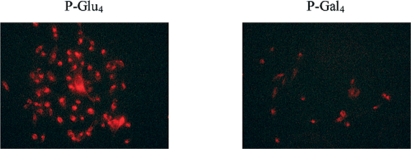
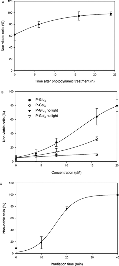
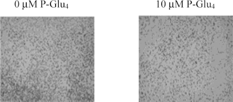
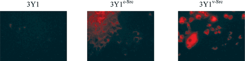
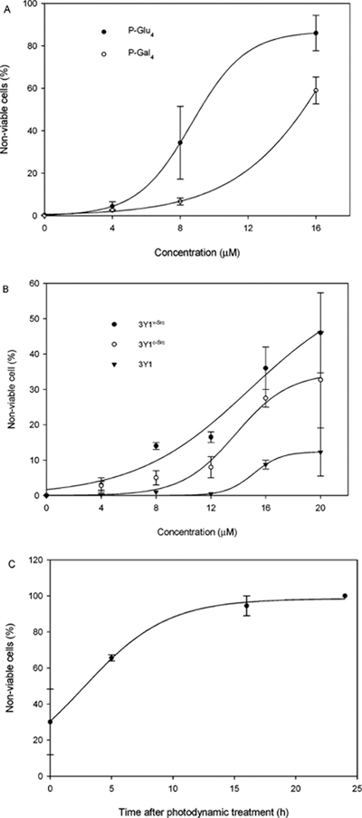
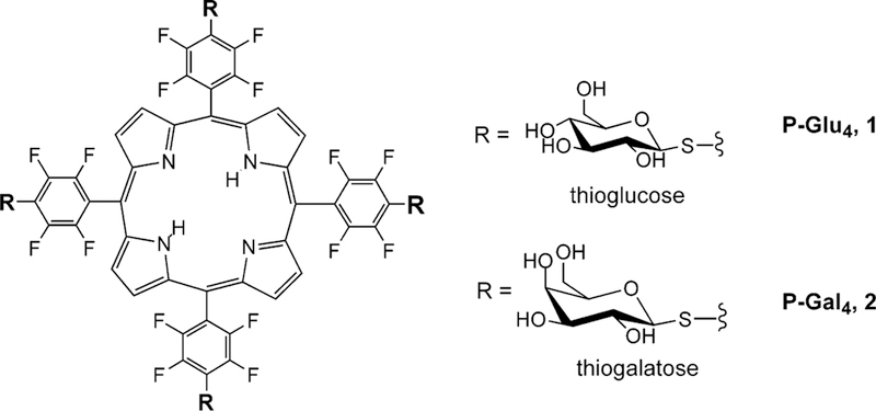
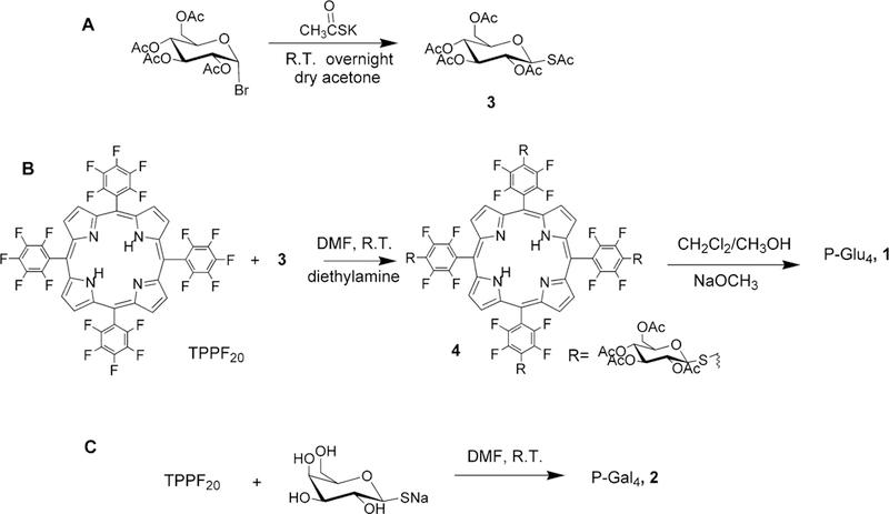
Since the role of saccharides in cell recognition, metabolism, and cell labeling is well-established, the conjugation of saccharides to drugs is an active area of research. Thus, one goal in the use of saccharide-drug conjugates is to impart a greater specificity toward a given cell type or other targets. Although widely used to treat some cancers and age related macular degeneration, the drugs used in photodynamic therapy (PDT) display poor chemical selectivity toward the intended targets, and uptake by cells most likely arises from passive, diffusional processes. Instead, the specific irradiation of the target tissues, and the formation of the toxic species in situ, are the primary factors that modulate the selectivity in the present mode of PDT. We report herein a two-step method to make nonhydrolyzable saccharide-porphyrin conjugates in high yields using a tetra(pentafluorophenyl)porphyrin and the thio derivative of the sugar. As a demonstration of their properties, the selective uptake (and/or binding) of these compounds to several cancer cell types was examined, followed by an investigation of their photodynamic properties. As expected, different malignant cell types take up one type of saccharide-porphyrin conjugate preferentially over others; for example, human breast cancer cells (MDA-MB-231) absorb a tetraglucose-porphyrin conjugate over the corresponding galactose derivative. Doseametric studies reveal that these saccharide-porphyrin conjugates exhibit varying PDT responses depending on drug concentration and irradiation energy. (1) Using 20 microM conjugate and greater irradiation energy induces cell death by necrosis. (2) When 10-20 microM conjugate and less irradiation energy are used, both necrosis and apoptosis are observed. (3) Using 10 microM and the least irradiation energy, a significant reduction in cell migration is observed, which indicates a reduction in aggressiveness of the cancer cells.


Figures

FIGURE 1:
FIGURE 1:
(A) UV-vis spectrum of ~2 µM P-Glu4 in methanol, where the inset is 5. (B) Fluorescence emission spectra of TPP, TPPF20, P-Glu4, and P-Gal4. The concentrations were 0.813, 0.718, 0.360, and 0.336 µM, respectively, in methanol, taken under identical instrumental conditions, and the spectra are normalized to the same molarity.
FIGURE 2:
FIGURE 2:
P-Glu4 is preferentially taken up by human breast cancer MDA-MB-231 cells over P-Gal4. Cells were treated with 10 µM glycosylated porphyrin for 24 h, rinsed, and fixed with a 4% paraformaldehyde solution. Fluorescence images were taken under identical conditions. R,G,B vector analysis of the unmodified images indicates the average relative fluorescence intensity, taken to be proportional to the extent of conjugate uptake by these cells, is 2.3:1 for P-Glu4/P-Gal4.
FIGURE 3:
FIGURE 3:
Photocytotoxic effects on human breast cancer MDA-MB-231 cells. Nonviable cells were counted with hemacytometer after staining with 0.4% w/v trypan blue. (A) MDA-MB-231 cells were treated with 20 µM P-Glu4 for 24 h, rinsed by exchanging the growth medium, and irradiated under a white 13 W fluorescent light (0.94 mW cm−2 for 20 min; 11.28 kJ m−2). The nonviable cells were counted at various lengths of time after photodynamic treatment. (B) MDA-MB-231 cells were treated with various concentrations of P-Glu4 (•) or P-Gal4 (ï) for 24 h, rinsed by exchanging the growth medium, and irradiated under a white 13 W fluorescent light (0.94 mW cm−2 for 20 min; 11.28 kJ m−2). Six hours after photodyamic treatment, nonviable cells were counted. Control experiments with no light show that MDA-MB-231 cells remain viable in the presence of the porphyrin-saccharide conjugates (▼, ▼). (C) MDA-MB-231 cells were treated with 20 µM P-Glu4 for 24 h, rinsed by exchanging the growth medium, and irradiated under a white 13 W fluorescent light (0.94 mW cm−2) for various lengths of time. Six hours after photodynamic treatment, nonviable cells were counted. Each data point represents an average ± SD from at least three independent measurements.
FIGURE 4:
FIGURE 4:
Detection of poly-ADP-ribose-polymerase (PARP) cleavage in human breast cancer MDA-MB-231 cells as an indication of apoptosis. MDA-MB-231 cells were treated with 20 µM P-Glu4 for 24 h and irradiated with a 13 W fluorescent light (0.27 mW cm−2 for 10 min; 1.62 kJ m−2), and 9 h after irradiation, cells were collected and lysed. The supernatant of the lysate was applied to Western blot to detect PARP cleavage. Lane 1: with no irradiation or P-Glu4; lane 2: with irradiation but no P-Glu4; lane 3: with P-Glu4 but no irradiation; and lane 4: with P-Glu4 and irradiation.
FIGURE 5:
FIGURE 5:
Low concentrations of P-Glu4 and low light irradiation inhibit cell migration. Reduced cell migration of human breast cancer MDA-MB-231 cells after nonlethal photodynamic treatment. Cells were treated with 0 or 10 µM P-Glu4 for 24 h and transferred to the Biocoat filters and irradiated under 13 W fluorescent light at 0.25 mW cm−2 for 5 min (0.75 kJ m−2). Cells were kept in dark, left to migrate for 18 h, and stained. The images were taken using a phase contrast light microscope.
FIGURE 6:
FIGURE 6:
P-Glu4 is preferentially taken up by transformed 3Y1v-Src cells as compared to partially transformed 3Y1c-Src cells and nontransformed 3Y1 cells. Cells were treated with 10 µM P-Glu4 for 24 h before fixing with a 4% paraformaldehyde solution. Fluorescence images were taken under identical conditions. The relative fluorescence intensity is 1:2.3:3.2.
FIGURE 7:
FIGURE 7:
(A) Photocytotoxic effects of various concentrations of P-Glu4 and P-Gal4 on 3Y1v-Src cells are compared using 5.76 kJ m−2 white light (0.96 mW cm−2 from a 13 W fluorescent bulb for 10 min), where nonviable cells were visualized with trypan blue staining 5 h after treatment. (B) The photocytotoxic effects of various concentrations of P-Glu4 on transformed 3Y1v-Src, partially transformed 3Y1c-Src, and normal 3Y1 cells using 3.53 kJ m−2 white light (0.84 mW cm−2 from a 13 W fluorescent bulb for 7 min), where necrotic cells were visualized with trypan blue staining immediately after treatment. (C) The photocytotoxic effects of 10 µM P-Glu4 on transformed 3Y1v-Src using 11.52 kJ m−2 white light (0.96 mW cm−2 from a 13 W fluorescent bulb for 20 min). Nonviable cells were counted at various lengths of time after photodynamic treatment. Each data point represents an average ± SD from at least three independent measurements.
FIGURE 8:
FIGURE 8:
Different degrees of PARP cleavage in fully transformed (3Y1v-Src), partially transformed (3Y1c-Src), and normal (3Y1) rat fibroblasts as indications of apoptosis. 3Y1, 3Y1c-Src, and 3Y1v-Src cells were treated with 8 µM P-Glu4 for 24 h. Cells were irradiated at 0.84 mW cm−2 for 3.5 min (1.76 kJ m−2), and 9 h later the cells were collected and lysed. The supernatant of the lysate was applied to Western blot to detect PARP cleavage. Lane 1: 3Y1 cells with no photodynamic treatment; lane 2: 3Y1c-Src cells with no photodynamic treatment; lane 3: 3Y1v-Src cells with no photodynamic treatment; lane 4: 3Y1 cells with irradiation but no P-Glu4; lane 5: 3Y1c-Src cells with irradiation but no P-Glu4; lane 6: 3Y1v-Src cells with irradiation but no P-Glu4; lane 7: 3Y1 cells with P-Glu4 but no irradiation; lane 8: 3Y1c-Src cells with P-Glu4 but no irradiation; lane 9: 3Y1v-Src cells with P-Glu4 but no irradiation; lane 10: 3Y1 cells with irradiation and P-Glu4; lane 11: 3Y1c-Src cells with irradiation and P-Glu4; and lane 12: 3Y1v-Src cells with irradiation and P-Glu4.
Scheme 1:
Scheme 1:
Structures of P-Glu4 and P-Gal4
Scheme 2:
Scheme 2:
Thioacetate Sugar Derivative (A) Is More Stable than the Free Thiol. (B) Synthesis of P-Glu4 and P-Gal4 Can Be Accomplished Using the Protected Sugar Followed by Deprotection or Directly by Using the Unprotected Sugar (C)

Similar articles

Cited by

References

    1. Drain CM, Christensen B, and Mauzerall DC (1989) Photogating of ionic currents across a lipid bilayer, Proc. Natl. Acad. Sci. U.S.A 86, 6959–6962. - PMC - PubMed
    1. Drain CM, and Mauzerall DC (1992) Photogating of ionic currents across lipid bilayers: Hydrophobic ion conductance by an ion chain mechanism, Biophys. J 63, 1556–1563. - PMC - PubMed
    1. MacDonald IJ, and Dougherty TJ (2001) Basic principles of photodynamic therapy, J. Porphyrins Phthalocyanines 5, 105–129.
    1. Neurath AR, Strick N, and Debnath AK (1995) Structural requirements for and consequences of an antiviral porphyrin binding to the V3 loop of the human immunodeficiency virus (HIV-1) envelope glycoprotein gp120, J. Mol. Recog 8, 345–357. - PubMed
    1. Vzorov AN, Dixon DW, Trommel JS, Marzilli LG, and Compans RW (2002) Inactivation of human immuno-deficiency virus type 1 by porphyrins, Antimicrob. Agents Chemother 46, 3917–3925. - PMC - PubMed

Publication types

MeSH terms